This website uses cookies to ensure you get the best experience on our website.


- Table of Contents
Plan CEBPE chromogenic IHC around nuclear staining in bone marrow hematopoietic cells (HPA tissue IHC). The IHC-validated antibody A03884-1 has a starting dilution range of 1:100–1:300 (datasheet); include a no-primary control to assess chromogenic background (standard IHC practice).

Expected localisation, validated protocols, controls and antibodies — the at-a-glance facts below, then the full design guide.
| Expected localisation | Nuclear in bone marrow hematopoietic cells (HPA tissue IHC) | |
| Staining pattern | Nuclear staining in bone marrow hematopoietic cells (HPA tissue IHC) | |
| Antigen retrieval | Tris-EDTA pH 9.0 HIER, 95–98 °C, 20 min (rule: nuclear antigen) | |
| Positive control | Bone marrow+1 more · see all | |
| Negative control | Adipose tissue+4 more · see all |
| Fixation | Keep paraffin-section fixation consistent across samples. (standard IHC practice; not target-specific) | |
| Caveat | Endogenous myeloid peroxidase may add background (HPA tissue IHC; standard IHC practice) | |
| Regulation | Strongest in promyelocyte-like cell lines (UniProt) | |
| Isoform / epitope | No isoforms or processing; one 1–281 chain (UniProt) |
The catalog antibody protocol is presented alongside published CEBPE IHC methods for human cartilage endplate and rat tissues (PMC12268938) and tumor and normal tissues (PMC11375656).
| Sample | Paraffin-embedded human brain tissue; fixative not specified (datasheet A03884-1) |
| Fixation | Image fixative and duration unreported (datasheet A03884-1); verify before use. |
| Sectioning | 4–5 µm sections on charged slides (standard) |
| Deparaffinisation | Xylene, graded ethanol series to water (standard) |
| Antigen retrieval | Heat-induced epitope retrieval in Tris-EDTA buffer, pH 9.0, 20 min at 95–98 °C (standard rule: nuclear antigen) |
| Peroxidase block | 3% H2O2, 10 min, room temperature (standard) |
| Blocking | 10% normal serum of the secondary host, 30 min, room temperature (standard) |
| Primary antibody | Rabbit anti-CEBPE, 1:100-1:300 (datasheet A03884-1) |
| Primary incubation | Overnight at 4 °C (standard) |
| Detection | HRP-polymer secondary, DAB chromogen 5–10 min (standard) |
| Counterstain | Hematoxylin, blue, dehydrate and mount (standard) |
| Expected result | CEBPE-positive staining in hematopoietic cells of bone marrow (HPA tissue IHC: High). HPA tissue profile: Nuclear expression in bone marrow poietic cells. No signal in the no-primary control. |
CEBPE should appear primarily in nuclei of bone marrow hematopoietic cells, where HPA reports high staining and an Enhanced tissue IHC reliability rating (HPA: High in bone marrow hematopoietic cells; Enhanced). Medium staining is reported in preleptotene spermatocytes (HPA: Medium in testis). A nuclear pattern fits this DNA-binding transcription factor, which has no transmembrane segment (UniProt Q15744: nucleus; no transmembrane segment).
| Clear nuclear chromogen in bone marrow hematopoietic cells, with surrounding tissue comparatively quiet. | This matches the principal tissue IHC pattern (HPA: nuclear expression in bone marrow poietic cells; High in hematopoietic cells). Interpret staining by cell and compartment, rather than treating every brown area as CEBPE (general IHC practice). |
| Medium nuclear staining in preleptotene spermatocytes. | This is another reported positive pattern (HPA: Medium in testis preleptotene spermatocytes). It need not match the intensity of the high bone marrow signal; compare like cells under the same staining conditions (general IHC practice). |
| Predominantly membranous or diffuse cytoplasmic chromogen in an IHC section. | This conflicts with the expected nuclear tissue pattern (HPA: nuclear expression in bone marrow poietic cells; UniProt Q15744: nucleus). Check morphology and controls before calling it specific; HPA's additional membrane and cytosol locations are uncertain ICC-IF observations (HPA: subcellular location). |
| Strong staining in cells reported as negative, such as adipocytes or bronchial respiratory epithelium. | These cells are listed as not detected (HPA: adipose adipocytes; bronchus respiratory epithelial cells). Consider cross-reactivity or endogenous detection activity, then compare a reagent control and an independently validated antibody (general IHC practice; HPA: two IHC Enhanced antibodies). |
| A widespread haze obscures nuclei, or known-positive bone marrow hematopoietic cells have no signal. | Neither gives an interpretable positive result (HPA: High in bone marrow hematopoietic cells; nuclear profile). Haze warrants background controls; absent signal warrants a check of retrieval, antibody and detection steps (general IHC practice). |
| Expected compartment | CEBPE is a nuclear DNA-binding transcription factor without a transmembrane segment (UniProt Q15744: nucleus; function; topology). Give nuclear staining priority when interpreting IHC; the uncertain additional ICC-IF locations do not establish a tissue IHC membrane pattern (HPA: subcellular location). |
| Choice of comparison tissue and cell | Bone marrow hematopoietic cells offer a reported high signal; preleptotene spermatocytes offer a reported medium signal (HPA: tissue IHC). Adipocytes and bronchial respiratory epithelial cells are reported as not detected and can help assess unexpected staining (HPA: tissue IHC). |
| Antibody evidence | HPA002928 and CAB005113 each have Enhanced IHC status (HPA: antibody validation). HPA describes its tissue IHC reliability as Enhanced because staining and RNA data show high consistency (HPA: tissue IHC). These ratings support the reported pattern; they do not specify a dilution or prove every stain in a new run is specific. |
| Antigen retrieval and fixation sensitivity | No CEBPE-specific fixation effect or retrieval condition is supplied (UniProt Q15744; HPA: tissue IHC). If signal needs optimization, compare retrieval conditions using reported positive cells and matched controls; treat this as general IHC practice, without inferring target-specific epitope masking. |
| Situation | Likely cause | Next action |
|---|---|---|
| Bone marrow section has little or no nuclear staining. | The expected high-positive cells are missing or a staining step underperformed (HPA: High in bone marrow hematopoietic cells; general IHC practice). | Confirm hematopoietic cells are present, then check antibody dilution, retrieval and detection with a positive control (general IHC practice). No CEBPE-specific retrieval setting is established here. |
| Chromogen spreads across the section and nuclear borders are hard to score. | Diffuse background may reflect nonspecific antibody binding or detection background (general IHC practice). | Review a reagent control, blocking, washes and detection exposure; score only resolved cellular staining (general IHC practice). Compare with the expected nuclear profile (HPA: tissue IHC). |
| Membrane or cytoplasm stains more strongly than nuclei. | The compartment differs from the tissue IHC expectation (HPA: nuclear bone marrow profile; UniProt Q15744: nucleus). | Check section morphology, reagent controls and staining with another IHC Enhanced antibody (general IHC practice; HPA: HPA002928 and CAB005113). Do not use uncertain ICC-IF membrane or cytosol calls to validate this IHC result (HPA: subcellular location). |
| Adipocytes or bronchial respiratory epithelial cells show prominent color. | Those cell types are reported as not detected (HPA: tissue IHC); cross-reactivity or endogenous detection activity is possible (general IHC practice). | Compare reagent controls, inspect the cellular compartment and repeat with an independently validated antibody if needed (general IHC practice; HPA: two IHC Enhanced antibodies). |
| Testis staining appears weaker than the bone marrow control. | This may reflect the reported difference between medium preleptotene spermatocyte staining and high bone marrow hematopoietic staining (HPA: tissue IHC). | Identify preleptotene spermatocytes and assess nuclear staining under matched conditions before treating lower intensity alone as assay failure (HPA: tissue IHC; general IHC practice). |
| Can an IF/ICC signal be read like the IHC result? | HPA supports nucleoplasmic ICC-IF localization, while its additional plasma membrane and cytosol assignments are uncertain (HPA: subcellular location). | Use nucleoplasmic signal as the main localization check and interpret extra-compartment IF signal cautiously (HPA: subcellular location). Follow the separate IF/ICC guide for its method; this section provides no IF protocol. |
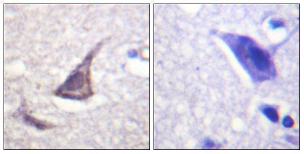
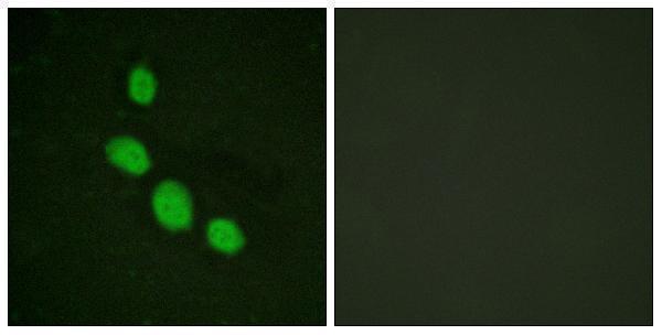
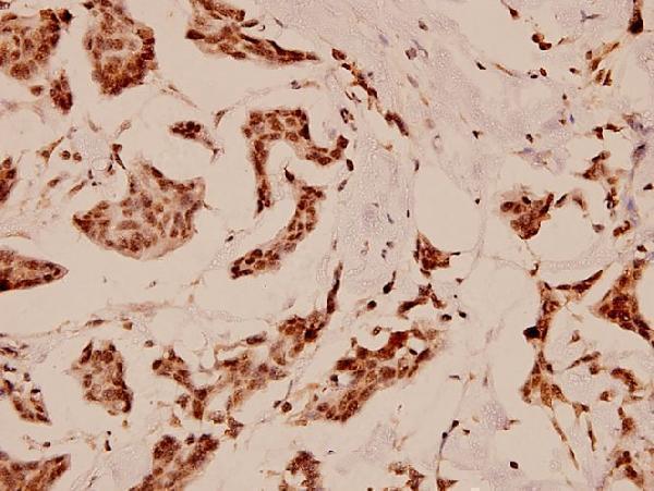

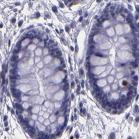


Comprehensive Human Protein Atlas IHC scoring per tissue (reliability: Enhanced — High consistency between antibody staining and RNA expression data.). Rows are taken directly from the HPA tissue chart — click any row's HPA link to view the source.
| Tissue | Cell type | Level | Evidence | Source |
|---|---|---|---|---|
| Bone marrow | Hematopoietic cells | High | Protein (IHC) | HPA → |
| Testis | Preleptotene spermatocytes | Medium | Protein (IHC) | HPA → |
| Tissue | Cell type | Level | Evidence | Source |
|---|---|---|---|---|
| Adipose tissue | Adipocytes | Not detected | Protein (IHC) | HPA → |
| Adrenal gland | Glandular cells | Not detected | Protein (IHC) | HPA → |
| Appendix | Glandular cells | Not detected | Protein (IHC) | HPA → |
| Breast | Adipocytes | Not detected | Protein (IHC) | HPA → |
| Bronchus | Respiratory epithelial cells | Not detected | Protein (IHC) | HPA → |
Troubleshoot CEBPE staining in paraffin sections by checking nuclear signal, hematopoietic cell identity, and assay controls (UniProt Q15744; HPA tissue IHC).
Two anti-CEBPE antibodies have paraffin-section IHC images (catalog IHC captions); one also has a HeLa-cell IF image (A03884-1 IF caption). Both list human, mouse and rat reactivity (catalog).
A03884-1 will render with human brain paraffin-section IHC, including a peptide-blocked comparison; its catalog also lists IF and shows HeLa-cell IF (A03884-1 image captions; catalog). A03884-2 will render with human breast carcinoma paraffin-section IHC at 1:100; its catalog lists IF but supplies no IF image (A03884-2 IHC caption; catalog).
Which to pick: For tissue IHC, choose A03884-1 when a peptide-blocked IHC comparison is useful, or A03884-2 when the breast carcinoma example is relevant; each caption establishes paraffin sections, and neither reports the fixative (IHC image captions). For IF/ICC planning, A03884-1 has a HeLa-cell IF image, while A03884-2 only lists IF; neither payload establishes an ICC protocol (IF image captions; catalog applications). Both list human, mouse and rat reactivity, so neither has a stated species advantage; A03884-1 is labeled polyclonal, while A03884-2 has no explicit clonality field (catalog).