This website uses cookies to ensure you get the best experience on our website.
- Table of Contents
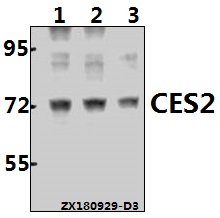
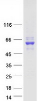
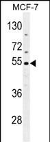
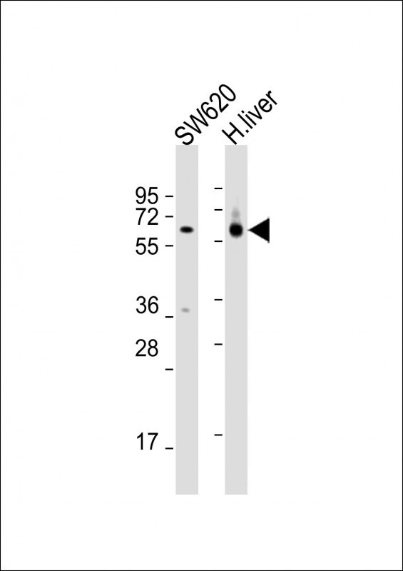
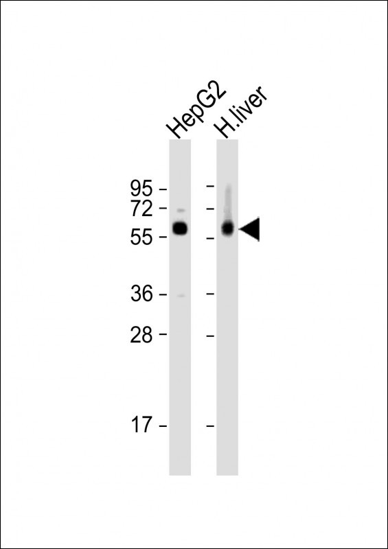
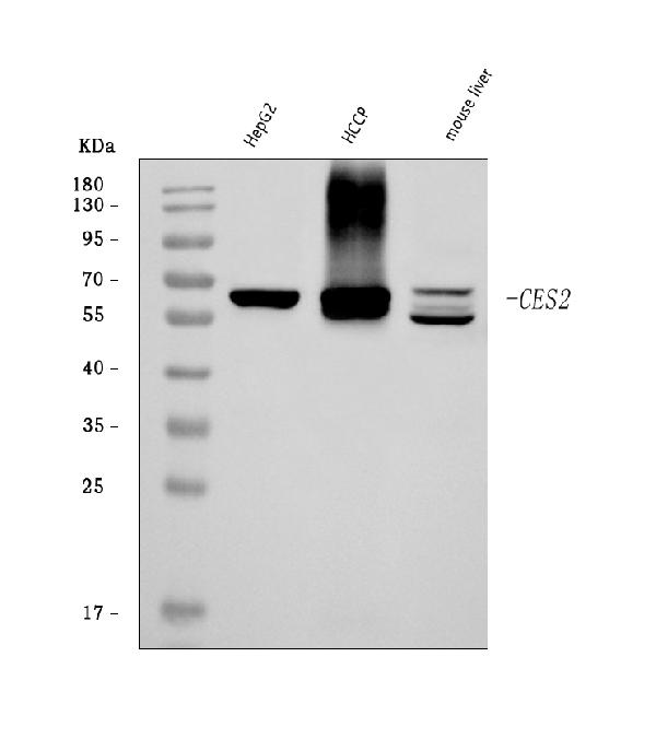
Facts about Cocaine esterase.

Hydrolyzes aspirin, substrates with big alcohol group and little acyl group and endogenous lipids like triacylglycerol (PubMed:28677105). .
| Human | |
|---|---|
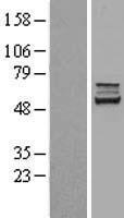
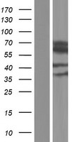
| Gene Name: | CES2 |
| Uniprot: | O00748 |
| Entrez: | 8824 |

| Belongs to: |
|---|
| type-B carboxylesterase/lipase family |

carboxylesterase 2 (intestine, liver); Carboxylesterase 2; carboxylesterase 2EC 3.1.1.1; CE-2; CE-2PCE-2; CES2; CES2A1; EC 3.1.1; EC 3.1.1.84; hCE-2; iCE; intestinal carboxylesterase; liver carboxylesterase-2; PCE-2
Mass (kDA):
61.807 kDA

| Human | |
|---|---|
| Location: | 16q22.1 |
| Sequence: | 16; NC_000016.10 (66934471..66945096) |
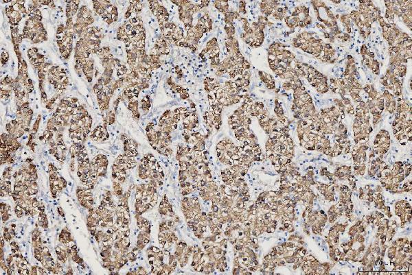
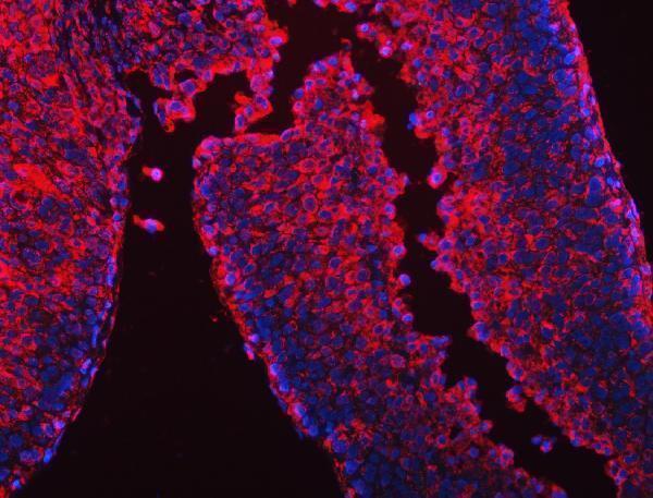
Preferentially expressed in intestine with moderate expression in liver. Within the intestine, highest expression is found in small intestine with lower expression in colon and rectum.
Endoplasmic reticulum lumen.


PMID: 9144407 by Schwer H., et al. Molecular cloning and characterization of a novel putative carboxylesterase, present in human intestine and liver.
PMID: 9169443 by Pindel E.V., et al. Purification and cloning of a broad substrate specificity human liver carboxylesterase that catalyzes the hydrolysis of cocaine and heroin.