This website uses cookies to ensure you get the best experience on our website.


- Table of Contents
Plan CFL2 paraffin-section IHC using muscle-rich positive tissue and an IHC-validated antibody (HPA tissue IHC; datasheet M04773). This guide covers expected cytoplasmic staining, controls and interpretation of isoform-related uncertainty (HPA tissue IHC; UniProt).

Expected localisation, validated protocols, controls and antibodies — the at-a-glance facts below, then the full design guide.
| Expected localisation | Cytoplasmic tissue staining; nuclear matrix also annotated (HPA tissue IHC; UniProt) | |
| Staining pattern | Muscle cells show abundant cytoplasmic staining (HPA tissue IHC) | |
| Antigen retrieval | Citrate pH 6 HIER, heat-mediated (datasheet M04773) | |
| Positive control | Cerebellum+4 more · see all | |
| Negative control | Bone marrow+4 more · see all |
| Fixation | Keep paraffin-section fixation consistent across samples. (standard IHC practice; not target-specific) | |
| Caveat | Staining and RNA have medium consistency (HPA tissue IHC) | |
| Regulation | CFL2b predominates in muscle and heart (UniProt) | |
| Isoform / epitope | Three isoforms; isoform-specific epitope coverage is unknown (UniProt) |
The catalog antibody’s IHC-P protocol is accompanied by published CFL2 IHC methods for colorectal, breast, and sarcoma tissues (PMC11443709; PMC3599769; PMC3440303).
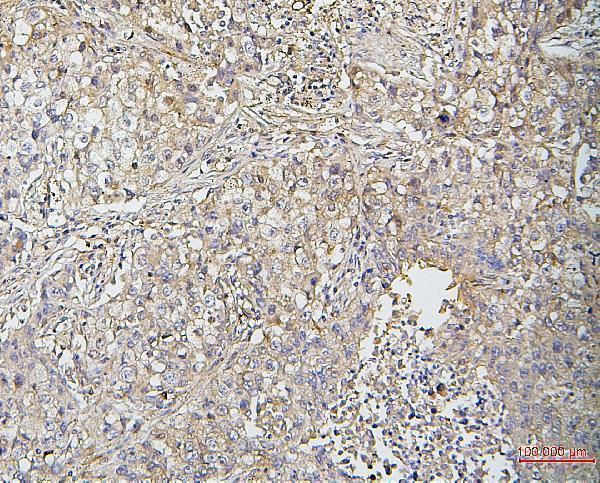
| Sample | Paraffin-embedded human lung cancer tissues; fixative not specified (datasheet M04773) |
| Fixation | Image fixative and duration unreported (datasheet M04773); verify before use. |
| Sectioning | 4–5 µm sections on charged slides (standard) |
| Deparaffinisation | Xylene, graded ethanol series to water (standard) |
| Antigen retrieval | Heat retrieval: Citrate pH 6, 20 min (datasheet M04773) |
| Peroxidase block | 3% H2O2, 10 min, room temperature (standard) |
| Blocking | 10% goat serum (datasheet M04773) |
| Primary antibody | Mouse monoclonal (clone 8C13) anti-CFL2, 0.5-1μg/ml (datasheet M04773) |
| Primary incubation | Overnight at 4 °C (datasheet M04773) |
| Detection | Streptavidin-biotin complex (SABC), DAB chromogen (datasheet M04773) |
| Counterstain | Hematoxylin, blue, dehydrate and mount (standard) |
| Expected result | CFL2-positive staining in granular cells - cytoplasm/membrane of cerebellum (HPA tissue IHC: High). HPA tissue profile: Cytoplasmic expression in several tissues, most abundant in muscle tissue. No signal in the no-primary control. |
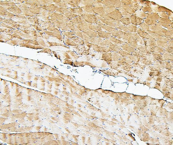
CFL2 should show predominantly cytoplasmic staining in tissue IHC, strongest in cardiac, skeletal, and smooth muscle cells; cerebellar granular cells may show cytoplasmic and membrane staining (HPA tissue IHC). UniProt also places CFL2 in the cytoskeleton and nuclear matrix, with Z-line colocalization in sarcomeres (UniProt Q9Y281). It has no transmembrane segment (UniProt Q9Y281). HPA rates its tissue IHC reliability Enhanced, while reporting medium agreement with RNA expression (HPA tissue IHC).
| Strong cytoplasmic staining in cardiomyocytes or skeletal myocytes, with stained smooth muscle cells. | This matches the High staining reported for all three cell types; muscle is the most abundant tissue expression in the HPA profile (HPA tissue IHC). Judge the stained cells and compartment together, since a dark section alone does not identify a correct pattern (general IHC practice). |
| Signal confined to an unexpected compartment, with little staining in the expected muscle-cell cytoplasm. | Treat an isolated extracellular or luminal deposit as a possible staining artefact and review the negative control (general IHC practice). Some membrane-associated signal in cerebellar granular cells is reported, however (HPA tissue IHC); compartment calls should therefore be tissue-specific. |
| Strong staining in lung alveolar cells or bone-marrow hematopoietic cells. | HPA reports CFL2 as Not detected in those cell populations (HPA tissue IHC). Review cell identification and controls; widespread signal there may reflect cross-reactivity or endogenous detection activity (general IHC practice). A discrepant stain alone does not establish which cause applies. |
| Diffuse colour across cells and surrounding tissue, obscuring cell boundaries. | This is background until a cell-specific pattern can be resolved (general IHC practice). Compare the primary-antibody-omission control and inspect blocking, washes, and detection chemistry (general IHC practice); HPA's tissue profile cannot assign a cause to background in an individual slide. |
| No detectable signal in heart or skeletal muscle despite an intact section. | These are known High-staining populations in HPA tissue IHC (HPA tissue IHC). Check whether the positive control and detection reagents worked, then review the antibody's IHC-P conditions (general IHC practice). Absence of colour alone does not show that CFL2 is biologically absent. |
| Tissue and cell identity | Cerebellar granular cells are High with cytoplasmic/membrane staining; kidney tubular cells and hepatocytes are Medium (HPA tissue IHC). Compare like cell types when judging intensity (general IHC practice). |
| Isoforms | CFL2b predominates in skeletal muscle and heart, whereas CFL2a occurs in various tissues (UniProt Q9Y281). These expression descriptions do not establish which isoform an unspecified antibody detects. |
| Protein location and processing | CFL2 has no transmembrane segment, signal peptide, or propeptide; the annotated chain spans residues 2–166 (UniProt Q9Y281). These annotations do not establish antibody epitope access in paraffin sections. |
| Tissue IHC confidence | HPA reports Enhanced tissue IHC reliability but medium consistency with RNA data (HPA tissue IHC). Antibody HPA045599 is IHC Enhanced (HPA antibodies); assess a new slide against controls as well as the reference pattern (general IHC practice). |
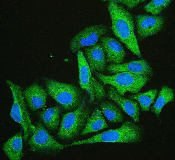
| Q: What should IF/ICC show? | A: HPA reports mainly plasma-membrane localization, with additional nucleoplasmic and cytosolic signal (HPA ICC-IF). Its ICC-approved antibodies are HPA077782 and CAB037078 (HPA antibodies). This observation comes from ICC-IF and does not replace the tissue IHC pattern. |
| Situation | Likely cause | Next action |
|---|---|---|
| Positive muscle control is blank. | A failed stain or detection step is possible (general IHC practice); HPA reports High signal in cardiac and skeletal myocytes (HPA tissue IHC). | Confirm the control contains the expected cells, then check the IHC-validated antibody, secondary detection, and chromogen against the established IHC-P workflow (general IHC practice). Do not infer CFL2 loss from this slide alone. |
| All tissue compartments appear brown. | Non-specific background or endogenous detection activity can obscure cell-specific staining (general IHC practice). | Compare a primary-antibody-omission control, review blocking and washes, and address endogenous activity appropriate to the detection chemistry (general IHC practice). Reassess only after muscle-cell cytoplasm can be distinguished. |
| A HPA Not detected cell population stains strongly. | Cross-reactivity, endogenous activity, or mistaken cell identification are possible explanations (general IHC practice); HPA lists lung alveolar cells and bone-marrow hematopoietic cells as Not detected (HPA tissue IHC). | Verify the cell type and compare negative and positive controls; investigate persistent discordance with an independently validated IHC antibody if available (general IHC practice). |
| Signal appears only in nuclei of muscle cells. | UniProt includes the nuclear matrix among CFL2 locations, but HPA describes tissue staining chiefly as cytoplasmic (UniProt Q9Y281; HPA tissue IHC). An exclusively nuclear muscle pattern is therefore discordant with the reported tissue profile. | Inspect matched controls and repeat localization assessment with identifiable myocytes (general IHC practice). Record the nuclear finding separately instead of scoring it as the expected cytoplasmic positive. |
| Kidney or liver stains less intensely than muscle. | HPA scores tubular cells and hepatocytes Medium, versus High in cardiac and skeletal myocytes (HPA tissue IHC). | Score the named cell populations against an on-run muscle positive control (general IHC practice). Lower intensity in these tissues alone is consistent with the HPA pattern. |
| ICC-IF membrane signal seems inconsistent with paraffin-section IHC. | The assays have different reported observations: HPA ICC-IF is mainly plasma-membrane localized, while HPA tissue IHC is chiefly cytoplasmic (HPA ICC-IF; HPA tissue IHC). | Interpret each result against its own assay and antibody validation (general IHC practice; HPA antibodies). Use the tissue IHC profile for the paraffin-section decision; consult the separate IF/ICC guide for that assay. |


Comprehensive Human Protein Atlas IHC scoring per tissue (reliability: Enhanced — Medium consistency between antibody staining and RNA expression data.). Rows are taken directly from the HPA tissue chart — click any row's HPA link to view the source.
| Tissue | Cell type | Level | Evidence | Source |
|---|---|---|---|---|
| Cerebellum | Granular cells - cytoplasm/membrane | High | Protein (IHC) | HPA → |
| Heart muscle | Cardiomyocytes | High | Protein (IHC) | HPA → |
| Skeletal muscle | Myocytes | High | Protein (IHC) | HPA → |
| Smooth muscle | Smooth muscle cells | High | Protein (IHC) | HPA → |
| Adrenal gland | Glandular cells | Medium | Protein (IHC) | HPA → |
| Tissue | Cell type | Level | Evidence | Source |
|---|---|---|---|---|
| Bone marrow | Hematopoietic cells | Not detected | Protein (IHC) | HPA → |
| Breast | Adipocytes | Not detected | Protein (IHC) | HPA → |
| Bronchus | Respiratory epithelial cells | Not detected | Protein (IHC) | HPA → |
| Esophagus | Squamous epithelial cells | Not detected | Protein (IHC) | HPA → |
| Hippocampus | Glial cells | Not detected | Protein (IHC) | HPA → |
Troubleshoot CFL2 staining in paraffin-section chromogenic IHC using the catalog antibody’s tissue example, with IF/ICC considerations kept separate.
Real CFL2 IHC images cover paraffin sections of human lung cancer and human, mouse, and rat skeletal muscle (M04773 IHC captions); an IF image covers U2OS cells (RP1107 IF caption).
M04773 is listed for IHC and IF (catalog applications), with paraffin-section images from human lung cancer and human, mouse, and rat skeletal muscle (M04773 IHC captions). RP1107 is listed for IF/ICC (catalog applications), with an IF image in U2OS cells (RP1107 IF caption).
Which to pick: Choose M04773 for paraffin-section tissue IHC: its monoclonal antibody is listed for IHC (M04773 catalog), and its IHC captions show 1 μg/ml staining after citrate retrieval (M04773 IHC captions); the fixative is unreported (M04773 IHC captions). Choose RP1107 for IF/ICC when its U2OS cell image fits the experiment (RP1107 catalog applications; RP1107 IF caption). For tissue IHC across human, mouse, and rat, M04773 has images in all three species (M04773 IHC captions); RP1107 lists all three as reactive, but its supplied IF image shows U2OS cells (RP1107 catalog reactivity; RP1107 IF caption).