This website uses cookies to ensure you get the best experience on our website.


- Table of Contents
Use proximal renal tubules as a high-staining reference for CHDH in paraffin-section IHC (HPA tissue IHC). Assess granular cytoplasmic staining (HPA tissue IHC), with mitochondrial membrane localisation as a molecular expectation (UniProt).

Expected localisation, validated protocols, controls and antibodies — the at-a-glance facts below, then the full design guide.
| Expected localisation | Granular cytoplasm (HPA tissue IHC) | |
| Staining pattern | Granular cytoplasmic staining, strongest in renal tubules (HPA tissue IHC) | |
| Antigen retrieval | Citrate pH 6.0 HIER, 95–98 °C, 20 min (rule: cytoplasmic / membrane antigen) | |
| Positive control | Kidney+4 more · see all | |
| Negative control | Adipose tissue+4 more · see all |
| Fixation | Keep fixation consistent across sections (standard IHC practice; not target-specific) | |
| Caveat | Colon stains less strongly than renal tubules (HPA tissue IHC) | |
| Regulation | No expression regulator annotated (UniProt) | |
| Isoform / epitope | No isoforms annotated; mature chain spans aa 30–594 (UniProt) |
Compare the catalog antibody’s IHC-P protocol with three published CHDH IHC methods (PMC12124313; PMC12421421; PMC9034393).
| Sample | Paraffin-embedded Mouse kidney tissue; fixative not specified (datasheet A10927) |
| Fixation | Image fixative and duration unreported (datasheet A10927); verify before use. |
| Sectioning | 4–5 µm sections on charged slides (standard) |
| Deparaffinisation | Xylene, graded ethanol series to water (standard) |
| Antigen retrieval | Heat-induced epitope retrieval in citrate buffer, pH 6.0, 20 min at 95–98 °C (standard rule: cytoplasmic / membrane antigen) |
| Peroxidase block | 3% H2O2, 10 min, room temperature (standard) |
| Blocking | 10% normal serum of the secondary host, 30 min, room temperature (standard) |
| Primary antibody | Rabbit anti-CHDH, 1:50-1:200 (datasheet A10927) |
| Primary incubation | Overnight at 4 °C (standard) |
| Detection | HRP-polymer secondary, DAB chromogen 5–10 min (standard) |
| Counterstain | Hematoxylin, blue, dehydrate and mount (standard) |
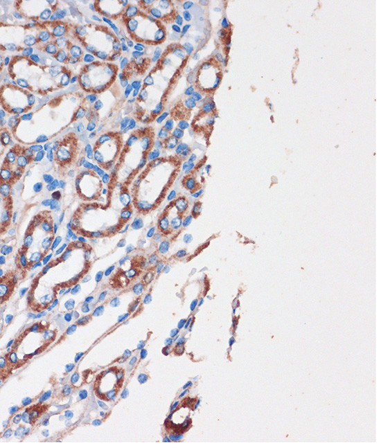
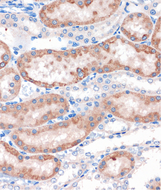
| Expected result | CHDH-positive staining in proximal tubules (cell body) of kidney (HPA tissue IHC: High). HPA tissue profile: Cytoplasmic expression with a granular pattern in several tissues, most abundant in renal tubules. No signal in the no-primary control. |
CHDH should appear as granular cytoplasmic staining, strongest in kidney proximal tubule cell bodies and also high in testis peritubular cells (HPA tissue IHC: Enhanced). The granular pattern fits its mitochondrial location; UniProt places CHDH at the inner membrane and, after mitochondrial damage, the outer membrane, with no annotated transmembrane segment (UniProt Q8NE62 topology). HPA’s Enhanced tissue reliability reflects consistency between staining and RNA expression (HPA tissue IHC).
| Granular cytoplasmic signal is strong in renal proximal tubule cell bodies. | This is the clearest tissue positive control: HPA reports High staining in these cells and describes the overall pattern as cytoplasmic and granular (HPA tissue IHC). Compare signal within the same section before treating every stained structure as CHDH-positive. |
| Testis peritubular cells stain strongly; glandular cells in appendix, breast, colon, duodenum, or gallbladder stain less strongly. | HPA reports High staining in testis peritubular cells and Medium staining in those glandular cells (HPA tissue IHC). An intensity difference between these listed cell populations can be expected; interpret each population against its own tissue context. |
| A paraffin section shows predominantly nuclear staining, with little granular cytoplasmic signal. | That conflicts with HPA’s tissue IHC profile and warrants an artefact check (HPA tissue IHC: cytoplasmic, granular). HPA separately reports approved nucleolar localization by ICC-IF, so isolated nucleolar fluorescence should be assessed in that application rather than assumed to explain a nuclear chromogenic pattern (HPA subcellular ICC-IF). |
| Strong staining appears in adipocytes, lung alveolar cells, or bone marrow hematopoietic cells. | HPA reports CHDH as Not detected in those cell populations (HPA tissue IHC). Check cell identification and controls; cross-reactivity or endogenous chromogenic activity are possible explanations. A reference result of Not detected is a comparison point, not proof that every specimen must be negative. |
| Color is diffuse across tissue structures, or kidney proximal tubules show no discernible signal. | Diffuse color does not match the HPA granular profile; absent proximal tubule staining misses an HPA High control (HPA tissue IHC). Review background controls, tissue preservation, retrieval and detection performance as general IHC checks before assigning a biological interpretation. |
| Cell population and tissue | HPA reports High proximal tubule and testis peritubular staining, Medium staining in several listed glandular populations, and Not detected staining in listed controls (HPA tissue IHC). Score the identified cells rather than assigning one intensity to an entire organ. |
| Subcellular interpretation | UniProt places CHDH at mitochondrial inner and outer membranes and notes outer membrane accumulation after mitochondrial damage (UniProt Q8NE62). HPA tissue IHC reports a granular cytoplasmic pattern; ordinary chromogenic granules do not resolve individual mitochondrial membranes (HPA tissue IHC; standard IHC interpretation). |
| Antibody evidence | HPA lists HPA036633 and HPA058130 as IHC Enhanced, while HPA036632 has ICC approval without an IHC status in the supplied record (HPA antibodies). Keep validation tied to the application and antibody used; one antibody’s ICC result does not validate another antibody’s paraffin IHC result. |
| Protein processing | UniProt annotates a mature CHDH chain spanning residues 30–594 and no annotated isoforms or glycosylation sites (UniProt Q8NE62). The supplied record gives no antibody epitope, so these annotations cannot predict which retrieval condition will expose it in a paraffin section. |
| IF/ICC localization | For the IF/ICC question, HPA approves mitochondria as the main location and nucleoli as an additional location, with images from CACO-2, Hep-G2 and OE19 (HPA subcellular ICC-IF). This supports an IF localization comparison, not a paraffin IHC protocol choice. |
| Situation | Likely cause | Next action |
|---|---|---|
| Known-positive kidney proximal tubules have no signal. | The result conflicts with HPA High staining in proximal tubule cell bodies; tissue processing, retrieval or detection failure is possible (HPA tissue IHC; general IHC practice). | Confirm the tubules are present, then review the antibody’s IHC-P instructions and run a suitable positive control through the same retrieval and detection workflow (general IHC practice). |
| The entire section develops diffuse chromogen. | Diffuse color obscures the HPA granular cytoplasmic pattern; excess background or endogenous enzyme activity may contribute (HPA tissue IHC; general chromogenic IHC practice). | Inspect a no-primary control, review blocking and washes, and check endogenous activity controls appropriate to the detection chemistry (general IHC practice). |
| Nuclei dominate the chromogenic result. | Predominant nuclear tissue IHC conflicts with HPA’s cytoplasmic granular profile, although HPA separately reports additional nucleolar localization in ICC-IF (HPA tissue IHC; HPA subcellular ICC-IF). | Recheck morphology and background controls, then compare an independently IHC-validated antibody if available; do not transfer the ICC-IF call directly to paraffin IHC (HPA antibodies; general IHC practice). |
| Cells listed as Not detected stain as strongly as proximal tubules. | Cell misidentification, cross-reactivity or endogenous chromogenic activity is possible; HPA lists adipocytes, alveolar cells and hematopoietic cells as Not detected (HPA tissue IHC). | Verify the stained cell type against the counterstain and inspect no-primary and detection controls before calling CHDH expression (general IHC practice). |
| Two antibodies give different tissue patterns. | Antibody performance can differ; HPA assigns IHC Enhanced to HPA036633 and HPA058130, while the supplied record gives HPA036632 no IHC status (HPA antibodies). | Check each antibody’s application validation and compare staining in the same HPA High and Not detected cell populations under matched IHC conditions (HPA antibodies; HPA tissue IHC). |
| IF/ICC shows puncta plus nucleolar signal. Is that compatible with CHDH? | Yes: HPA approves mitochondria as the main ICC-IF location and nucleoli as an additional location (HPA subcellular ICC-IF). | Interpret both compartments against IF controls and keep the conclusion specific to ICC-IF; use the HPA granular cytoplasmic profile when judging tissue IHC (HPA subcellular ICC-IF; HPA tissue IHC). |


Comprehensive Human Protein Atlas IHC scoring per tissue (reliability: Enhanced — High consistency between antibody staining and RNA expression data.). Rows are taken directly from the HPA tissue chart — click any row's HPA link to view the source.
| Tissue | Cell type | Level | Evidence | Source |
|---|---|---|---|---|
| Kidney | Proximal tubules (cell body) | High | Protein (IHC) | HPA → |
| Testis | Peritubular cells | High | Protein (IHC) | HPA → |
| Appendix | Glandular cells | Medium | Protein (IHC) | HPA → |
| Breast | Glandular cells | Medium | Protein (IHC) | HPA → |
| Cerebral cortex | Glial cells | Medium | Protein (IHC) | HPA → |
| Tissue | Cell type | Level | Evidence | Source |
|---|---|---|---|---|
| Adipose tissue | Adipocytes | Not detected | Protein (IHC) | HPA → |
| Bone marrow | Hematopoietic cells | Not detected | Protein (IHC) | HPA → |
| Bronchus | Basal cells | Not detected | Protein (IHC) | HPA → |
| Cervix | Glandular cells | Not detected | Protein (IHC) | HPA → |
| Epididymis | Glandular cells | Not detected | Protein (IHC) | HPA → |
Troubleshoot CHDH staining in paraffin sections by checking retrieval, cellular distribution, controls and scoring against the expected mitochondrial pattern (UniProt Q8NE62; HPA tissue IHC).
A10927 has IHC images of paraffin-embedded mouse and rat kidney and human colon (catalog image captions); no IF/ICC validation is supplied (catalog applications and images).
A10927 is listed for IHC in human, mouse and rat (catalog applications and reactivity). Its IHC images show paraffin-embedded mouse kidney, rat kidney and human colon stained at 1:100 after microwave retrieval in 10 mM PBS, pH 7.2 (catalog image captions).
Which to pick: Choose A10927 for paraffin-section IHC: its rabbit polyclonal antibody has images for mouse and rat kidney and human colon, with 1:100 staining and microwave retrieval specified in each caption (catalog host and image captions); the fixative is unreported (catalog image captions). For cross-species IHC, A10927 lists human, mouse and rat reactivity (catalog reactivity). No SKU in this payload is validated for IF/ICC, so A10927 cannot be recommended for that application (catalog applications and images).