This website uses cookies to ensure you get the best experience on our website.


- Table of Contents
Plan chromogenic CHIT1 IHC on paraffin sections using the catalog antibody at 1:50–1:200 (datasheet: IHC). Compare staining with the cytoplasmic signal reported in subsets of spleen, bone marrow and lymph node cells (HPA tissue IHC).

Expected localisation, validated protocols, controls and antibodies — the at-a-glance facts below, then the full design guide.
| Expected localisation | Cytoplasmic in a subset of tissue cells (HPA tissue IHC) | |
| Staining pattern | Cytoplasmic in subsets of spleen, marrow and lymph node cells (HPA tissue IHC) | |
| Antigen retrieval | Citrate pH 6.0 HIER, 95–98 °C, 20 min (rule: cytoplasmic / membrane antigen) | |
| Positive control | Lymph node | |
| Negative control | Adipose tissue+4 more · see all |
| Fixation | Matched tissue-IHC evidence does not establish the fixation claim. Validate the specimen-specific method before use. (selected-SKU IHC image A03406) | |
| Caveat | Secreted CHIT1 may yield different RNA and protein tissue patterns (HPA tissue IHC) | |
| Regulation | Staining regulation unreported (UniProt) | |
| Isoform / epitope | 4 isoforms; epitope effects are unreported (UniProt) |
The catalog antibody’s IHC-P protocol is accompanied by a published rat cartilage CHIT1 protocol (PMC4194398).
| Sample | Paraffin-embedded human normal stomach tissue; fixative not specified (datasheet A03406) |
| Fixation | Image fixative and duration unreported (datasheet A03406); verify before use. |
| Sectioning | 4–5 µm sections on charged slides (standard) |
| Deparaffinisation | Xylene, graded ethanol series to water (standard) |
| Antigen retrieval | Heat-induced epitope retrieval in citrate buffer, pH 6.0, 20 min at 95–98 °C (standard rule: cytoplasmic / membrane antigen) |
| Peroxidase block | 3% H2O2, 10 min, room temperature (standard) |
| Blocking | 10% normal serum of the secondary host, 30 min, room temperature (standard) |
| Primary antibody | Rabbit anti-CHIT1, 1:50-1:200 (datasheet A03406) |
| Primary incubation | Overnight at 4 °C (standard) |
| Detection | HRP-polymer secondary, DAB chromogen 5–10 min (standard) |
| Counterstain | Hematoxylin, blue, dehydrate and mount (standard) |
| Expected result | CHIT1-positive staining in germinal center cells of lymph node (HPA tissue IHC: Medium). HPA tissue profile: Cytoplasmic expression in subset of cells in spleen, bone marrow and lymph node. No signal in the no-primary control. |
CHIT1 is a secreted protein with no transmembrane segment; a small proportion is lysosomal (UniProt Q13231). In paraffin-section IHC, expect cytoplasmic staining in a subset of cells in spleen, bone marrow and lymph node (HPA: tissue IHC, Enhanced reliability). HPA reports medium staining in lymph-node germinal center cells and low staining in splenic red-pulp and bone-marrow hematopoietic cells (HPA: tissue IHC).
| A subset of lymph-node germinal center cells shows medium cytoplasmic staining. | This matches the clearest supplied positive-cell example (HPA: lymph-node germinal center cells, Medium). Judge the stained cells and their cytoplasmic compartment, rather than expecting uniform staining throughout the section (HPA: subset expression). |
| Scattered cells in splenic red pulp or bone marrow show weak cytoplasmic staining. | A low-level result can fit the reference pattern: HPA reports Low in red-pulp cells and hematopoietic cells, within its broader cytoplasmic subset profile (HPA: tissue IHC). Weak signal needs comparison with background on the same slide (general IHC practice). |
| Staining appears mainly nuclear or as a continuous cell-membrane rim. | That distribution does not match HPA's cytoplasmic tissue pattern or the absence of a transmembrane segment (HPA: tissue IHC; UniProt Q13231 topology). Treat it as a possible artifact until controls and antibody specificity support it (general IHC practice). |
| Strong staining appears in adipocytes or bronchial respiratory epithelium, or color covers many cell types. | HPA reports CHIT1 as Not detected in those named cells (HPA: adipose tissue and bronchus IHC). Check for cross-reactivity, nonspecific binding or endogenous detection activity; the staining alone cannot distinguish these causes (general IHC practice). |
| There is no signal in lymph-node germinal center cells while the assay otherwise appears clean. | This conflicts with HPA's Medium positive example, but one negative section does not establish that CHIT1 is absent (HPA: lymph-node IHC; general IHC practice). Check tissue representation, detection performance and the antibody's IHC validation before interpreting a biological negative. |
| Secreted protein with a minor lysosomal fraction (UniProt Q13231). | Interpret the documented cytoplasmic staining as the tissue reference pattern (HPA: tissue IHC). Do not require a discrete lysosomal pattern or use diffuse extracellular color alone to identify CHIT1; neither is established as a diagnostic IHC pattern by the supplied HPA record. |
| Cell distribution and signal level vary across the supplied tissues (HPA: tissue IHC). | Lymph-node germinal center cells are Medium, while splenic red-pulp and bone-marrow hematopoietic cells are Low (HPA: tissue IHC). Compare the same named cells when judging a positive control; an entire tissue need not stain uniformly (HPA: subset expression). |
| HPA rates tissue staining Enhanced and lists two rabbit polyclonal antibodies with Enhanced and Supported IHC status (HPA: tissue IHC; HPA: antibodies). | These summaries support the reported tissue pattern, but do not validate every catalog antibody or every staining run (HPA: antibody-validation summaries). Confirm the antibody used is IHC validated and interpret its result against the named positive and negative cells (general IHC practice). |
| CHIT1 has a signal peptide at residues 1–21, four isoforms and one annotated glycosylation site at residue 100 (UniProt Q13231). | The mature chain is annotated as residues 22–466 (UniProt Q13231). The supplied record gives no antibody epitope or isoform recognition data, so these features cannot predict which species an individual IHC antibody detects or justify a retrieval change. |
| HPA lists tissue-enhanced RNA in bone marrow, lung and lymphoid tissue, and notes that secreted-protein RNA and tissue protein locations may differ (HPA: tissue IHC). | Use the reported protein staining to set slide expectations; RNA enrichment alone does not establish staining in every cell of those tissues (HPA: tissue IHC). In particular, the supplied HPA protein pattern remains cytoplasmic and subset restricted (HPA: tissue IHC). |
| Situation | Likely cause | Next action |
|---|---|---|
| Lymph-node positive control has little or no staining. | The expected Medium germinal-center signal is missing (HPA: lymph-node IHC); section content or an IHC workflow step may be responsible (general IHC practice). | Verify that germinal centers are present, then check the IHC-validated antibody, retrieval settings and detection controls using the assay's documented procedure (general IHC practice). The supplied sources give no CHIT1-specific retrieval condition or fixation-sensitivity result. |
| All sections have diffuse chromogen or staining beyond cell boundaries. | This exceeds HPA's subset cytoplasmic pattern (HPA: tissue IHC). Nonspecific binding, incomplete washing or detection-system background are possibilities (general IHC practice). | Inspect a section processed without primary antibody; review blocking, wash steps and detection chemistry (general IHC practice). Do not score diffuse color as CHIT1-positive cells without a resolved cellular pattern (HPA: tissue IHC). |
| Adipocytes or bronchial respiratory epithelial cells stain prominently. | Both cell types are reported as Not detected (HPA: adipose tissue and bronchus IHC); cross-reactivity or endogenous activity is possible (general IHC practice). | Compare with a no-primary control and the named lymph-node positive cells; if enzyme-based detection is used, assess endogenous enzyme background with an appropriate detection control (general IHC practice). |
| Signal is mainly nuclear or outlines plasma membranes. | The compartment disagrees with the HPA cytoplasmic profile (HPA: tissue IHC); CHIT1 has no transmembrane segment (UniProt Q13231 topology). | Review morphology, counterstain and no-primary control, then reassess antibody specificity on a tissue with the documented cytoplasmic pattern (general IHC practice; HPA: lymph-node IHC). |
| Spleen or bone marrow looks negative despite a working lymph-node control. | HPA reports only Low staining in splenic red-pulp and bone-marrow hematopoietic cells, versus Medium in lymph-node germinal center cells (HPA: tissue IHC). | Examine the named cell populations and compare them with local background before scoring absence; report the tissue and cell type with the result (general IHC practice). Avoid treating equal intensity across these tissues as the expected pattern (HPA: tissue IHC). |
| Can an IF/ICC image be expected to show CHIT1 in a specific organelle? | HPA summarizes CHIT1 as Secreted but provides no main ICC-IF location or cell-line images; UniProt says only a small proportion is lysosomal (HPA: subcellular; UniProt Q13231). | Do not assign an IF/ICC organelle pattern from these records. Use the separate IF/ICC guide for that application; the supplied HPA record offers no image-based intracellular reference (HPA: subcellular). |


Comprehensive Human Protein Atlas IHC scoring per tissue (reliability: Enhanced — Secreted protein, tissue location of RNA and protein is expected to differ. External characterization data supports antibody staining.). Rows are taken directly from the HPA tissue chart — click any row's HPA link to view the source.
| Tissue | Cell type | Level | Evidence | Source |
|---|---|---|---|---|
| Lymph node | Germinal center cells | Medium | Protein (IHC) | HPA → |
| Tissue | Cell type | Level | Evidence | Source |
|---|---|---|---|---|
| Adipose tissue | Adipocytes | Not detected | Protein (IHC) | HPA → |
| Adrenal gland | Glandular cells | Not detected | Protein (IHC) | HPA → |
| Appendix | Glandular cells | Not detected | Protein (IHC) | HPA → |
| Breast | Adipocytes | Not detected | Protein (IHC) | HPA → |
| Bronchus | Respiratory epithelial cells | Not detected | Protein (IHC) | HPA → |
Troubleshoot CHIT1 staining in paraffin-section IHC by checking retrieval, cellular distribution, controls and scoring against the available tissue evidence.
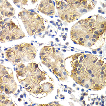
A03406 has real CHIT1 IHC data from paraffin-embedded human normal stomach (A03406 image caption); listed reactivity covers human and mouse (catalog: A03406 reactivity).
A03406 will render with an IHC image of paraffin-embedded human normal stomach stained at 1:200 (A03406 image caption). Its listed applications are IHC and WB, with human and mouse reactivity (catalog: A03406 applications/reactivity).
Which to pick: Choose A03406 for tissue IHC: this rabbit antibody lists IHC, and its own image shows paraffin-embedded human normal stomach after microwave retrieval in 10 mM PBS, pH 7.2 (catalog: A03406 host/applications; A03406 image caption); the fixative is unreported (A03406 image caption). For cross-species work, A03406 lists human and mouse reactivity, although its IHC image documents human tissue only (catalog: A03406 reactivity; A03406 image caption). No IF/ICC pick is supported because A03406 has no listed IF application, IF image or IF dilution (catalog: A03406 applications/IF fields).