This website uses cookies to ensure you get the best experience on our website.


- Table of Contents
Plan CHPF2 staining in paraffin sections using the reported cytoplasmic tissue pattern (HPA tissue IHC) and the catalog antibody’s 1:100–1:300 IHC dilution (datasheet). Compare positive and negative tissue compartments when interpreting staining (HPA tissue IHC).

Expected localisation, validated protocols, controls and antibodies — the at-a-glance facts below, then the full design guide.
| Expected localisation | Cytoplasmic staining (HPA tissue IHC); Golgi membrane protein (UniProt) | |
| Staining pattern | Cytoplasmic staining in most tissues (HPA tissue IHC) | |
| Antigen retrieval | Tris-EDTA pH 8.0 HIER, heat-mediated (datasheet A30609) | |
| Positive control | Adipose tissue+4 more · see all | |
| Negative control | Esophagus+4 more · see all |
| Fixation | Matched tissue-IHC evidence does not establish the fixation claim. Validate the specimen-specific method before use. (selected-SKU IHC image A30609) | |
| Caveat | Some squamous epithelia have no detectable staining (HPA tissue IHC) | |
| Regulation | Stimulus regulation unreported (UniProt) | |
| Isoform / epitope | 2 isoforms; epitope coverage is unspecified (UniProt) |
The catalog antibody’s IHC-P protocol (datasheet A30609) is accompanied by a published colon cancer tissue protocol (PMC8377503 methods).
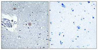
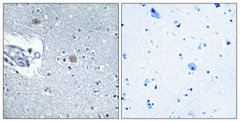
| Sample | Paraffin-embedded Human brain tissue; fixative not specified (datasheet A30609) |
| Fixation | Image fixative and duration unreported (datasheet A30609); verify before use. |
| Sectioning | 4–5 µm sections on charged slides (standard) |
| Deparaffinisation | Xylene, graded ethanol series to water (standard) |
| Antigen retrieval | Heat retrieval: Tris-EDTA pH 8.0 (datasheet A30609); 20 min, 95–100 °C (standard) |
| Peroxidase block | 3% H2O2, 10 min, room temperature (standard) |
| Blocking | 10% normal serum of the secondary host, 30 min, room temperature (standard) |
| Primary antibody | Rabbit anti-CHPF2, 1:100 - 1:300 (datasheet A30609) |
| Primary incubation | Overnight at 4 °C (standard) |
| Detection | HRP-polymer secondary, DAB chromogen 5–10 min (standard) |
| Counterstain | Hematoxylin, blue, dehydrate and mount (standard) |
| Expected result | CHPF2-positive staining in adipocytes of adipose tissue (HPA tissue IHC: Medium). HPA tissue profile: Cytoplasmic expression in most tissues. No signal in the no-primary control. |
In paraffin-section IHC, expect cytoplasmic CHPF2 staining in many cell types, including adipocytes, glandular cells, hematopoietic cells and glia (HPA: cytoplasmic expression in most tissues; listed cells Medium). A Golgi-associated component is plausible because CHPF2 is a membrane protein with a short cytoplasmic tail and a lumenal region (UniProt Q9P2E5: Golgi membrane; topology 1–6 and 30–772). The HPA tissue IHC profile is Approved, pending external verification (HPA: reliability description).
| Cytoplasmic chromogen in adipocytes, glandular cells, hematopoietic cells or glia. | This agrees with the reported compartment and sampled positive cells; the listed examples are Medium, so intensity need not be strong (HPA: tissue IHC). Assess identifiable cells and tissue architecture together. A compact perinuclear component can be compatible with Golgi residence, but HPA describes the tissue IHC pattern broadly as cytoplasmic (UniProt Q9P2E5: Golgi membrane; HPA: tissue profile). |
| Predominantly nuclear chromogen in a tissue section, with little cytoplasmic signal. | Treat this as discordant with the HPA tissue IHC profile and investigate section or detection artefact before assigning CHPF2 (HPA: cytoplasmic expression in most tissues). Nuclear signal is not a universal exclusion: HPA separately reports approved nucleoplasm and cytosol staining in ICC-IF, which uses a different specimen and readout (HPA: subcellular ICC-IF). |
| Strong staining in squamous epithelium of esophagus, oral mucosa or vagina. | These specific cell populations were Not detected in the supplied tissue IHC observations (HPA: esophagus, oral mucosa and vagina). Check whether the chromogen follows cells or a general tissue feature; possible explanations include antibody cross-reactivity or endogenous detection activity. The HPA observations are comparisons, not proof that every individual section must be blank. |
| Diffuse chromogen across cells, stroma and blank regions, obscuring cell boundaries. | A field-wide deposit cannot be scored reliably as the reported cytoplasmic pattern (HPA: tissue profile). In chromogenic IHC, nonspecific antibody binding, inadequate blocking or endogenous detection activity can raise background (standard IHC practice). Compare a reagent-control section and inspect whether any signal remains specifically associated with cells. |
| No visible signal in a sampled HPA Medium population. | An absent result in adipocytes or a listed glandular, hematopoietic or glial population conflicts with that particular HPA observation (HPA: tissue IHC, Medium), but does not alone show that CHPF2 is absent. Confirm that the relevant cells are present, then evaluate retrieval, antibody concentration, detection and controls using standard IHC practice; no CHPF2-specific retrieval condition is supplied. |
| Subcellular context | Golgi membrane residence supports an intracellular expectation (UniProt Q9P2E5: subcellular location), while HPA tissue IHC reports cytoplasmic staining without resolving a Golgi-only pattern (HPA: tissue profile). Do not require a visible Golgi outline. |
| Topology and unknown epitope | CHPF2 has residues 1–6 cytoplasmic, 7–29 transmembrane and 30–772 lumenal (UniProt Q9P2E5: topology). The supplied record does not place the antibody epitope, so topology alone cannot specify retrieval or permeabilisation settings. |
| Isoforms and glycosylation | Two isoforms and glycosylation sites at residues 121 and 342 are annotated (UniProt Q9P2E5: isoforms; glycosylation). Without an epitope map or target-specific comparison, neither annotation predicts a different IHC staining pattern. |
| Evidence scope | The tissue antibody is IHC Approved, with external verification pending (HPA: reliability); its status is not Enhanced (HPA: antibody HPA020992, IHC Approved). No target-specific fixation sensitivity or antigen-retrieval effect is provided. |
| Situation | Likely cause | Next action |
|---|---|---|
| Known-positive cells appear blank in the paraffin section. | The run may have insufficient staining sensitivity, or the expected cells may be absent from the section; the supplied examples are Medium, not uniformly strong (HPA: tissue IHC). | Confirm cell identity and a working positive control. Review the catalog antibody's IHC-P instructions, retrieval, dilution and detection as general workflow checks; do not infer a CHPF2-specific fixation effect from HPA. |
| Chromogen spreads over the section or appears in reagent-control areas. | Background from nonspecific binding or endogenous detection activity can obscure a cellular result (standard chromogenic IHC practice). | Inspect the reagent control, blocking and detection chemistry; adjust one general workflow variable at a time, then rescore whether signal is confined to cells with cytoplasmic staining (HPA: tissue profile). |
| The strongest signal is nuclear in IHC. | That distribution differs from the reported tissue IHC pattern (HPA: cytoplasmic expression in most tissues). The approved nuclear ICC-IF observation cannot by itself validate nuclear chromogen in paraffin tissue (HPA: subcellular ICC-IF). | Compare with the cytoplasmic positive control and reagent control; document the discordance before interpreting the nuclear signal as CHPF2. |
| Squamous epithelial cells stain strongly while expected cells do not. | The supplied squamous populations are Not detected (HPA: esophagus, oral mucosa and vagina); cross-reactivity or endogenous detection activity is possible. | Recheck cell identity and controls, then compare another HPA Medium cell population in the same run (HPA: tissue IHC). Treat the unexpected pattern as unconfirmed. |
| Some tissues stain more weakly than the chosen positive control. | The tissue profile spans Medium, Low and Not detected observations, despite cytoplasmic expression in most tissues (HPA: tissue IHC). | Score the specified cell population against its reported level: for example, lung alveolar cells are Low, whereas adipocytes are Medium (HPA: tissue IHC). Avoid treating a weak Low population as a failed run. |
| Does an IF/ICC image need the same pattern as paraffin IHC? | The assays have distinct observations: HPA reports nucleoplasm and cytosol in ICC-IF, and a broadly cytoplasmic tissue IHC profile (HPA: subcellular ICC-IF; tissue IHC). | Interpret the IF/ICC result on its own guide page and keep this section's paraffin-section decision anchored to tissue IHC; the ICC status of HPA020992 is Approved (HPA: antibody validation). |


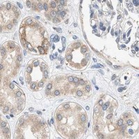


Comprehensive Human Protein Atlas IHC scoring per tissue (reliability: Approved — Pending external verification.). Rows are taken directly from the HPA tissue chart — click any row's HPA link to view the source.
| Tissue | Cell type | Level | Evidence | Source |
|---|---|---|---|---|
| Adipose tissue | Adipocytes | Medium | Protein (IHC) | HPA → |
| Adrenal gland | Glandular cells | Medium | Protein (IHC) | HPA → |
| Appendix | Glandular cells | Medium | Protein (IHC) | HPA → |
| Bone marrow | Hematopoietic cells | Medium | Protein (IHC) | HPA → |
| Breast | Glandular cells | Medium | Protein (IHC) | HPA → |
| Tissue | Cell type | Level | Evidence | Source |
|---|---|---|---|---|
| Esophagus | Squamous epithelial cells | Not detected | Protein (IHC) | HPA → |
| Oral mucosa | Squamous epithelial cells | Not detected | Protein (IHC) | HPA → |
| Ovary | Ovarian stroma cells | Not detected | Protein (IHC) | HPA → |
| Parathyroid gland | Glandular cells | Not detected | Protein (IHC) | HPA → |
| Vagina | Squamous epithelial cells | Not detected | Protein (IHC) | HPA → |
For CHPF2 chromogenic IHC in paraffin sections, assess retrieval, compartment, and controls together before interpreting staining intensity.
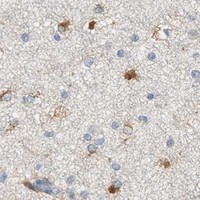
Both catalog antibodies have paraffin-section IHC images from human brain (catalog image captions); IF is listed for both, and ICC for A30609 (catalog applications).
A30609 has a human brain paraffin-section IHC image and lists Human, Mouse, and Rat reactivity plus IHC, IF, and ICC applications (A30609 image caption; catalog applications/reactivity). A14020-1 has a human brain paraffin-section IHC image and lists Human reactivity plus IHC and IF applications (A14020-1 image caption; catalog applications/reactivity).
Which to pick: For human brain paraffin-section IHC, either SKU has its own image at 1:100; A30609’s caption also reports Tris-EDTA retrieval at pH 8.0 and a peptide-absorbed control (A30609 and A14020-1 image captions). For IF/ICC, choose A30609 when ICC is needed because ICC and IF are listed for it, while A14020-1 lists IF only; neither has an IF image in the payload (catalog applications; IF image alts). For Mouse or Rat work, A30609 is the listed cross-species option and is described as polyclonal; both IHC captions show paraffin sections, with fixative unreported (catalog reactivity; A30609 dilution_raw; IHC image captions).