This website uses cookies to ensure you get the best experience on our website.


- Table of Contents
Plan CHRM1 chromogenic IHC in paraffin sections using human cerebellum as a positive tissue (HPA tissue IHC; datasheet A04081). Interpret neuronal cytoplasmic staining alongside CHRM1’s annotated cell membrane location (HPA tissue IHC; UniProt).

Expected localisation, validated protocols, controls and antibodies — the at-a-glance facts below, then the full design guide.
| Expected localisation | Neuronal cytoplasm (HPA tissue IHC); cell membrane (UniProt) | |
| Staining pattern | Pyramidal neurons and Purkinje cells: cytoplasmic staining (HPA tissue IHC) | |
| Antigen retrieval | EDTA pH 8.0 HIER, heat-mediated (datasheet A04081) | |
| Positive control | Cerebellum+2 more · see all | |
| Negative control | Adipose tissue+4 more · see all |
| Fixation | Keep fixation consistent across sections (standard IHC practice; not target-specific). Selected-image fixative and duration unreported (datasheet A04081); verify before use. | |
| Caveat | Cytoplasmic staining can complicate membrane interpretation (HPA tissue IHC; UniProt) | |
| Regulation | Expression regulation is not annotated (UniProt) | |
| Isoform / epitope | 2 isoforms; check whether the epitope is extracellular or cytoplasmic (UniProt) |
The catalog antibody has an IHC-P protocol with EDTA pH 8.0 retrieval (datasheet: A04081). The published IHC protocols below provide prostate tumor and rat hippocampal tissue examples (PMC6219293; PMC9203210; PMC7811316).
| Sample | Paraffin-embedded mouse brain tissue; fixative not specified (datasheet A04081) |
| Fixation | Image fixative and duration unreported (datasheet A04081); verify before use. |
| Sectioning | 4–5 µm sections on charged slides (standard) |
| Deparaffinisation | Xylene, graded ethanol series to water (standard) |
| Antigen retrieval | Heat retrieval: EDTA pH 8.0 (datasheet A04081); 20 min, 95–100 °C (standard) |
| Peroxidase block | 3% H2O2, 10 min, room temperature (standard) |
| Blocking | 10% goat serum (datasheet A04081) |
| Primary antibody | Rabbit anti-CHRM1, 0.5-1μg/ml (datasheet A04081) |
| Primary incubation | Overnight at 4 °C (datasheet A04081) |
| Detection | Streptavidin-biotin complex (SABC), DAB chromogen (datasheet A04081) |
| Counterstain | Hematoxylin, blue, dehydrate and mount (standard) |
| Expected result | CHRM1-positive staining in molecular layer cells - cytoplasm/membrane of cerebellum (HPA tissue IHC: High). HPA tissue profile: Cytoplasmic expression in pyramidal neurons and Purkinje cells. No signal in the no-primary control. |
CHRM1 is a seven-pass receptor at the cell and postsynaptic membranes (UniProt P11229 topology; UniProt P11229 subcellular location). In paraffin-section IHC, HPA reports cytoplasmic staining in pyramidal neurons and Purkinje cells, with high cytoplasm/membrane staining in cerebellar molecular-layer cells and medium staining in cortical and hippocampal neurons (HPA tissue IHC). HPA rates the tissue pattern Enhanced, while noting medium consistency with RNA data (HPA tissue IHC).
| Neuronal staining with a membrane-associated outline and some cytoplasmic signal. | This fits CHRM1 membrane topology and HPA’s observed neuronal cytoplasmic staining; the two descriptions need not look identical in a tissue section (UniProt P11229 topology; HPA tissue IHC). Compare the distribution across cells, not the membrane edge alone (general IHC practice). |
| Strong nuclear staining dominates the section. | A predominantly nuclear pattern does not match the reported membrane location or neuronal cytoplasmic IHC profile (UniProt P11229 subcellular location; HPA tissue IHC). Treat it as suspect until a control and a different detection condition reproduce it (general IHC practice). |
| Adipocytes stain as strongly as the neuronal positive control. | HPA reports no detectable CHRM1 staining in adipocytes from adipose tissue (HPA tissue IHC). This pattern raises concern for cross-reactivity or endogenous detection activity; it does not by itself identify which is responsible (general IHC practice). |
| Color is spread evenly across cells and surrounding tissue. | Diffuse staining that obscures cell boundaries cannot establish the HPA neuronal pattern (HPA tissue IHC). Compare a no-primary control and inspect blocking, washes, and chromogen development for nonspecific background (general IHC practice). |
| No staining appears in a cerebral cortex or hippocampus control. | HPA reports medium neuronal staining in these tissues, so a blank section calls for a run-level check before interpreting test samples as negative (HPA tissue IHC). Examine tissue preservation, primary-antibody use, retrieval, and detection controls (general IHC practice). |
| Which compartment should guide scoring? | CHRM1 has seven transmembrane segments and a postsynaptic membrane annotation, while HPA describes cytoplasmic neuronal IHC signal (UniProt P11229 topology; UniProt P11229 subcellular location; HPA tissue IHC). Score the observed cell pattern alongside membrane association; do not require a continuous membrane rim. |
| How strong is the tissue reference? | HPA labels its IHC pattern Enhanced and cites external characterization, but reports only medium consistency between staining and RNA expression (HPA tissue IHC). Use its positive and negative cell examples as benchmarks, not as proof that every section or antibody will match exactly. |
| Can sequence features predict retrieval or isoform staining? | UniProt lists two isoforms and extracellular glycosylation sites at residues 2 and 12 (UniProt P11229). No antibody epitope or target-specific retrieval response is supplied; these annotations cannot establish which isoform stains or which retrieval condition works. |
| IF/ICC: is the same pattern independently confirmed? | HPA summarizes CHRM1 as membrane-localized for ICC-IF but provides no main-location call or cell-line images in the supplied record (HPA subcellular ICC-IF). This supports a membrane expectation, not an image-verified IF pattern for a particular cell type. |
| Situation | Likely cause | Next action |
|---|---|---|
| The neuronal positive control is blank. | The run may have lost detectable signal despite medium cortical or hippocampal neuronal staining reported by HPA (HPA tissue IHC). | Check the control section, primary-antibody step, retrieval conditions, and chromogenic detection with the run controls (general IHC practice). Do not assign the blank test section a biological negative call yet. |
| Cerebellar staining is weaker than expected. | HPA reports high staining specifically in molecular-layer cells, rather than a uniform high signal throughout cerebellum (HPA tissue IHC). | Confirm that the scored region contains the relevant cells, then compare it with the positive control from the same run (HPA tissue IHC; general IHC practice). |
| Nuclei dominate the stain. | That compartment conflicts with the membrane annotation and HPA neuronal cytoplasmic profile (UniProt P11229 subcellular location; HPA tissue IHC). | Review the no-primary control and staining distribution; optimize blocking and detection if background is present (general IHC practice). Avoid counting nuclear-only cells as CHRM1 positive. |
| Adipocytes or unrelated cells are strongly colored. | HPA lists adipocytes in adipose tissue as not detected, so this warrants a specificity check (HPA tissue IHC). Cross-reactivity and endogenous detection activity are possible explanations (general IHC practice). | Compare a no-primary control with the stained section, assess endogenous detection activity, and verify the neuronal control pattern before scoring (general IHC practice). |
| The section has widespread hazy color. | Broad background can conceal the neuronal pattern documented by HPA (HPA tissue IHC). Excess detection signal or incomplete washing are possible workflow causes (general IHC practice). | Compare no-primary and tissue controls; review blocking, washing, primary dilution, and chromogen development (general IHC practice). Reassess cell-level localization after background is reduced. |
| Membrane outlines are hard to distinguish from cytoplasm. | HPA reports cytoplasmic neuronal staining, while UniProt places the receptor in membranes; a crisp rim is therefore an unreliable sole criterion (HPA tissue IHC; UniProt P11229 subcellular location). | Score the named neuronal populations and relative signal against run controls. Record cytoplasmic and membrane-associated appearances separately when visible (HPA tissue IHC; general IHC practice). |
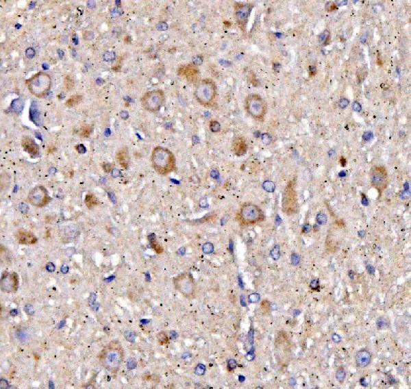
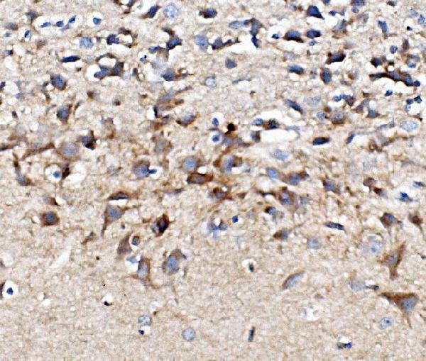
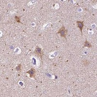
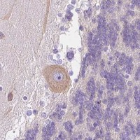


Comprehensive Human Protein Atlas IHC scoring per tissue (reliability: Enhanced — Medium consistency between antibody staining and RNA expression data. External characterization data supports antibody staining.). Rows are taken directly from the HPA tissue chart — click any row's HPA link to view the source.
| Tissue | Cell type | Level | Evidence | Source |
|---|---|---|---|---|
| Cerebellum | Molecular layer cells - cytoplasm/membrane | High | Protein (IHC) | HPA → |
| Cerebral cortex | Neuronal cells | Medium | Protein (IHC) | HPA → |
| Hippocampus | Neuronal cells | Medium | Protein (IHC) | HPA → |
| Tissue | Cell type | Level | Evidence | Source |
|---|---|---|---|---|
| Adipose tissue | Adipocytes | Not detected | Protein (IHC) | HPA → |
| Adrenal gland | Glandular cells | Not detected | Protein (IHC) | HPA → |
| Appendix | Endocrine cells | Not detected | Protein (IHC) | HPA → |
| Bone marrow | Hematopoietic cells | Not detected | Protein (IHC) | HPA → |
| Breast | Adipocytes | Not detected | Protein (IHC) | HPA → |
Use the catalog antibody’s paraffin-section workflow as the starting point, then assess staining against CHRM1 topology and the reported neuronal pattern.
A04081 has CHRM1 IHC images from paraffin sections of mouse brain, rat brain, and human cerebellum (A04081 IHC captions). No IF images are supplied (catalog: IF image alts).
A04081 is listed for IHC and for Human, Mouse, and Rat reactivity (catalog: A04081 applications and reactivity). Its IHC captions document paraffin sections of mouse brain, rat brain, and human cerebellum (A04081 IHC captions).
Which to pick: Choose A04081 for paraffin-section tissue IHC; its captions show mouse and rat brain and human cerebellum, but do not report the fixative (A04081 IHC captions). For IF/ICC, no listed SKU has an IF/ICC application or IF image (catalog: A04081 applications and IF image alts). For work across the three listed species, A04081 is the available choice based on its Human, Mouse, and Rat reactivity; it is a rabbit antibody with no clone specified (catalog: A04081 reactivity, host, and clone).