This website uses cookies to ensure you get the best experience on our website.


- Table of Contents
Plan CHST12 IHC-P with adrenal glandular cells as a high-staining reference and parathyroid glandular cells as a not-detected comparator (HPA tissue IHC). This guide covers fixation, the catalog antibody’s 1:25 starting dilution (datasheet A10456-1), and interpretation of cytoplasmic and nuclear tissue staining (HPA tissue IHC).

Expected localisation, validated protocols, controls and antibodies — the at-a-glance facts below, then the full design guide.
| Expected localisation | Tissue: cytoplasmic/nuclear (HPA tissue IHC); protein: Golgi membrane (UniProt) | |
| Staining pattern | Cytoplasmic and nuclear staining in most tissues (HPA tissue IHC) | |
| Antigen retrieval | Citrate pH 6 HIER, heat-mediated (datasheet A10456-1) | |
| Positive control | Adrenal gland+4 more · see all | |
| Negative control | Cerebellum+2 more · see all |
| Fixation | Use formaldehyde-fixed paraffin sections and consistent fixation (selected-SKU IHC image A10456-1) | |
| Caveat | Antibody staining has medium consistency with RNA data (HPA tissue IHC) | |
| Regulation | Widely expressed; no regulator specified (UniProt) | |
| Isoform / epitope | No isoforms annotated; N-tail cytoplasmic, bulk lumenal (UniProt) |
The catalog antibody’s IHC protocol is accompanied by one published CHST12 protocol for glioblastoma and adjacent tissues (PMC8806823: Immunohistochemistry).
| Sample | Formaldehyde-fixed, paraffin-embedded Human small intestine tissue (datasheet A10456-1) |
| Fixation | Image formalin-fixed; duration unreported (datasheet A10456-1); verify before use. |
| Sectioning | 4–5 µm sections on charged slides (standard) |
| Deparaffinisation | Xylene, graded ethanol series to water (standard) |
| Antigen retrieval | Heat retrieval: Citrate pH 6 (datasheet A10456-1); 20 min, 95–100 °C (standard) |
| Peroxidase block | 3% H2O2, 10 min, room temperature (standard) |
| Blocking | 10% normal serum of the secondary host, 30 min, room temperature (standard) |
| Primary antibody | Rabbit anti-CHST12, 1:25 (datasheet A10456-1) |
| Primary incubation | 1 hours at 37°C (datasheet A10456-1) |
| Detection | HRP-polymer secondary, DAB chromogen 5–10 min (standard) |
| Counterstain | Hematoxylin, blue, dehydrate and mount (standard) |
| Expected result | CHST12-positive staining in glandular cells of adrenal gland (HPA tissue IHC: High). HPA tissue profile: Cytoplasmic and nuclear expression in most tissues. No signal in the no-primary control. |
CHST12 is a Golgi membrane protein with residues 1–5 cytoplasmic, residues 6–26 spanning the membrane, and residues 27–414 lumenal (UniProt Q9NRB3 topology). In paraffin sections, look for staining in glandular cells of adrenal gland and gallbladder and in skin melanocytes (HPA: High). HPA also reports cytoplasmic and nuclear staining in most tissues; its tissue IHC reliability is Approved, with medium consistency between staining and RNA data (HPA tissue IHC).
| Granular or compact perinuclear staining appears in adrenal or gallbladder glandular cells, or skin melanocytes. | This is compatible with Golgi localization (UniProt Q9NRB3; HPA ICC-IF: Golgi approved) in cell types with High tissue IHC staining (HPA tissue IHC). Judge the cell type and subcellular pattern separately: paraffin-section staining alone does not prove that every positive structure is Golgi. |
| Predominantly nuclear staining appears without a convincing cytoplasmic or perinuclear component. | Treat this as unresolved, rather than automatically calling it specific or artifactual. HPA reports cytoplasmic and nuclear staining in most tissues (HPA tissue IHC), while UniProt places CHST12 in the Golgi membrane and HPA ICC-IF assigns its main location to Golgi (UniProt Q9NRB3; HPA ICC-IF). Compare the pattern with a known-positive section and controls. |
| The strongest stain is in a cell population other than the expected positive population. | Check cell identity and the matched negative control before assigning CHST12. For example, HPA reports High staining in skin melanocytes, but does not thereby establish High staining in every skin cell (HPA tissue IHC). Unexpected staining may reflect cross-reactivity or endogenous chromogen-producing activity (general IHC practice); neither cause follows from the image alone. |
| Color spreads across tissue, stroma, or blank areas and obscures cell boundaries. | A diffuse deposit cannot be scored as the expected cellular pattern. HPA describes cytoplasmic and nuclear expression in most tissues, while its ICC-IF localization is Golgi (HPA tissue IHC; HPA ICC-IF). Review the negative control, blocking, washes, and detection chemistry for nonspecific or endogenous signal (general IHC practice). |
| A putative positive section has no detectable stain in its expected cells. | First confirm the sampled cell type: adrenal and gallbladder glandular cells and skin melanocytes are reported High by HPA (HPA tissue IHC). An absent signal could reflect assay performance or specimen variation; HPA staining levels are reference observations, not guarantees for every section. Check controls and the antibody's IHC instructions before interpreting absence biologically. |
| Membrane orientation and epitope location | CHST12 has one membrane-spanning segment at residues 6–26, a short cytoplasmic segment at 1–5, and a lumenal region at 27–414 (UniProt Q9NRB3 topology). Epitope location could matter when selecting or interpreting an antibody, but the supplied record gives no antibody epitope, so it cannot predict antigen-retrieval conditions. |
| Glycosylation | Four glycosylation sites are annotated at residues 134, 209, 280, and 370 (UniProt Q9NRB3). This identifies a potential consideration if an epitope is later mapped near those sites; the record does not show an effect on this antibody's IHC staining or justify a retrieval change. |
| Tissue reference and validation scope | HPA rates tissue IHC Approved with medium consistency between antibody staining and RNA expression; antibody HPA041680 is IHC Approved (HPA tissue IHC; HPA antibody record). Use the reported cell-level staining to choose comparison sections. Approval supports use as a reference, but does not make every stained compartment independently confirmed. |
| IF/ICC: what pattern should I expect? | HPA assigns an approved Golgi apparatus location in ICC-IF and lists images from A-431, U-251MG, and U2OS (HPA ICC-IF). This supports a Golgi-localization expectation for IF/ICC; it does not establish that a paraffin-section chromogenic image will resolve the same fine structure. |
| Situation | Likely cause | Next action |
|---|---|---|
| No stain in adrenal or gallbladder glandular cells. | The section may lack suitable target cells, or the IHC run may have failed; both tissues have High staining in those cells in HPA's reference images (HPA tissue IHC). | Verify cell identity and section quality, inspect the run control, then follow the IHC-validated antibody's documented staining conditions. Do not infer CHST12 absence from a failed control (general IHC practice). |
| Only broad nuclear staining is visible. | Its meaning is ambiguous: HPA reports nuclear as well as cytoplasmic tissue staining, whereas UniProt and HPA ICC-IF support Golgi localization (HPA tissue IHC; UniProt Q9NRB3; HPA ICC-IF). | Compare a known-positive section, assess the negative control, and record nuclear and cytoplasmic scores separately. Avoid calling nuclear signal either definitive CHST12 or definitive artifact from this evidence alone. |
| Strong stain appears in expected-negative cells. | HPA reports no detection in cerebellar granular-layer cells, hippocampal glial cells, and parathyroid glandular cells (HPA tissue IHC). A conflicting result may be biological variation, cross-reactivity, or assay background. | Confirm the exact cell population and examine negative controls before scoring. Compare with a High-staining reference cell population in the same run (HPA tissue IHC; general IHC practice). |
| The entire section has diffuse chromogen. | Nonspecific binding, insufficient washing, or endogenous detection activity can produce broad color (general IHC practice). A diffuse field cannot establish the cell-specific HPA pattern. | Review the detection-only control, blocking, washes, and endogenous-activity control appropriate to the chromogen system; repeat scoring only after cell boundaries and compartments can be distinguished (general IHC practice). |
| Staining is patchy within a supposedly uniform positive tissue. | A tissue label does not define every cell's staining: HPA's High calls specify glandular cells or melanocytes (HPA tissue IHC). Section quality and uneven reagent coverage can also produce patchiness (general IHC practice). | Score the named cell population, inspect morphology and section coverage, and compare another area or section before treating patchiness as a CHST12 distribution change. |
| Perinuclear staining is difficult to distinguish from general cytoplasmic stain. | HPA's paraffin-tissue profile includes cytoplasmic staining, while the finer Golgi assignment comes from ICC-IF and UniProt localization (HPA tissue IHC; HPA ICC-IF; UniProt Q9NRB3). | Report the compartment visible at the section's resolution, with its cell type and intensity. Use the HPA ICC-IF result as localization context, without requiring a sharply resolved Golgi pattern for every chromogenic section. |


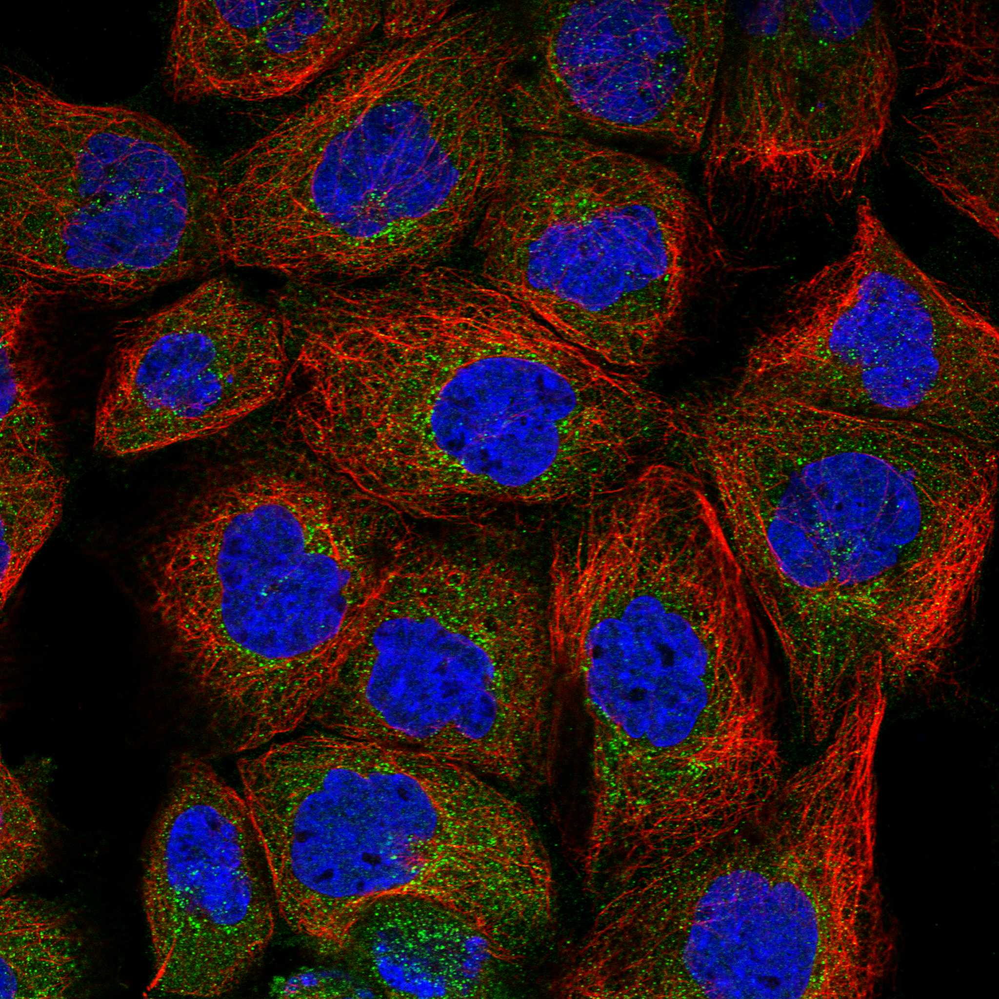
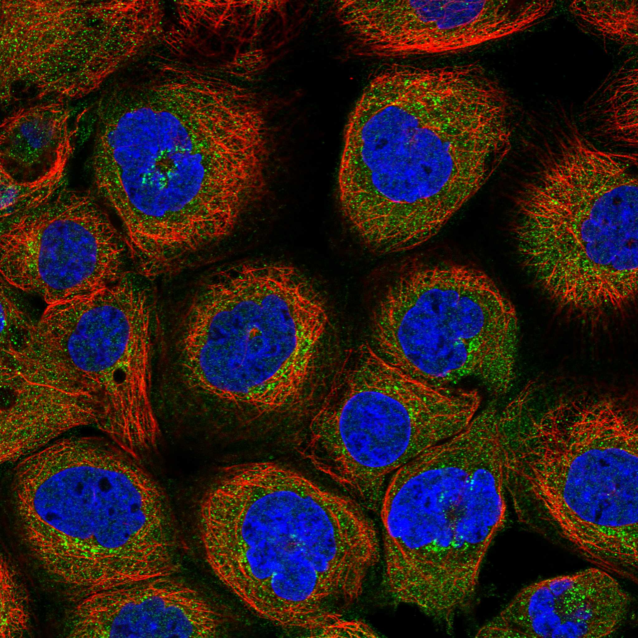
Comprehensive Human Protein Atlas IHC scoring per tissue (reliability: Approved — Medium consistency between antibody staining and RNA expression data.). Rows are taken directly from the HPA tissue chart — click any row's HPA link to view the source.
| Tissue | Cell type | Level | Evidence | Source |
|---|---|---|---|---|
| Adrenal gland | Glandular cells | High | Protein (IHC) | HPA → |
| Gallbladder | Glandular cells | High | Protein (IHC) | HPA → |
| Skin | Melanocytes | High | Protein (IHC) | HPA → |
| Stomach | Glandular cells | Medium | Protein (IHC) | HPA → |
| Adipose tissue | Adipocytes | Medium | Protein (IHC) | HPA → |
Troubleshoot CHST12 staining in paraffin sections by checking retrieval, cell type and compartment before interpreting chromogenic signal.
The catalog provides real CHST12 IHC data from human small-intestine paraffin sections (A10456-1 image caption); it provides no IF figure (catalog: IF image alts).
A10456-1 is listed for human IHC-P (catalog: applications and reactivity). Its figure shows staining in human small-intestine paraffin sections fixed with formaldehyde, with citrate pH 6 heat retrieval (A10456-1 image caption).
Which to pick: For tissue IHC, choose A10456-1: this rabbit polyclonal antibody is listed for human IHC-P, and its own figure documents paraffin sections (catalog: host, clonality, applications and reactivity; A10456-1 image caption). No listed SKU has IF/ICC validation or an IF figure (catalog: applications and IF image alts). No listed SKU supports a cross-species choice because A10456-1 lists human reactivity only (catalog: reactivity).