This website uses cookies to ensure you get the best experience on our website.


- Table of Contents
Plan CIAPIN1 paraffin-section IHC around predominantly cytoplasmic staining, with nuclear staining in some tissues (HPA tissue IHC). Use testis pachytene spermatocytes as a strong reference and assess antibody staining alongside controls (HPA tissue IHC).

Expected localisation, validated protocols, controls and antibodies — the at-a-glance facts below, then the full design guide.
| Expected localisation | Cytoplasmic; nuclear in some tissues (HPA tissue IHC) | |
| Staining pattern | Mostly cytoplasmic, with nuclear staining in some tissues (HPA tissue IHC) | |
| Antigen retrieval | Tris-EDTA pH 9.0 HIER, 95–98 °C, 20 min (rule: nuclear antigen) | |
| Positive control | Testis+4 more · see all | |
| Negative control | Adipose tissue+4 more · see all |
| Fixation | Keep fixation consistent across paraffin sections. (standard IHC practice; not target-specific) | |
| Caveat | Antibody staining and RNA show medium consistency (HPA tissue IHC) | |
| Regulation | Tissue abundance varies; testis is highest (HPA tissue IHC) | |
| Isoform / epitope | 3 isoforms; epitope coverage is unspecified (UniProt) |
Compare the catalog antibody’s IHC-P protocol (datasheet) with four published CIAPIN1 tissue IHC protocols (PMC6481027; PMC9267148; PMC3502349; PMC2516489).
| Sample | Paraffin-embedded human liver injury tissue; fixative not specified (datasheet A04333) |
| Fixation | Image fixative and duration unreported (datasheet A04333); verify before use. |
| Sectioning | 4–5 µm sections on charged slides (standard) |
| Deparaffinisation | Xylene, graded ethanol series to water (standard) |
| Antigen retrieval | Heat-induced epitope retrieval in Tris-EDTA buffer, pH 9.0, 20 min at 95–98 °C (standard rule: nuclear antigen) |
| Peroxidase block | 3% H2O2, 10 min, room temperature (standard) |
| Blocking | 10% normal serum of the secondary host, 30 min, room temperature (standard) |
| Primary antibody | Rabbit anti-CIAPIN1, 1:50-1:200 (datasheet A04333) |
| Primary incubation | Overnight at 4 °C (standard) |
| Detection | HRP-polymer secondary, DAB chromogen 5–10 min (standard) |
| Counterstain | Hematoxylin, blue, dehydrate and mount (standard) |
| Expected result | CIAPIN1-positive staining in pachytene spermatocytes of testis (HPA tissue IHC: High). HPA tissue profile: Cytoplasmic expression in most tissues which in some tissues was combined with nuclear expression, most abundant in testis. No signal in the no-primary control. |
CIAPIN1 is mainly cytoplasmic by tissue IHC, with nuclear staining in some tissues; UniProt also places it in the mitochondrial intermembrane space and reports no transmembrane segment (HPA tissue IHC; UniProt Q6FI81 localization and topology). Expect strongest staining in testis pachytene spermatocytes and moderate staining in several glandular cell populations (HPA tissue IHC). HPA rates the tissue IHC evidence Enhanced, with medium consistency between staining and RNA expression (HPA tissue IHC reliability).
| Pachytene spermatocytes show strong cytoplasmic staining; some nuclei also stain. | This fits the strongest listed tissue result and the reported cytoplasmic pattern with nuclear staining in some tissues (HPA tissue IHC). Judge nuclear staining alongside the cytoplasmic signal and tissue context; nuclear signal alone does not reproduce the typical tissue pattern (HPA tissue IHC). |
| Staining is confined to cell borders or extracellular spaces, with little intracellular signal. | This is inconsistent with the reported cytoplasmic and occasional nuclear tissue pattern and with the absence of a transmembrane segment (HPA tissue IHC; UniProt Q6FI81 topology). Suspect nonspecific staining or a detection artefact; check a known-positive section and detection controls (general IHC practice). |
| Adipocytes stain strongly while expected positive cells show little signal. | HPA reports adipocytes as not detected in adipose tissue and breast, while testis pachytene spermatocytes are High (HPA tissue IHC). Consider cross-reactivity or endogenous detection activity; interpret the named cell populations, rather than treating every cell in a tissue as positive or negative (general IHC practice; HPA tissue IHC). |
| Most of the section has an even brown haze that obscures cell boundaries. | Uniform haze cannot establish the cell-specific cytoplasmic pattern reported by HPA (HPA tissue IHC). Assess the no-primary control and review blocking, washes, antibody concentration and chromogen development as general IHC troubleshooting steps (general IHC practice); the supplied sources do not identify a CIAPIN1-specific cause. |
| A testis section has no detectable signal in pachytene spermatocytes. | That conflicts with HPA's High result for this cell population (HPA tissue IHC). First confirm the cells are present and the run's detection controls worked; then review the catalog antibody's IHC-P instructions and the run's retrieval and dilution settings (general IHC practice). No target-specific fixation sensitivity is established here. |
| Which tissue and cells should anchor interpretation? | Use testis pachytene spermatocytes as a strong reference; HPA calls them High, while appendix and duodenum glandular cells are Medium (HPA tissue IHC). Pancreatic exocrine glandular cells are Low despite UniProt's high pancreas expression statement; distinguish a named cell's IHC level from whole-tissue expression (HPA tissue IHC; UniProt Q6FI81 tissue specificity). |
| How much confidence does the tissue pattern support? | HPA assigns Enhanced IHC validation to HPA041350 and HPA042182, while describing overall staining-to-RNA consistency as medium (HPA antibodies; HPA tissue IHC reliability). Treat this as support for the reported pattern, with controls and cell-level interpretation still needed in a new experiment (general IHC practice). |
| Does topology predict a membrane stain? | UniProt reports no transmembrane segment and lists cytoplasm, nucleus and mitochondrial intermembrane space (UniProt Q6FI81 localization and topology). The tissue IHC profile is predominantly cytoplasmic, sometimes nuclear (HPA tissue IHC). A crisp plasma-membrane outline therefore needs independent validation before assignment to CIAPIN1 (HPA tissue IHC; UniProt Q6FI81 topology). |
| Can the supplied record resolve isoforms or fixation effects? | Target-specific fixation effects are not established by the supplied assay evidence. Verify with a matched IHC source before attributing a result to fixation. |
| Q: What should IF/ICC show? | A: HPA reports mainly mitochondrial localization with additional nucleoplasmic localization, both supported (HPA subcellular ICC-IF). UniProt also lists the mitochondrial intermembrane space, cytoplasm and nucleus (UniProt Q6FI81 localization). This is an interpretation reference for IF/ICC; it does not turn the tissue IHC pattern into a resolved mitochondrial IHC readout. |
| Situation | Likely cause | Next action |
|---|---|---|
| Testis pachytene spermatocytes are weak or blank. | Possible run failure or a mismatch between the applied conditions and the catalog antibody's IHC-P instructions; HPA reports these cells as High (HPA tissue IHC; general IHC practice). | Verify the cell population and run controls, then review the antibody's IHC-P retrieval, dilution and detection instructions (general IHC practice). Do not infer a CIAPIN1-specific fixation problem from this result. |
| A weak signal appears in pancreatic exocrine glandular cells. | That cell population is Low by HPA tissue IHC, although UniProt describes pancreas as highly expressed at the tissue level (HPA tissue IHC; UniProt Q6FI81 tissue specificity). | Compare with a stronger reference such as testis pachytene spermatocytes before calling the run insensitive (HPA tissue IHC; general IHC practice). Record the precise cells scored. |
| Adipocytes show prominent brown signal. | HPA lists adipocytes in adipose tissue and breast as not detected; nonspecific binding or endogenous detection activity are possible general IHC causes (HPA tissue IHC; general IHC practice). | Check a no-primary control and the detection system's endogenous-activity blocking steps; compare the signal's distribution with a positive cell population (general IHC practice; HPA tissue IHC). |
| Staining is mostly nuclear with no discernible cytoplasm. | Nuclear expression can accompany cytoplasmic expression in some tissues, but HPA describes the overall tissue pattern as cytoplasmic (HPA tissue IHC). | Review the matched tissue reference and controls before scoring it as expected; document both compartments separately (HPA tissue IHC; general IHC practice). |
| Staining forms sharp cell-border outlines. | This differs from the reported intracellular tissue pattern and the lack of a transmembrane segment (HPA tissue IHC; UniProt Q6FI81 topology). | Reassess specificity with a known-positive section and detection controls; avoid assigning border signal to CIAPIN1 without further evidence (general IHC practice). |
| Brown background obscures medium-level glandular staining. | Diffuse background can conceal the Medium glandular-cell signals listed for appendix, duodenum, gallbladder, rectum, small intestine and stomach (HPA tissue IHC; general IHC practice). | Check the no-primary control, then review blocking, washes, antibody concentration and chromogen development under the IHC-P workflow (general IHC practice). Score only interpretable cells. |
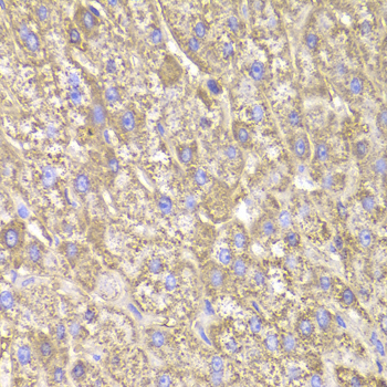
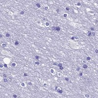

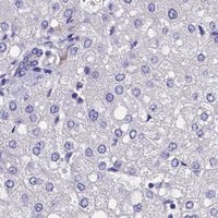
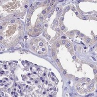


Comprehensive Human Protein Atlas IHC scoring per tissue (reliability: Enhanced — Medium consistency between antibody staining and RNA expression data.). Rows are taken directly from the HPA tissue chart — click any row's HPA link to view the source.
| Tissue | Cell type | Level | Evidence | Source |
|---|---|---|---|---|
| Testis | Pachytene spermatocytes | High | Protein (IHC) | HPA → |
| Appendix | Glandular cells | Medium | Protein (IHC) | HPA → |
| Duodenum | Glandular cells | Medium | Protein (IHC) | HPA → |
| Gallbladder | Glandular cells | Medium | Protein (IHC) | HPA → |
| Ovary | Follicle cells | Medium | Protein (IHC) | HPA → |
| Tissue | Cell type | Level | Evidence | Source |
|---|---|---|---|---|
| Adipose tissue | Adipocytes | Not detected | Protein (IHC) | HPA → |
| Bone marrow | Hematopoietic cells | Not detected | Protein (IHC) | HPA → |
| Breast | Adipocytes | Not detected | Protein (IHC) | HPA → |
| Caudate | Glial cells | Not detected | Protein (IHC) | HPA → |
| Cerebellum | Cells in granular layer | Not detected | Protein (IHC) | HPA → |
Troubleshoot CIAPIN1 staining in paraffin sections by checking retrieval, compartment pattern, controls and scoring before interpreting biological differences.
A04333 lists IHC and IF use in human and mouse samples (catalog: applications/reactivity). Its supplied image shows IHC in paraffin-embedded human liver-injury tissue (A04333 image alt); no IF image is supplied (catalog: IF image alts).
A04333 is the only card (catalog: SKU), with an IHC image of paraffin-embedded human liver-injury tissue (A04333 image alt). IF is listed and human/mouse reactivity is claimed (catalog: applications/reactivity), but no IF or mouse image is supplied (catalog: image alts).
Which to pick: For tissue IHC, choose A04333: its own caption documents paraffin-embedded human liver-injury tissue, but does not report the fixative (A04333 image alt). For IF, A04333 lists a 1:50–1:200 dilution (catalog: IF dilution); ICC validation and an IF image are unreported (catalog: applications/IF image alts). A04333 lists human and mouse reactivity, has a rabbit host, and does not specify clonality (catalog: reactivity/host/clone); the supplied IHC image documents human tissue only (A04333 image alt).