CISD1 / CDGSH iron-sulfur domain-containing protein 1 · IHC design guide

Design Immunohistochemistry for CISD1

Plan chromogenic CISD1 IHC on paraffin sections with the catalog antibody at 2–5 μg/mL (datasheet A04360-2). Compare granular cytoplasmic staining in appendix or colon glandular cells with adipocytes, where staining was not detected (HPA tissue IHC).

Evidence assembled Oct 2026 · For research use; verify linked source records and product datasheet before use
Immunohistochemistry protocol sheet for CISD1 (IHC for CISD1): expected localisation Granular cytoplasm (HPA tissue IHC); outer mitochondrial membrane (UniProt), antibody A04360-2, validated IHC image, and IHC protocol steps
Printable CISD1 IHC protocol sheet — expected localisation Granular cytoplasm (HPA tissue IHC); outer mitochondrial membrane (UniProt), antibody A04360-2, controls and protocol steps. Open the full CISD1 IHC guide →

CISD1 Immunohistochemistry Experimental Design Guide

Expected localisation, validated protocols, controls and antibodies — the at-a-glance facts below, then the full design guide.

Must know before staining
Expected localisation Granular cytoplasm (HPA tissue IHC); outer mitochondrial membrane (UniProt)
Staining pattern Granular cytoplasmic staining in most tissues (HPA tissue IHC)
Antigen retrieval EDTA pH 8.0 HIER, heat-mediated (datasheet A04360-2)
Positive control ⓘ Appendix+4 more · see all
Negative control ⓘ Adipose tissue+4 more · see all
Important caveats
Reasons your staining may differ from the expected pattern.
Fixation Matched tissue-IHC evidence does not establish the fixation claim. Validate the specimen-specific method before use. (selected-SKU IHC image A04360-2)
Caveat Tissue staining has medium agreement with RNA data (HPA tissue IHC)
Regulation Reduced in cystic fibrosis patient cells (UniProt)
Isoform / epitope No isoforms annotated; residues 32–108 face cytoplasm (UniProt)
Section 1

Recommended CISD1 IHC & IF Protocols

The catalog antibody’s IHC-P protocol is accompanied by four published CISD1 IHC methods covering breast cancer, paraffin sections, cancer tissues, and pancreatic cancer (PMC9729735; PMC11948984; PMC8936422; PMC8760727).

Recommended immunohistochemistry (IHC-P) protocol parameters
SampleParaffin-embedded human gastric cancer tissue; fixative not specified (datasheet A04360-2)
FixationImage fixative and duration unreported (datasheet A04360-2); verify before use.
Sectioning4–5 µm sections on charged slides (standard)
DeparaffinisationXylene, graded ethanol series to water (standard)
Antigen retrievalHeat retrieval: EDTA pH 8.0 (datasheet A04360-2); 20 min, 95–100 °C (standard)
Peroxidase block3% H2O2, 10 min, room temperature (standard)
Blocking10% goat serum (datasheet A04360-2)
Primary antibodyRabbit anti-CISD1, 2-5μg/ml (datasheet A04360-2)
Primary incubationOvernight at 4 °C (datasheet A04360-2)
DetectionStreptavidin-biotin complex (SABC), DAB chromogen (datasheet A04360-2)
CounterstainHematoxylin, blue, dehydrate and mount (standard)
Expected resultCISD1-positive staining in glandular cells of appendix (HPA tissue IHC: High). HPA tissue profile: Granular cytoplasmic expression in most tissues. No signal in the no-primary control.
💡Decision noteStart with heat-mediated EDTA pH 8.0 retrieval for the catalog antibody (datasheet A04360-2). Use the published retrieval conditions when reproducing those specific methods (PMC11948984; PMC8760727).
Section 2

What Is the Expected CISD1 Staining Pattern?

CISD1 is an outer mitochondrial membrane protein with a cytoplasm-facing region (UniProt Q9NZ45 topology). In paraffin-section IHC, expect granular cytoplasmic staining in selected cells, including colon glandular cells and cardiomyocytes (HPA: tissue IHC). HPA rates the tissue pattern Supported, with medium agreement between staining and RNA data; external verification is pending (HPA: reliability).

What am I looking at on my slide?
Granular cytoplasmic staining in colon or duodenal glandular cells.Fits the reported pattern and a High-staining cell population (HPA: tissue IHC). Chromogenic granules alone cannot prove outer-membrane localisation (UniProt Q9NZ45 topology).
Predominantly nuclear staining in otherwise positive cells.Does not fit the reported mitochondrial location (UniProt Q9NZ45; HPA: subcellular). Review controls and scoring before treating it as CISD1-specific.
Strong staining in adipocytes or lung alveolar cells.Those cell types were Not detected by HPA (HPA: tissue IHC). Check for cross-reactivity or endogenous detection activity; the discrepancy alone does not identify its cause.
Uniform colour across cells and surrounding tissue, obscuring granules.This is difficult to score against HPA’s granular cytoplasmic pattern (HPA: tissue IHC). Assess background in the no-primary control (general IHC practice).
No staining in colon glandular cells or cardiomyocytes.Unexpected for populations reported High (HPA: tissue IHC). Check the positive control and detection workflow before interpreting the sample as CISD1-negative (general IHC practice).
💡Expected CISD1 appearanceCall a result consistent when selected positive cells show clear granular cytoplasmic staining, potentially strong in colon glands or cardiomyocytes (HPA: High); dominant nuclear colour or uniform background is suspect (UniProt Q9NZ45; HPA: tissue IHC).
How each factor affects the staining
Where should signal sit?CISD1 spans the outer mitochondrial membrane at residues 14–31; residues 32–108 face the cytoplasm (UniProt Q9NZ45 topology). IHC does not resolve that membrane boundary.
Which cells are useful comparators?Colon glandular cells and cardiomyocytes are High; adipocytes and lung alveolar cells are Not detected (HPA: tissue IHC). Compare the named cell populations, not whole tissues.
How firm is the tissue pattern?HPA calls it Supported, citing medium staining–RNA consistency and pending external verification (HPA: reliability). Treat a mismatch as a finding to investigate.
What does IF/ICC add?HPA reports supported mitochondrial localisation in ICC-IF and lists PC-3 and U2OS images (HPA: subcellular). That supports compartment interpretation; IF/ICC methods belong in its separate guide.
Can RNA select an IHC positive control?HPA calls parathyroid gland tissue-enhanced by RNA, but supplies no parathyroid IHC level here (HPA: RNA specificity; tissue IHC). Use an observed High-staining cell population for the IHC comparator.
Why is my staining missing, weak or wrong?
SituationLikely causeNext action
Granular staining is absent in a High-staining comparator.The staining run may have failed; the HPA level describes observed tissue IHC, not guaranteed signal in every preparation (HPA: tissue IHC).Check positive-control tissue, antibody and detection steps, then review retrieval conditions using general IHC practice; target-specific retrieval sensitivity is unreported.
A Not detected cell population stains strongly.Possible nonspecific antibody binding or endogenous detection activity; HPA reports no detectable staining in that named population (HPA: tissue IHC).Compare a no-primary control and the expected cell distribution; adjust blocking or detection controls as indicated (general IHC practice).
Signal is mainly nuclear.The compartment conflicts with mitochondrial localisation (UniProt Q9NZ45; HPA: subcellular).Verify the staining controls and inspect whether cytoplasmic granules are present before assigning the nuclear signal to CISD1 (general IHC practice).
Diffuse colour masks cell boundaries or granules.Background prevents assessment of HPA’s granular cytoplasmic pattern (HPA: tissue IHC).Inspect the no-primary control and review blocking, washes and chromogen development (general IHC practice); avoid scoring obscured cells.
Staining varies between cells in the same section.HPA reports staining by cell type, including High glandular cells and Not detected populations (HPA: tissue IHC). Variation need not indicate a failed run.Score the specified cells separately and compare each with its HPA level; retain a positive control to assess the run (general IHC practice).

Sample controls for CISD1 IHC & IF

🧪Run colon first: its glandular cells must show CISD1 staining (HPA: High in colon glandular cells). Use cervix glandular cells as the negative tissue (HPA: Not detected); on the colon slide, assess stromal cells as internal background comparators and call them negative only if they lack specific staining.
Positive control tissue: Appendix (Glandular cells, HPA High)
Negative control tissue: Adipose tissue (HPA Not detected)
ICC-IF cell lines (HPA subcellular resource): HPA ICC-IF images show CISD1 in PC-3, U2OS, with annotated localisation: Mitochondria (supported) (HPA subcellular).
Technical controls: Include a no-primary, secondary-only slide; a host- and clonality-matched rabbit IgG isotype control (selected-SKU caption: rabbit primary antibody); and CISD1-knockout tissue or cells as a biological negative. For the colon DAB assay, block endogenous peroxidase and check for endogenous biotin background because the caption uses biotinylated secondary antibody and streptavidin–biotin detection (selected-SKU tissue-IHC caption).
⚠️Feasibility: No target-specific fixation window or fixation effect is reported, and the selected-SKU paraffin-section caption does not state a fixative (selected-SKU tissue-IHC caption). That caption uses heat retrieval in EDTA at pH 8.0, but retrieval dependence is unreported (selected-SKU tissue-IHC caption). Frozen sections have no stated advantage; ICC-IF images support mitochondrial localization in PC-3 and U2OS, while colon glandular staining should be judged against possible luminal or endogenous-biotin background (HPA: mitochondria supported; HPA: High in colon glandular cells; selected-SKU tissue-IHC caption: biotin-based DAB detection).

HPA tissue IHC evidence for CISD1

Comprehensive Human Protein Atlas IHC scoring per tissue (reliability: Supported — Medium consistency between antibody staining and RNA expression data. Pending external verification.). Rows are taken directly from the HPA tissue chart — click any row's HPA link to view the source.

Positive expression · recommended positive controls

TissueCell typeLevelEvidenceSource
Appendix Glandular cells High Protein (IHC) HPA →
Cerebellum Cells in molecular layer High Protein (IHC) HPA →
Cerebral cortex Neuropil High Protein (IHC) HPA →
Colon Glandular cells High Protein (IHC) HPA →
Duodenum Glandular cells High Protein (IHC) HPA →

Undetected expression · recommended negative controls

TissueCell typeLevelEvidenceSource
Adipose tissue Adipocytes Not detected Protein (IHC) HPA →
Bone marrow Hematopoietic cells Not detected Protein (IHC) HPA →
Caudate Glial cells Not detected Protein (IHC) HPA →
Cervix Glandular cells Not detected Protein (IHC) HPA →
Lung Alveolar cells Not detected Protein (IHC) HPA →
Section 3

Advanced CISD1 IHC Tips

Troubleshoot CISD1 staining in paraffin sections by checking retrieval, cell type and granular cytoplasmic localisation before scoring the chromogenic signal.

What should I adjust when CISD1 staining is weak after antigen retrieval?
Start with heat-mediated retrieval in EDTA at pH 8.0 for paraffin sections, matching the selected tissue image (datasheet A04360-2). If staining is weak, standardise heating, cooling and section thickness across slides before testing another buffer as a documented fallback (standard IHC practice). Keep the primary at 2 μg/ml overnight at 4 °C while comparing retrieval conditions, since these were the image conditions (caption A04360-2). Assess granular cytoplasmic signal, background and tissue morphology together, because harsh retrieval can compromise interpretation (HPA: granular cytoplasmic profile; standard IHC practice).
Could fixation explain weak or uneven CISD1 staining in paraffin sections?
Target-specific fixation sensitivity is unknown: the selected paraffin-section image does not state which fixative was used (caption A04360-2). Record the fixative, fixation duration and processing history for each specimen, then compare matched sections using identical retrieval and antibody conditions (standard IHC practice). Uneven staining may reflect processing differences, so inspect morphology and compare interior tissue with edges before changing the primary concentration (standard IHC practice). Use the reported 2 μg/ml primary incubation overnight at 4 °C as a starting point for that comparison, without treating it as evidence of fixation tolerance (caption A04360-2).
What staining pattern should I expect for CISD1 in chromogenic IHC?
Expect predominantly granular cytoplasmic staining in appropriate cells, consistent with the tissue staining profile and mitochondrial localisation (HPA: granular cytoplasmic profile; HPA: mitochondria supported). CISD1 is an outer mitochondrial membrane protein with a transmembrane segment at residues 14–31 and a cytoplasmic region at 32–108 (UniProt Q9NZ45 topology). Routine DAB microscopy cannot establish which side of the mitochondrial membrane contains the antibody epitope, so avoid inferring epitope orientation from granules alone (standard IHC practice). Compare cell types within the section; HPA reports high staining in colon glandular cells but no detection in adipocytes (HPA: colon glandular cells High; HPA: adipocytes Not detected).
How should I troubleshoot an unexpected pattern when the CISD1 epitope is unknown?
Do not assign the antibody an epitope from the staining pattern: this payload supplies no immunogen sequence or mapped binding site (catalog antibody evidence supplied). The record lists 0 isoforms, a chain spanning residues 2–108, and a transmembrane segment at 14–31 (UniProt Q9NZ45 record). The cytoplasmic region spans 32–108, while alternate acetyllysine sites are annotated at 55, 68 and 104; their effect on antibody binding is unestablished (UniProt Q9NZ45 topology and modified residues). Check the antibody's epitope documentation and compare appropriate tissue controls before attributing unusual staining to processing or modification (standard IHC practice).
How can IF help assess whether the granular IHC signal is cellular and mitochondrial?
On a separate IF/ICC assay, multiplex CISD1 with a marker for the expected cell type, such as an epithelial marker when examining colon glandular cells (HPA: colon glandular cells High; standard IF practice). Choose spectrally separated fluorophores and favour a red or far-red CISD1 channel if tissue autofluorescence obscures shorter wavelengths; check unstained and single-stain controls (standard IF practice). CISD1 has a cytoplasmic region at residues 32–108, so assess permeabilisation for access to that side of the outer mitochondrial membrane without assuming the antibody epitope lies there (UniProt Q9NZ45 topology; standard IF practice). Mitochondrial IF localisation has independent support, including images from PC-3 and U2OS cells (HPA subcellular: mitochondria supported; HPA subcellular: ICC/IF image cell lines).
How can I separate CISD1 staining from chromogenic background?
Begin with the reported 10% goat serum block and 2 μg/ml primary concentration when assessing the selected paraffin-section method (caption A04360-2). Its biotinylated secondary, streptavidin-biotin complex and DAB detection make reagent-only and no-primary controls useful for investigating unwanted colour development (caption A04360-2; standard IHC practice). Include an endogenous peroxidase block and, if background persists, evaluate endogenous biotin and pigment before increasing antibody dilution (standard chromogenic IHC practice). Inspect whether signal forms cytoplasmic granules in expected cells or instead follows tissue edges and damaged areas (HPA: granular cytoplasmic profile; standard IHC practice).
How should I score CISD1 staining across tissue sections? ⚠ ANSWER MARKED FOR VERIFICATION
Define the cell population and analysis area before scoring, because CISD1 staining varies among cell types and tissues (HPA tissue IHC profile; standard IHC practice). For cellular staining, record the percentage at each intensity and calculate an H-score from 0–300, or report the percentage of positive cells using a fixed threshold (standard IHC practice). If counting discrete positive cells, normalise density to analysed tissue area in mm²; otherwise normalise positive counts to eligible nucleated cells (standard IHC practice). Exclude folds, necrosis and edge artefacts consistently, and keep illumination, DAB development and scoring rules matched across comparisons (standard IHC practice).
When is a CISD1-positive IHC result convincing rather than artefactual?
A convincing result shows granular cytoplasmic staining in an appropriate cell population, consistent with mitochondrial localisation rather than dominant nuclear or extracellular colour (HPA: granular cytoplasmic profile; UniProt Q9NZ45 localisation). Compare a positive reference such as colon glandular cells with a low or undetected reference chosen for the study context, while recognising that HPA rates its tissue staining only Supported (HPA: colon glandular cells High; HPA: adipocytes Not detected; HPA: reliability Supported). Edge accentuation, necrotic deposits and colour in no-primary controls favour artefact or endogenous detection chemistry (standard IHC practice). The gastric cancer image demonstrates staining with A04360-2 in a paraffin section, but does not by itself establish specificity in every specimen (caption A04360-2; standard IHC practice).
Boster reagents

Best CISD1 / CDGSH iron-sulfur domain-containing protein 1 IHC Antibodies

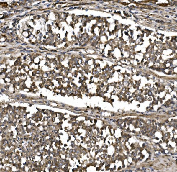
A04360-2 has real IHC data from a human paraffin-embedded gastric cancer section (catalog image caption). Human, mouse, and rat reactivity is listed; no IF data is supplied (catalog reactivity; catalog IF images).

Real IHC data IHC analysis of MitoNEET/CISD1 using anti-MitoNEET/CISD1 antibody (A04360-2). MitoNEET/CISD1 was detected in paraffin-embedded section of human gastric cancer tissue. Heat mediated antigen retrieval was performed in EDTA buffer (pH8.0, epitope retrieval solution). The tissue section was blocked with 10% goat serum. The tissue section was then incubated with 2μg/ml rabbit anti-MitoNEET/CISD1 Antibody (A04360-2) overnight at 4°C. Biotinylated goat anti-rabbit IgG was used as secondary antibody and incubated for 30 minutes at 37°C. The tissue section was developed using Strepavidin-Biotin-Complex (SABC) (Catalog # SA1022) with DAB as the chromogen.
Anti-MitoNEET/CISD1 Antibody ®
Cat # A04360-2

A04360-2 is the only card and shows chromogenic IHC on a paraffin-embedded human gastric cancer section (A04360-2 image caption). Its application list includes IHC, and its listed reactivity covers human, mouse, and rat (catalog applications; catalog reactivity).

Which to pick: Choose A04360-2 for paraffin-section IHC: its caption documents EDTA pH 8 retrieval, 2 μg/ml primary antibody, and DAB detection; the fixative is unreported (A04360-2 image caption). For mouse or rat tissue, the catalog lists reactivity, but the supplied IHC image shows human tissue only; clonality is unreported (catalog reactivity; A04360-2 image caption; catalog clone field). No IF/ICC recommendation is supported because IF/ICC is absent from the application list and no IF image is supplied (catalog applications; catalog IF images).

Each figure is that product's own IHC / IF validation image from its datasheet.

References

  1. UniProt Consortium. UniProt entry Q9NZ45 (CISD1_HUMAN, CDGSH iron-sulfur domain-containing protein 1).
  2. Human Protein Atlas. CISD1 tissue IHC expression (reliability: Supported).
  3. Human Protein Atlas. CISD1 subcellular location (ICC-IF): Localized to the mitochondria..
  4. Human Protein Atlas. CISD1 antibody validation summary (1 antibodies).
  5. Identification of CISD1 as a Prognostic Biomarker for Breast Cancer. International journal of general medicine 2022 — PMC9729735.
  6. Predictive and therapeutic value of the ferroptosis gene CISD1 in non?small cell lung cancer. Oncology letters 2025 — PMC11948984.
  7. Systematic Analysis and Validation of the Prognosis, Immunological Role and Biology Function of the Ferroptosis-Related lncRNA GSEC/miRNA-101-3p/CISD1 Axis in Lung Adenocarcinoma. Frontiers in molecular biosciences 2021 — PMC8936422.
  8. Development and validation of a novel 3-gene prognostic model for pancreatic adenocarcinoma based on ferroptosis-related genes. Cancer cell international 2022 — PMC8760727.
  9. PubMed PMID:18047834 — UniProt-cited evidence.
  10. PubMed PMID:14702039 — UniProt-cited evidence.
  11. PubMed PMID:15489334 — UniProt-cited evidence.