This website uses cookies to ensure you get the best experience on our website.


- Table of Contents
Plan CKAP5 staining in paraffin sections around the observed cytoplasmic tissue pattern (HPA tissue IHC). This guide covers consistent fixation, the catalog antibody's 2–5 μg/mL IHC range (datasheet A05324), and mitotic localization when interpreting staining (UniProt).

Expected localisation, validated protocols, controls and antibodies — the at-a-glance facts below, then the full design guide.
| Expected localisation | Observed tissue: cytoplasm (HPA tissue IHC); molecular: centrosome/spindle (UniProt) | |
| Staining pattern | Widespread cytoplasmic staining across cell types (HPA tissue IHC) | |
| Antigen retrieval | EDTA pH 8.0 HIER, heat-mediated (datasheet A05324) | |
| Positive control | Adipose tissue+4 more · see all | |
| Negative control | None in HPA (detected in all 45 tissues); use no-primary + isotype controls |
| Fixation | Keep fixation consistent across sections (standard IHC practice; not target-specific) | |
| Caveat | Mitotic cells may show spindle enrichment (UniProt) | |
| Regulation | Elevated in hepatomas and colonic tumors (UniProt) | |
| Isoform / epitope | 3 isoforms; epitope coverage is undetermined (UniProt) |
The catalog antibody protocol is paired with a published CKAP5 IHC protocol for xenograft tumors (PMC10075965).
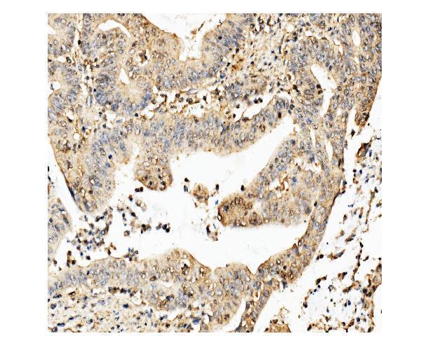
| Sample | Paraffin-embedded human liver cancer tissue; fixative not specified (datasheet A05324) |
| Fixation | Image fixative and duration unreported (datasheet A05324); verify before use. |
| Sectioning | 4–5 µm sections on charged slides (standard) |
| Deparaffinisation | Xylene, graded ethanol series to water (standard) |
| Antigen retrieval | Heat retrieval: EDTA pH 8.0 (datasheet A05324); 20 min, 95–100 °C (standard) |
| Peroxidase block | 3% H2O2, 10 min, room temperature (standard) |
| Blocking | 10% goat serum (datasheet A05324) |
| Primary antibody | Rabbit anti-CKAP5, 2-5μg/ml (datasheet A05324) |
| Primary incubation | Overnight at 4 °C (datasheet A05324) |
| Detection | HRP-conjugated secondary, DAB chromogen (datasheet A05324) |
| Counterstain | Hematoxylin, blue, dehydrate and mount (standard) |
| Expected result | CKAP5-positive staining in adipocytes of adipose tissue (HPA tissue IHC: High). HPA tissue profile: Ubiquitous cytoplasmic expression. No signal in the no-primary control. |
CKAP5 should produce a predominantly cytoplasmic tissue IHC pattern, with prominent staining in HPA-listed cells such as adipocytes, cortical neurons, bronchial ciliated cell bodies and colon endothelial cells (HPA: ubiquitous cytoplasmic expression; High in these cells). Centrosomal and spindle-associated localisation is biologically plausible, although those small structures may be difficult to resolve in chromogenic sections (UniProt Q14008: subcellular localisation). The HPA tissue assessment is Supported, with medium consistency between staining and RNA data (HPA: tissue reliability). CKAP5 has no transmembrane segment (UniProt Q14008: topology).
| Broad cytoplasmic colour in intact cells, with stronger staining in an HPA-listed population. | This fits the reported ubiquitous cytoplasmic profile; examples rated High include adipocytes, cortical neurons and bronchial ciliated cell bodies (HPA: tissue IHC). Judge the signal in cell context: a small pericentrosomal focus can also fit CKAP5 localisation, but chromogenic IHC may not resolve it reliably (UniProt Q14008: centrosome; general IHC practice). |
| Signal is confined to nuclei, cell surfaces or extracellular material, without convincing cytoplasmic staining. | Treat this as a compartment mismatch requiring review, because the tissue profile is cytoplasmic and CKAP5 has no transmembrane segment (HPA: tissue IHC; UniProt Q14008: topology). Apparent nuclear colour alone does not establish CKAP5: the HPA nucleolar observations are uncertain subcellular findings, not a validated tissue IHC pattern (HPA: subcellular localisation). |
| Strong colour appears only in an unexpected cell population, while the HPA-described cells lack convincing signal. | Cross-reactivity or endogenous detection activity is possible; inspect a detection control and compare the staining with intact cell morphology (general IHC practice). HPA reports low tissue specificity and ubiquitous cytoplasmic expression, so an unlisted cell type is not automatically CKAP5-negative (HPA: tissue IHC). |
| Colour spreads across tissue, lumen and surrounding spaces, obscuring cell boundaries. | This is diffuse background rather than a readable cellular pattern (general IHC practice). It cannot support the reported cytoplasmic distribution or a comparison between HPA High cell populations and surrounding cells (HPA: tissue IHC). Reassess the background before scoring CKAP5 expression (general IHC practice). |
| A section containing an HPA-listed High cell population has no convincing cellular colour. | First confirm that the expected cells are present and identifiable; examples include adrenal glandular cells, duodenal glandular cells and cerebral cortical neurons (HPA: High in these cells). An absent signal can reflect a failed staining run or detection problem (general IHC practice). HPA's Supported rating does not guarantee positivity in every individual specimen (HPA: tissue reliability). |
| Choice of tissue and cell population | HPA rates adipocytes, adrenal and duodenal glandular cells, bronchial ciliated cell bodies, cortical neurons and colon endothelial cells High; endometrial glandular cells are Medium (HPA: tissue IHC). These are useful comparison populations, while the reported ubiquitous profile limits the value of assuming an unlisted cell type is negative (HPA: tissue IHC). |
| Antibody evidence | The two listed rabbit polyclonal antibodies, HPA039377 and HPA040375, each have Supported IHC status (HPA: antibody validation). The overall tissue assessment has medium consistency with RNA data (HPA: tissue reliability). These ratings support comparison with the reported pattern; they do not validate every staining feature in an individual section (general IHC practice). |
| Intracellular distribution | UniProt places CKAP5 at centrosomes, spindle poles, spindle microtubules and kinetochores during relevant cell-cycle stages (UniProt Q14008: subcellular localisation). HPA tissue IHC describes the broader observable pattern as cytoplasmic (HPA: tissue IHC). Score routine chromogenic sections at a resolution the cells permit; do not require a visible mitotic structure in every positive cell (general IHC practice). |
| Isoforms and processing | Three isoforms are listed, and the annotated protein chain spans residues 1–2032, with no signal peptide or propeptide (UniProt Q14008: isoforms and processing). The supplied evidence does not map either IHC antibody to a specific epitope, so it cannot establish which isoforms an antibody detects or predict a staining difference between them (HPA: listed antibody validation; UniProt Q14008: isoforms). |
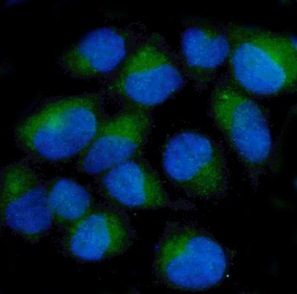
| What should IF/ICC show? | A centrosomal signal is the supported main subcellular observation; basal body, nucleolar, plasma membrane, spindle and primary-cilium observations are marked uncertain by HPA (HPA: subcellular localisation). UniProt also describes CKAP5 at centrosomes and mitotic spindle structures (UniProt Q14008: subcellular localisation). Interpret those observations within the separate IF/ICC guide. |
| Situation | Likely cause | Next action |
|---|---|---|
| No signal in an HPA High comparison population. | The expected cells may be absent from the section, or the staining run may have failed (HPA: tissue IHC; general IHC practice). | Confirm cell identity and section integrity, then check the existing IHC antibody and detection controls before interpreting a biological absence (general IHC practice). |
| Uniform colour covers cells and empty spaces. | Excess background or incomplete removal of detection reagents can obscure cellular staining (general IHC practice). | Review the detection control, blocking and washes; score CKAP5 only after cell boundaries and cytoplasmic colour can be distinguished (general IHC practice). |
| Colour is confined to nuclei or tissue edges. | This distribution conflicts with the reported cytoplasmic tissue profile and may be artefactual (HPA: tissue IHC; general IHC practice). | Compare intact internal cells with the affected regions and inspect a detection control before calling the stain positive (general IHC practice). |
| Only an unexpected cell population stains strongly. | Cross-reactivity or endogenous detection activity is possible, but an unlisted cell type is not proven negative by HPA's ubiquitous profile (HPA: tissue IHC; general IHC practice). | Check morphology and the detection control, then compare the result with HPA-described cells in the same tissue (HPA: tissue IHC; general IHC practice). |
| Cytoplasmic staining is present, but no centrosomal dots are visible. | Small centrosomal structures may be unresolved in chromogenic tissue IHC (UniProt Q14008: centrosome; general IHC practice). | Judge the tissue result against the supported cytoplasmic pattern and identified positive cells; do not require a resolvable centrosome (HPA: tissue IHC; general IHC practice). |
| A suspected mitotic cell shows staining that differs from neighbouring cells. | CKAP5 occupies spindle poles, spindle microtubules and kinetochores during mitosis (UniProt Q14008: subcellular localisation). | Check mitotic morphology and surrounding cytoplasmic staining before treating the difference as artefact; avoid assigning a specific spindle structure that the section cannot resolve (UniProt Q14008: subcellular localisation; general IHC practice). |


Comprehensive Human Protein Atlas IHC scoring per tissue (reliability: Supported — Medium consistency between antibody staining and RNA expression data.). Rows are taken directly from the HPA tissue chart — click any row's HPA link to view the source.
| Tissue | Cell type | Level | Evidence | Source |
|---|---|---|---|---|
| Adipose tissue | Adipocytes | High | Protein (IHC) | HPA → |
| Adrenal gland | Glandular cells | High | Protein (IHC) | HPA → |
| Bronchus | Ciliated cells (cell body) | High | Protein (IHC) | HPA → |
| Cerebral cortex | Neuronal cells | High | Protein (IHC) | HPA → |
| Cervix | Glandular cells | High | Protein (IHC) | HPA → |
| Tissue | Cell type | Level | Evidence | Source |
|---|---|---|---|---|
| None in HPA: CKAP5 is detected in all 45 scored tissues. Use a no-primary (secondary-only) and an isotype control instead. | ||||
Troubleshoot CKAP5 staining in paraffin sections by checking retrieval, cell compartment, detection background, and scoring before interpreting chromogenic signal.
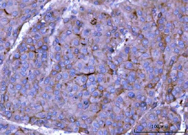
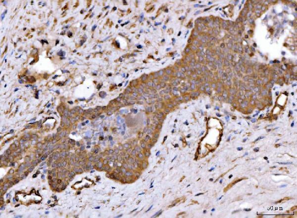
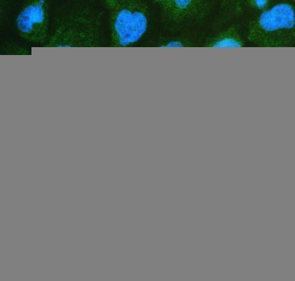
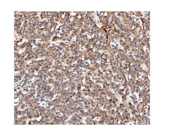
Two anti-CKAP5 antibodies have IHC images from human tumor and rodent brain paraffin sections, plus IF/ICC images from human cells (catalog IHC and IF captions).
A05324 has IHC images from human liver and prostate cancer and mouse and rat brain, plus an IF/ICC image from A431 cells (A05324 image captions). M05324 has IHC images from human liver, rectal, and renal cancer and rat brain, plus an IF/ICC image from HeLa cells (M05324 image captions).
Which to pick: For tissue IHC across human, mouse, and rat, choose rabbit antibody A05324, which has paraffin-section images in all three species (A05324 host and IHC captions). Choose mouse monoclonal M05324, clone 3C13, when its human or rat paraffin-section images and mouse-primary detection suit the experiment; mouse reactivity is listed, but no mouse IHC image is provided (M05324 catalog and IHC captions). For IF/ICC, A05324 is shown in A431 cells and M05324 in HeLa cells; the fixative used for either antibody’s paraffin-section IHC images is unreported (catalog IF and IHC captions).