This website uses cookies to ensure you get the best experience on our website.


- Table of Contents
Plan paraffin-section CKB IHC using the catalog antibody at 0.5–1 μg/mL (datasheet: A01695-1). Assess cytoplasmic staining in stomach parietal cells and CNS cells, with low staining expected in heart and skeletal myocytes (HPA tissue IHC).

Expected localisation, validated protocols, controls and antibodies — the at-a-glance facts below, then the full design guide.
| Expected localisation | Cytoplasmic in stomach parietal and CNS cells (HPA tissue IHC) | |
| Staining pattern | Parietal-cell cytoplasm; Purkinje cytoplasm/membrane (HPA tissue IHC) | |
| Antigen retrieval | Citrate pH 6 HIER, heat-mediated (datasheet A01695-1) | |
| Positive control | Cerebellum+4 more · see all | |
| Negative control | Adrenal gland+4 more · see all |
| Fixation | Keep fixation consistent across sections. (standard IHC practice; not target-specific) | |
| Caveat | Heart and skeletal myocytes show low CKB staining (HPA tissue IHC) | |
| Regulation | Regulation not annotated (UniProt) | |
| Isoform / epitope | No annotated isoforms; mature chain spans residues 2–381 (UniProt) |
Compare the catalog antibody’s IHC-P protocol (datasheet A01695-1) with four published CKB IHC protocols (PMC6221385; PMC10197964; PMC8494442; PMC7617069).
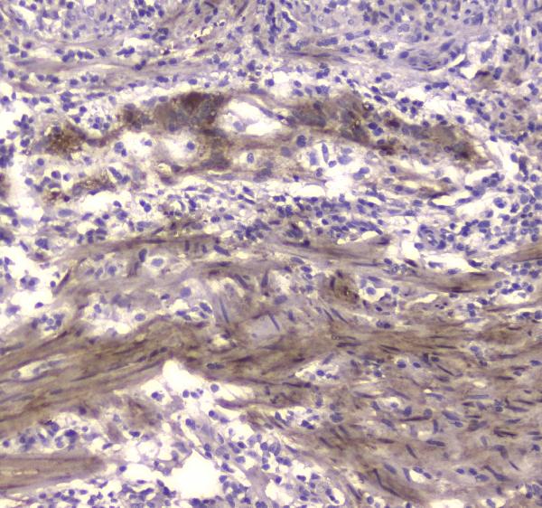
| Sample | Paraffin-embedded human colon cancer tissue; fixative not specified (datasheet A01695-1) |
| Fixation | Image fixative and duration unreported (datasheet A01695-1); verify before use. |
| Sectioning | 4–5 µm sections on charged slides (standard) |
| Deparaffinisation | Xylene, graded ethanol series to water (standard) |
| Antigen retrieval | Heat retrieval: Citrate pH 6, 20 min (datasheet A01695-1) |
| Peroxidase block | 3% H2O2, 10 min, room temperature (standard) |
| Blocking | 10% goat serum (datasheet A01695-1) |
| Primary antibody | Rabbit anti-CKB, 0.5-1μg/ml (datasheet A01695-1) |
| Primary incubation | Overnight at 4 °C (datasheet A01695-1) |
| Detection | Streptavidin-biotin complex (SABC), DAB chromogen (datasheet A01695-1) |
| Counterstain | Hematoxylin, blue, dehydrate and mount (standard) |
| Expected result | CKB-positive staining in purkinje cells - cytoplasm/membrane of cerebellum (HPA tissue IHC: High). HPA tissue profile: Cytoplasmic expression in parietal cells in stomach, and cells in the CNS. No signal in the no-primary control. |
CKB is mainly cytosolic; UniProt also lists mitochondrial and cell membrane localization, with mitochondrial localization specified for thermogenic fat cells (UniProt P12277). In paraffin IHC, expect cytoplasmic staining in CNS cells and stomach parietal cells (HPA tissue IHC). HPA rates the tissue staining pattern Enhanced, citing consistency between antibody staining and RNA expression (HPA tissue IHC). CKB has no transmembrane segment (UniProt P12277 topology).
| Strong Purkinje cell cytoplasmic/membrane staining or strong cerebral cortex neuropil staining (HPA tissue IHC). | These match HPA's High observations. Medium glial staining in caudate or hippocampus can also fit; compare the identified cell population, not just overall section darkness (HPA tissue IHC; general IHC practice). |
| Predominantly nuclear signal, with little cytoplasmic signal, in an expected positive cell population. | A nuclear-only pattern conflicts with the supported cytosolic location and reported tissue pattern (HPA subcellular ICC-IF; HPA tissue IHC). Check localization against the counterstain and staining controls before scoring it as CKB (general IHC practice). |
| Strong staining in adrenal gland glandular cells or bone marrow hematopoietic cells. | HPA reports CKB as Not detected in those specific cell populations (HPA tissue IHC). Unexpected signal warrants checks for antibody cross-reactivity and endogenous detection activity; a single discrepant section cannot establish which caused it (general IHC practice). |
| Diffuse chromogen across tissue and spaces, obscuring cell boundaries. | This is difficult to assign to the cytoplasmic cell patterns reported by HPA (HPA tissue IHC). Compare a no-primary control, blocking, washes and development time to assess nonspecific or detection background (general IHC practice). |
| No visible signal in cerebellar Purkinje cells or cerebral cortex neuropil. | Both are High HPA observations, so a blank expected-positive region calls for a run-level check (HPA tissue IHC). Confirm tissue identity and examine retrieval, antibody incubation and detection controls before interpreting other negatives (general IHC practice). |
| Tissue and cell context | HPA reports Medium staining in kidney collecting ducts, prostate glandular cells, rectal enterocytes and stomach glandular cells, alongside High CNS observations (HPA tissue IHC). Score the named cells separately from surrounding tissue (general IHC practice). |
| Localization and topology | Cytosol is the supported HPA ICC-IF location; UniProt additionally lists mitochondrion and cell membrane and specifies mitochondrial localization in thermogenic fat cells (HPA subcellular ICC-IF; UniProt P12277). No transmembrane segment is annotated (UniProt P12277 topology). |
| Isoenzyme context | CKB forms dimers with B or M chains; UniProt describes MM as major in skeletal muscle and myocardium, with MB in myocardium (UniProt P12277). HPA reports Low CKB staining in cardiomyocytes and skeletal myocytes (HPA tissue IHC). |
| Antibody evidence | Target-specific fixation effects are not established by the supplied assay evidence. Verify with a matched IHC source before attributing a result to fixation. |
| What should IF/ICC show? | Predominantly cytosolic signal is the supported HPA ICC-IF localization; HPA001254 has Supported ICC status (HPA subcellular ICC-IF; HPA antibody validation). Treat organelle or membrane assignments as context dependent under the UniProt annotation (UniProt P12277). |
| Situation | Likely cause | Next action |
|---|---|---|
| Expected-positive CNS region is blank. | A blank Purkinje cell or cortical neuropil region conflicts with HPA's High observations (HPA tissue IHC); a failed staining run is possible (general IHC practice). | Verify the region and a same-run positive control, then review retrieval, primary incubation and chromogenic detection steps (general IHC practice). |
| Only nuclei appear positive. | Nuclear-only staining does not match supported cytosolic localization (HPA subcellular ICC-IF). | Use the counterstain to confirm compartment boundaries; compare the no-primary control and reassess antibody concentration and detection background (general IHC practice). |
| Adrenal gland glandular cells stain strongly. | HPA records these cells as Not detected (HPA tissue IHC). Cross-reactivity or endogenous chromogenic activity may explain the discrepancy (general IHC practice). | Run a no-primary control and check the detection system's endogenous-activity block; repeat alongside an HPA High CNS region (general IHC practice; HPA tissue IHC). |
| Brown color is diffuse across the section. | Cell-independent color does not resemble HPA's localized cell observations (HPA tissue IHC); incomplete blocking, washing or excessive development may contribute (general IHC practice). | Compare no-primary and tissue controls, then review blocking, washes and chromogen development before scoring cells (general IHC practice). |
| Muscle is weaker than brain. | HPA reports Low staining in cardiomyocytes and skeletal myocytes, versus High staining in Purkinje cells and cortical neuropil (HPA tissue IHC). | Check the specified cell populations and same-run controls; do not use total creatine kinase activity to infer CKB staining, because B and M chains form different dimers (general IHC practice; UniProt P12277). |
| A negative cell population appears positive after longer development. | HPA's Not detected calls apply to specified cells, not every cell in an organ (HPA tissue IHC). Prolonged development can increase nonspecific color (general IHC practice). | Identify the stained cell type, compare no-primary and positive controls, and review development time before changing the interpretation (general IHC practice). |


Comprehensive Human Protein Atlas IHC scoring per tissue (reliability: Enhanced — High consistency between antibody staining and RNA expression data.). Rows are taken directly from the HPA tissue chart — click any row's HPA link to view the source.
| Tissue | Cell type | Level | Evidence | Source |
|---|---|---|---|---|
| Cerebellum | Purkinje cells - cytoplasm/membrane | High | Protein (IHC) | HPA → |
| Cerebral cortex | Neuropil | High | Protein (IHC) | HPA → |
| Caudate | Glial cells | Medium | Protein (IHC) | HPA → |
| Hippocampus | Glial cells | Medium | Protein (IHC) | HPA → |
| Kidney | Collecting ducts | Medium | Protein (IHC) | HPA → |
| Tissue | Cell type | Level | Evidence | Source |
|---|---|---|---|---|
| Adrenal gland | Glandular cells | Not detected | Protein (IHC) | HPA → |
| Bone marrow | Hematopoietic cells | Not detected | Protein (IHC) | HPA → |
| Breast | Adipocytes | Not detected | Protein (IHC) | HPA → |
| Bronchus | Respiratory epithelial cells | Not detected | Protein (IHC) | HPA → |
| Cervix | Glandular cells | Not detected | Protein (IHC) | HPA → |
Use the catalog antibody’s paraffin-section IHC evidence to troubleshoot CKB staining, and interpret the pattern against documented cell and compartment distributions (datasheet A01695-1; HPA tissue IHC; UniProt P12277).
The catalog covers human, mouse and rat CKB (catalog reactivity). IHC images show paraffin sections; no IF image is supplied (A01695-1 image captions; catalog IF images).
A01695-1 has IHC images from human colon cancer and mouse and rat kidney paraffin sections (A01695-1 IHC image captions). M01695 lists IHC and IF/ICC for human, mouse and rat, but supplies no IHC or IF image (M01695 catalog applications, reactivity and image fields).
Which to pick: For tissue IHC, choose A01695-1: its own caption documents citrate pH 6 retrieval and staining of a human colon cancer paraffin section; the fixative is unreported (A01695-1 IHC caption). For IF/ICC, M01695 is the listed option, but its IF/ICC performance needs validation because no image is supplied (M01695 catalog applications and image fields). Both list human, mouse and rat reactivity; A01695-1 also has mouse and rat kidney IHC images, whereas M01695 has no IHC image (catalog reactivity; A01695-1 and M01695 image fields).