This website uses cookies to ensure you get the best experience on our website.


- Table of Contents
Plan chromogenic CLCF1 IHC around the reported cytoplasmic tissue pattern and plasma positivity (HPA tissue IHC). Bone marrow hematopoietic cells and lung macrophages show medium staining, but HPA rates the tissue profile uncertain because secreted protein may differ from local RNA expression (HPA tissue IHC).

Expected localisation, validated protocols, controls and antibodies — the at-a-glance facts below, then the full design guide.
| Expected localisation | Cytoplasmic staining in several tissues (HPA tissue IHC) | |
| Staining pattern | Cytoplasmic staining in several tissues; plasma positive (HPA tissue IHC) | |
| Antigen retrieval | Citrate pH 6.0 HIER, 95–98 °C, 20 min (rule: cytoplasmic / membrane antigen) | |
| Positive control | Bone marrow+4 more · see all | |
| Negative control | Caudate+4 more · see all |
| Fixation | Keep formalin fixation consistent across sections (standard IHC practice; not target-specific) | |
| Caveat | Secretion can separate tissue RNA from protein staining (HPA tissue IHC) | |
| Regulation | CRLF1/CNTFR-dependent secretion (UniProt) | |
| Isoform / epitope | 2 isoforms; signal peptide 1–27; epitope impact unknown (UniProt) |
The catalog antibody IHC-P protocol is accompanied by published CLCF1 IHC methods for mouse IRI tissue (PMC9073803) and human kidney tissue (PMC12305993).
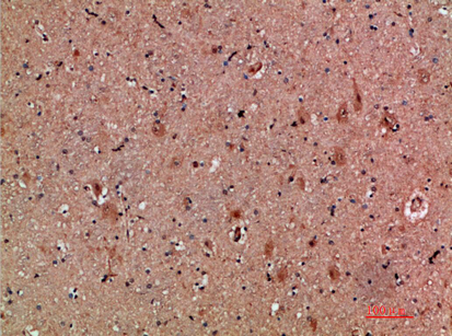
| Sample | Paraffin-embedded human brain tissue; fixative not specified (datasheet A08886) |
| Fixation | Image fixative and duration unreported (datasheet A08886); verify before use. |
| Sectioning | 4–5 µm sections on charged slides (standard) |
| Deparaffinisation | Xylene, graded ethanol series to water (standard) |
| Antigen retrieval | Heat-induced epitope retrieval in citrate buffer, pH 6.0, 20 min at 95–98 °C (standard rule: cytoplasmic / membrane antigen) |
| Peroxidase block | 3% H2O2, 10 min, room temperature (standard) |
| Blocking | 10% normal serum of the secondary host, 30 min, room temperature (standard) |
| Primary antibody | Rabbit anti-CLCF1, 1:50 recommended; image 1:200 (datasheet A08886) |
| Primary incubation | Overnight at 4 °C (standard) |
| Detection | HRP-polymer secondary, DAB chromogen 5–10 min (standard) |
| Counterstain | Hematoxylin, blue, dehydrate and mount (standard) |
| Expected result | CLCF1-positive staining in hematopoietic cells of bone marrow (HPA tissue IHC: Medium). HPA tissue profile: Cytoplasmic expression in several tissues and positivity in plasma. No signal in the no-primary control. |
CLCF1 is a secreted cytokine with no transmembrane segment (UniProt Q9UBD9 topology). In paraffin sections, expect mainly cytoplasmic staining in selected cells, including bone marrow hematopoietic cells and lung macrophages; HPA also reports positivity in plasma (HPA tissue IHC). Treat this as a provisional pattern: HPA rates its tissue staining Uncertain and notes that a secreted protein’s RNA and protein locations may differ (HPA tissue IHC).
| Cytoplasmic signal in bone marrow hematopoietic cells or lung macrophages. | This fits HPA’s Medium staining in both cell populations (HPA tissue IHC). Judge the labelled cells and their compartment separately from overall slide colour; the reported pattern has Uncertain reliability and awaits external verification (HPA tissue IHC). |
| Signal is confined to nuclei, with little cytoplasmic staining in an IHC section. | That departs from the reported tissue IHC profile of cytoplasmic expression (HPA tissue IHC). Check morphology and detection controls before calling it CLCF1. Nuclear bodies are an approved ICC-IF location, so the IF observation alone cannot validate nuclear IHC staining (HPA subcellular; HPA tissue IHC). |
| Strong staining appears in caudate glia or prostate glandular cells. | HPA reports CLCF1 as Not detected in those respective cell populations (HPA tissue IHC). Investigate antibody cross-reactivity, endogenous detection activity, or tissue identification before accepting an unexpected positive; an HPA negative is a reference observation, not proof that every specimen must be negative (HPA tissue IHC). |
| Colour spreads across unrelated cells, extracellular spaces, and blank areas. | A uniform haze is less persuasive than cell-associated cytoplasmic staining (HPA tissue IHC). Secreted CLCF1 and reported plasma positivity complicate extracellular interpretation (UniProt Q9UBD9 subcellular; HPA tissue IHC); compare background controls and morphology before assigning the signal to producing cells (general IHC practice). |
| No signal appears in bone marrow hematopoietic cells on a stained section. | HPA reports Medium staining there, so inspect the section, antibody and detection controls, and staining conditions (HPA tissue IHC; general IHC practice). This is a useful reference tissue, but HPA’s Uncertain reliability does not make a negative result conclusive (HPA tissue IHC). |
| Secretion and tissue location (UniProt Q9UBD9; HPA tissue IHC) | CLCF1 is secreted, and HPA notes plasma positivity and possible RNA–protein location differences (UniProt Q9UBD9 subcellular; HPA tissue IHC). Cytoplasmic staining can support a cellular pattern, but extracellular signal cannot by itself identify the producing cell (general IHC interpretation). |
| Mature protein and signal peptide (UniProt Q9UBD9 processing) | The precursor has a signal peptide at residues 1–27; the annotated mature chain spans 28–225 (UniProt Q9UBD9 processing). Epitope location would affect which form an antibody recognizes, but the supplied evidence does not identify this antibody’s epitope. |
| Isoforms and glycosylation (UniProt Q9UBD9) | UniProt lists 2 isoforms and one glycosylation site at residue 29 (UniProt Q9UBD9 isoforms; UniProt Q9UBD9 glycosylation). Their effects on this antibody’s paraffin-section staining are unreported; avoid assigning an unexpected pattern to either feature without further evidence. |
| Tissue reference and confidence (HPA tissue IHC) | HPA reports Medium staining in several distinct cell populations and Not detected staining in others, but rates the tissue profile Uncertain pending external verification (HPA tissue IHC). Use these observations as comparison points rather than definitive acceptance criteria. |
| IF/ICC pattern? (HPA subcellular; HPA antibodies) | For IF/ICC, HPA approves nuclear bodies and vesicles as locations and lists antibody HPA042444 as ICC Approved (HPA subcellular; HPA antibodies). Its IHC status is Uncertain; the IF/ICC locations do not establish a paraffin IHC nuclear pattern (HPA antibodies; HPA tissue IHC). |
| Situation | Likely cause | Next action |
|---|---|---|
| Reference cells show no detectable chromogen. | The tissue may lack identifiable reference cells, or the IHC staining workflow may have failed (general IHC practice). HPA’s Medium bone marrow signal is an uncertain reference, not a guaranteed result (HPA tissue IHC). | Confirm hematopoietic cells in the section, then check antibody application, detection reagents, and a working assay control (general IHC practice). Interpret any persistent negative alongside HPA’s Uncertain tissue reliability (HPA tissue IHC). |
| Most cells and blank regions stain similarly. | Nonspecific reagent binding or detection background can obscure a cellular pattern (general IHC practice). HPA describes cytoplasmic staining in several tissues and plasma positivity, not uniform staining of the entire section (HPA tissue IHC). | Inspect a no-primary control and review blocking, washes, and chromogen development (general IHC practice). Score cell-associated signal only where morphology and controls permit; avoid treating generalized haze as CLCF1 (general IHC interpretation). |
| An HPA Not detected population stains strongly. | Possible explanations include cross-reactivity, endogenous detection activity, or misidentified cells (general IHC practice). The comparison is against HPA observations for the named population, not a universal absence claim (HPA tissue IHC). | Verify cell identity and compare no-primary and detection controls; assess whether staining tracks the expected cytoplasmic profile in HPA Medium populations (general IHC practice; HPA tissue IHC). |
| Only nuclei stain in paraffin IHC. | A nuclear-only IHC result conflicts with HPA’s cytoplasmic tissue profile (HPA tissue IHC). HPA’s approved nuclear-body localisation comes from ICC-IF, a different application (HPA subcellular; HPA antibodies). | Check counterstain interpretation, tissue morphology, and detection controls before assigning nuclear CLCF1 (general IHC practice). Record the IHC–ICC difference as unresolved if controls are clean (HPA tissue IHC; HPA subcellular). |
| Extracellular or plasma signal dominates the section. | CLCF1 is secreted, and HPA reports plasma positivity; signal away from cells therefore needs careful attribution (UniProt Q9UBD9 subcellular; HPA tissue IHC). Diffuse background remains another possibility (general IHC practice). | Compare signal with vessel and cell morphology and a no-primary control (general IHC practice). Report extracellular staining separately from cytoplasmic staining; do not infer its cell of origin from location alone (UniProt Q9UBD9 subcellular). |
| IF/ICC shows puncta while paraffin IHC appears cytoplasmic. | HPA approves nuclear bodies and vesicles in ICC-IF, while its tissue IHC summary describes cytoplasmic staining (HPA subcellular; HPA tissue IHC). Those application-specific observations need not yield identical images. | Interpret each result using its own application and controls (general IHC/IF practice). Use the dedicated IF/ICC guide for IF design; do not transfer an ICC-approved location into an IHC positive call without supporting IHC evidence (HPA antibodies; HPA tissue IHC). |

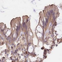


Comprehensive Human Protein Atlas IHC scoring per tissue (reliability: Uncertain — Secreted protein, tissue location of RNA and protein is expected to differ. Pending external verification.). Rows are taken directly from the HPA tissue chart — click any row's HPA link to view the source.
| Tissue | Cell type | Level | Evidence | Source |
|---|---|---|---|---|
| Bone marrow | Hematopoietic cells | Medium | Protein (IHC) | HPA → |
| Breast | Myoepithelial cells | Medium | Protein (IHC) | HPA → |
| Bronchus | Respiratory epithelial cells | Medium | Protein (IHC) | HPA → |
| Heart muscle | Cardiomyocytes | Medium | Protein (IHC) | HPA → |
| Kidney | Cells in tubules | Medium | Protein (IHC) | HPA → |
| Tissue | Cell type | Level | Evidence | Source |
|---|---|---|---|---|
| Caudate | Glial cells | Not detected | Protein (IHC) | HPA → |
| Cerebellum | Cells in granular layer | Not detected | Protein (IHC) | HPA → |
| Esophagus | Squamous epithelial cells | Not detected | Protein (IHC) | HPA → |
| Prostate | Glandular cells | Not detected | Protein (IHC) | HPA → |
| Salivary gland | Glandular cells | Not detected | Protein (IHC) | HPA → |
Troubleshoot CLCF1 staining in paraffin sections by checking retrieval, compartment, cell identity and controls before scoring chromogenic signal.
A08886 is listed for IHC and IF in human, mouse, and rat; its supplied image shows IHC in paraffin-embedded human brain (catalog applications and reactivity; IHC image caption).
A08886 will render with an IHC image of paraffin-embedded human brain at 1:200 (IHC image caption). IF is listed, but no IF image is supplied; human, mouse, and rat reactivity is listed (catalog applications and reactivity; IF image alts).
Which to pick: For tissue IHC, choose A08886 when working with paraffin sections; its image shows human brain, and the fixative is unreported (IHC image caption). For IF, A08886 is listed at 1:50, but ICC validation is unreported (catalog applications and IF dilution). A08886 lists human, mouse, and rat reactivity, though the supplied IHC image documents human tissue only (catalog reactivity; IHC image caption).