This website uses cookies to ensure you get the best experience on our website.


- Table of Contents
Plan CLDN10 paraffin IHC around membranous staining in renal tubules and glandular cells (HPA tissue IHC). The catalog antibody lists an IHC dilution of 1:50–1:200 (datasheet); interpret renal segments cautiously because UniProt reports TAL expression and HPA lists proximal tubule cell bodies (UniProt; HPA tissue IHC).

Expected localisation, validated protocols, controls and antibodies — the at-a-glance facts below, then the full design guide.
| Expected localisation | Membranous staining in renal and glandular epithelia (HPA tissue IHC) | |
| Staining pattern | Membranous renal tubules and exocrine or glandular cells (HPA tissue IHC) | |
| Antigen retrieval | Citrate pH 6.0 HIER, 95–98 °C, 20 min (rule: cytoplasmic / membrane antigen) | |
| Positive control | Gallbladder+4 more · see all | |
| Negative control | Adipose tissue+4 more · see all |
| Fixation | Keep fixation consistent across paraffin sections. (standard IHC practice; not target-specific) | |
| Caveat | Renal segment calls differ: TAL versus proximal tubules (UniProt; HPA tissue IHC) | |
| Regulation | Regulation not specified (UniProt) | |
| Isoform / epitope | 3 isoforms; check epitope side and variant coverage (UniProt) |
The catalog antibody’s IHC-P protocol is accompanied by four published CLDN10 IHC protocols (provided article excerpts).
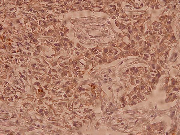
| Sample | Paraffin-embedded human breast carcinoma tissue; fixative not specified (datasheet A10653-1) |
| Fixation | Image fixative and duration unreported (datasheet A10653-1); verify before use. |
| Sectioning | 4–5 µm sections on charged slides (standard) |
| Deparaffinisation | Xylene, graded ethanol series to water (standard) |
| Antigen retrieval | Heat-induced epitope retrieval in citrate buffer, pH 6.0, 20 min at 95–98 °C (standard rule: cytoplasmic / membrane antigen) |
| Peroxidase block | 3% H2O2, 10 min, room temperature (standard) |
| Blocking | 10% normal serum of the secondary host, 30 min, room temperature (standard) |
| Primary antibody | Rabbit anti-CLDN10, 1:50-1:200 (datasheet A10653-1) |
| Primary incubation | Overnight at 4 °C (standard) |
| Detection | HRP-polymer secondary, DAB chromogen 5–10 min (standard) |
| Counterstain | Hematoxylin, blue, dehydrate and mount (standard) |
| Expected result | CLDN10-positive staining in glandular cells of gallbladder (HPA tissue IHC: Medium). HPA tissue profile: Membranous expression in several tissues, most abundant in renal tubules, exocrine pancreas, gall bladder and salivary gland. No signal in the no-primary control. |
CLDN10 is a four-pass membrane protein at tight junctions (UniProt P78369 topology and location). In paraffin-section IHC, expect membranous staining in renal tubules and glandular epithelium, especially exocrine pancreas, gallbladder and salivary gland (HPA tissue IHC). HPA rates the tissue staining “Enhanced,” with medium consistency between antibody staining and RNA expression (HPA tissue IHC reliability).
| Membrane-associated staining outlines renal tubular or glandular epithelial cells. | This fits CLDN10 localisation at cell membranes and tight junctions (UniProt P78369 location) and HPA’s membranous tissue profile (HPA tissue IHC). Judge the pattern alongside tissue architecture: a positive chromogenic deposit should be attributable to epithelial cell borders, without requiring every cell or every border to stain equally. |
| Strong, widespread nuclear or purely cytoplasmic staining replaces a discernible membrane pattern. | That compartment disagrees with the annotated membrane and tight-junction location (UniProt P78369 location) and the observed membranous IHC profile (HPA tissue IHC). Treat it as suspect staining and review morphology, detection controls and antibody conditions before assigning it to CLDN10; the sources do not validate a nuclear pattern. |
| Signal dominates a cell population outside the reported epithelial pattern. | HPA reports staining in renal tubules, exocrine pancreatic cells and glandular cells of gallbladder and salivary gland (HPA tissue IHC). Unexpected staining may reflect cross-reactivity or endogenous detection activity; morphology and a detection-only control can help distinguish these possibilities. It does not, by itself, establish new CLDN10 expression. |
| A uniform haze or deposit covers epithelium, stroma and empty areas. | A field-wide pattern cannot be assigned confidently to the membrane-associated signal reported for CLDN10 (HPA tissue IHC; UniProt P78369 location). As general IHC practice, assess background with an appropriate negative control, then review blocking, wash steps and detection conditions. Preserve cell-border detail when judging any remaining signal. |
| No convincing staining appears in a known-positive control section. | HPA reports medium staining in renal proximal tubules and in exocrine pancreatic, gallbladder and salivary glandular cells (HPA tissue IHC). A blank result in a suitable control leaves the run unvalidated. Check control tissue identity and preservation, antibody and detection steps, and section quality before interpreting an experimental specimen as negative. |
| Membrane topology and epitope access | CLDN10 has 4 transmembrane segments, with extracellular and cytoplasmic regions (UniProt P78369 topology). Epitope position for the antibodies listed here is unspecified. For a particular antibody, consult its own epitope information before predicting whether retrieval or permeabilisation will expose the recognised site; topology alone cannot settle that choice. |
| Renal site of staining | UniProt reports CLDN10 in the thick ascending limb of Henle’s loop, while HPA’s listed IHC positive is proximal-tubule cell body at medium level (UniProt P78369 tissue specificity; HPA tissue IHC). Identify renal structures morphologically and report the observed compartment. The supplied records do not resolve the difference between those descriptions. |
| Isoform coverage | UniProt lists 3 CLDN10 isoforms (UniProt P78369 isoforms). The supplied antibody entries provide no isoform-specific epitope information (HPA antibody records). A positive result establishes staining with the chosen reagent under its tested conditions; it cannot identify an isoform from these sources alone. |
| IHC validation and expected intensity | HPA lists HPA042348 and CAB012969 as rabbit polyclonal antibodies with “Enhanced” IHC validation (HPA antibody records). Its tissue profile still describes medium consistency with RNA data and medium staining in the listed positive cell types (HPA tissue IHC). Use the validation as support for the pattern, not a promise of uniform staining in every specimen. |
| Q: Can the same pattern be assumed for IF/ICC? | A: HPA summarises a membrane location but supplies no ICC-IF image-bearing cell line or antibody ICC validation in this payload (HPA subcellular; HPA antibody records). UniProt supports a tight-junction and membrane location (UniProt P78369 location). Interpret IF/ICC against its own controls and guide; these IHC observations do not establish an IF/ICC protocol or intensity. |
| Situation | Likely cause | Next action |
|---|---|---|
| Known-positive renal or glandular control is blank. | The run may have failed, or the selected section may lack the reported positive cells; HPA’s listed positive cells stain at medium level (HPA tissue IHC). | Confirm control morphology and the presence of the relevant epithelium. As general IHC practice, verify primary-antibody application, retrieval and chromogenic detection against the reagent instructions before scoring study tissue. |
| Staining appears mainly nuclear or broadly cytoplasmic. | That distribution conflicts with the membrane and tight-junction annotation and HPA’s membranous IHC profile (UniProt P78369 location; HPA tissue IHC). | Inspect cell borders at higher magnification and compare a negative control. If the unexpected compartment persists without credible membrane staining, treat the result as unconfirmed rather than assigning it to CLDN10. |
| Color develops across multiple unrelated cell types or the whole section. | Cross-reactivity, endogenous detection activity or general background are possible IHC explanations; HPA’s reported pattern is epithelial and membranous (HPA tissue IHC). | Use a detection-only control to assess reagent-derived signal, then review blocking and washing as general IHC practice. Reassess whether any remaining deposit follows the reported cells and their membranes. |
| A renal section stains, but the labelled tubule differs from the expected segment. | The source descriptions differ: UniProt names the thick ascending limb, whereas HPA lists proximal-tubule cell body staining (UniProt P78369 tissue specificity; HPA tissue IHC). | Check segment assignment against section morphology and document the observed cells and compartment. Do not resolve the source difference from stain intensity alone. |
| A listed HPA-negative tissue shows convincing-looking color. | HPA marks adipocytes in adipose tissue and adrenal glandular cells, among other listed examples, as “Not detected” (HPA tissue IHC); staining there warrants scrutiny. | Confirm the cell identity and membrane pattern, then compare negative and known-positive controls. Record the discrepancy as an observation rather than extending the HPA expression profile from one slide. |
| Membrane staining is present, but an isoform-specific conclusion is needed. | UniProt records 3 isoforms, while the supplied antibody records do not identify isoform-specific recognition (UniProt P78369 isoforms; HPA antibody records). | Report CLDN10-associated staining and the antibody identifier. Reserve isoform attribution for separate evidence that establishes which isoform the antibody recognises. |


Comprehensive Human Protein Atlas IHC scoring per tissue (reliability: Enhanced — Medium consistency between antibody staining and RNA expression data.). Rows are taken directly from the HPA tissue chart — click any row's HPA link to view the source.
| Tissue | Cell type | Level | Evidence | Source |
|---|---|---|---|---|
| Gallbladder | Glandular cells | Medium | Protein (IHC) | HPA → |
| Kidney | Proximal tubules (cell body) | Medium | Protein (IHC) | HPA → |
| Pancreas | Exocrine glandular cells | Medium | Protein (IHC) | HPA → |
| Salivary gland | Glandular cells | Medium | Protein (IHC) | HPA → |
| Tonsil | Squamous epithelial cells | Medium | Protein (IHC) | HPA → |
| Tissue | Cell type | Level | Evidence | Source |
|---|---|---|---|---|
| Adipose tissue | Adipocytes | Not detected | Protein (IHC) | HPA → |
| Adrenal gland | Glandular cells | Not detected | Protein (IHC) | HPA → |
| Appendix | Glandular cells | Not detected | Protein (IHC) | HPA → |
| Bone marrow | Hematopoietic cells | Not detected | Protein (IHC) | HPA → |
| Breast | Adipocytes | Not detected | Protein (IHC) | HPA → |
Troubleshoot CLDN10 staining in paraffin section IHC by checking retrieval, membrane localisation, tissue context, and controls before scoring.
A10653-1 has a paraffin-section IHC image from human breast carcinoma (IHC image caption); IF is listed without an IF image (catalog applications; IF image alts). Human, mouse and rat reactivity is listed (catalog reactivity).
A10653-1 is shown by IHC on paraffin-embedded human breast carcinoma at 1:100 (IHC image caption). The catalog lists IHC and IF applications and human, mouse and rat reactivity for A10653-1 (catalog applications; catalog reactivity).
Which to pick: For tissue IHC, choose A10653-1 as a rabbit polyclonal antibody with a paraffin-section human breast carcinoma IHC image; the fixative is unreported (catalog host; IHC image caption). For IF, A10653-1 is listed at 1:50–1:200, but no IF image or ICC validation is supplied (catalog applications; catalog IF dilution; IF image alts). For mouse or rat samples, A10653-1 has listed reactivity, while its supplied IHC image shows human tissue only (catalog reactivity; IHC image caption).