This website uses cookies to ensure you get the best experience on our website.


- Table of Contents
Plan CLDN2 paraffin-section IHC around membranous staining in gallbladder, kidney and GI (HPA tissue IHC). This guide covers tissue controls, fixation, detection and interpretation, with a starting antibody concentration of 0.5–1 μg/mL (datasheet: RP1042).

Expected localisation, validated protocols, controls and antibodies — the at-a-glance facts below, then the full design guide.
| Expected localisation | Membranous staining in gallbladder, kidney and GI (HPA tissue IHC) | |
| Staining pattern | Membranous glandular and proximal tubule staining (HPA tissue IHC) | |
| Antigen retrieval | Citrate pH 6.0 HIER, 95–98 °C, 20 min (rule: cytoplasmic / membrane antigen) | |
| Positive control | Gallbladder+1 more · see all | |
| Negative control | Adipose tissue+4 more · see all |
| Fixation | Keep fixation consistent across sections. (standard IHC practice; not target-specific) | |
| Caveat | Presumed off-target binding may confound staining (HPA tissue IHC) | |
| Regulation | Expression regulation not annotated (UniProt) | |
| Isoform / epitope | No isoforms; extracellular and cytoplasmic epitopes differ (UniProt) |
Compare the catalog antibody’s IHC-P protocol with published CLDN2 protocols for goat jejunum (PMC4847856), human lung tissue microarrays (PMC10366158), and pancreatic FFPE samples (PMC13222116).
| Sample | Tissue sections; selected-image fixative not specified (standard IHC workflow) |
| Fixation | Image fixative and duration unreported (datasheet RP1042); verify before use. |
| Sectioning | 4–5 µm sections on charged slides (standard) |
| Deparaffinisation | Xylene, graded ethanol series to water (standard) |
| Antigen retrieval | Heat-induced epitope retrieval in citrate buffer, pH 6.0, 20 min at 95–98 °C (standard rule: cytoplasmic / membrane antigen) |
| Peroxidase block | 3% H2O2, 10 min, room temperature (standard) |
| Blocking | 10% normal serum of the secondary host, 30 min, room temperature (standard) |
| Primary antibody | Rabbit anti-CLDN2, 0.5-1μg/ml (datasheet RP1042) |
| Primary incubation | Overnight at 4 °C (standard) |
| Detection | HRP-polymer secondary, DAB chromogen 5–10 min (standard) |
| Counterstain | Hematoxylin, blue, dehydrate and mount (standard) |
| Expected result | CLDN2-positive staining in glandular cells of gallbladder (HPA tissue IHC: High). HPA tissue profile: Membranous expression in gall bladder, kidney and GI. No signal in the no-primary control. |
CLDN2 is a 4-pass membrane protein at tight junctions (UniProt P57739 topology; UniProt P57739 subcellular). In paraffin-section IHC, expect membranous staining in gallbladder glandular cells and kidney proximal tubules (HPA: gallbladder High; kidney Medium). HPA rates its tissue IHC profile Enhanced, while noting medium agreement with RNA data and presumed off-target staining that was disregarded (HPA: tissue IHC reliability).
| Distinct staining outlines gallbladder glandular cells, with less intense staining in kidney proximal tubules. | This fits HPA's membranous profile and its High gallbladder versus Medium kidney levels (HPA: tissue IHC). Judge the relevant cells and their borders; a whole-section average can obscure the expected cell pattern. |
| Strong nuclear or broadly cytoplasmic chromogen dominates the expected epithelial borders. | That is discordant with the main tight-junction and membrane localization (UniProt P57739 subcellular; HPA: tissue IHC). Treat it as possible nonspecific staining until controls support it; HPA's additional nuclear and vesicular locations come from ICC-IF, not tissue IHC (HPA: subcellular). |
| The same conspicuous signal appears in the expected cells and in unrelated cells, including cells in a comparison section. | Consider cross-reactivity or endogenous detection activity before scoring those extra cells as CLDN2. HPA reports presumed off-target tissue staining that it disregarded (HPA: tissue IHC reliability). Appendix glandular cells are a useful comparison because HPA reports them as not detected (HPA: appendix). |
| A diffuse tint covers tissue, lumina, or the slide rather than tracing cell borders. | This cannot establish the membranous pattern reported by HPA (HPA: tissue IHC). Review the negative detection control and background across the section; in routine IHC, excess reagent signal or incomplete washing can obscure localization. |
| Gallbladder glandular cells lack detectable staining in a section expected to be positive. | The result conflicts with HPA's High gallbladder finding (HPA: gallbladder). Check the assay and section before interpreting absence as biology. A negative result from one preparation does not by itself overturn a tissue-level reference profile. |
| Membrane topology | CLDN2 has 4 transmembrane segments, extracellular loops, and cytoplasmic regions (UniProt P57739 topology). Interpret a border-associated pattern in light of this location; topology alone does not specify an antibody's epitope or an antigen-retrieval condition. |
| Cell and tissue distribution | HPA reports High gallbladder glandular staining, Medium kidney proximal-tubule staining, and Low staining in stomach, duodenum, small intestine, and seminal-vesicle glandular cells (HPA: tissue IHC). Match the named cell type when choosing comparisons. |
| Tissue IHC confidence | HPA calls the tissue profile Enhanced but describes medium agreement between antibody staining and RNA expression; presumed off-target staining was disregarded (HPA: tissue IHC reliability). Use localization and comparison tissue together when judging an unexpected signal. |
| Antibody-specific validation | HPA lists CAB002609 as IHC Enhanced; its other listed antibodies have no supplied IHC rating (HPA: antibodies). That rating does not establish the performance of a different catalog antibody, so evaluate that antibody against the expected tissue pattern. |
| Processing and fixation evidence | UniProt lists one chain spanning residues 1–230, with no signal peptide, propeptide, or annotated glycosylation site (UniProt P57739 processing). These features do not establish fixation sensitivity; no target-specific fixation effect is supplied. |
| Situation | Likely cause | Next action |
|---|---|---|
| Gallbladder glandular cells are blank. | A technical failure is possible because HPA reports High staining in those cells (HPA: gallbladder). The supplied sources do not identify a CLDN2-specific retrieval failure. | Confirm the expected cells are present; review antibody dilution, detection reagents, and the run's positive control. If optimizing retrieval, document it as general IHC optimization rather than a known CLDN2 requirement. |
| Only diffuse cytoplasmic or nuclear stain is visible. | The compartment conflicts with the main membrane and junction location (UniProt P57739 subcellular; HPA: tissue IHC). ICC-IF additional locations do not validate dominant nuclear tissue-IHC staining (HPA: subcellular). | Compare cell borders with the counterstain and a negative detection control; adjust general background controls before assigning a CLDN2-positive score. |
| Appendix glandular cells stain as strongly as gallbladder glandular cells. | HPA reports appendix glandular cells as not detected and gallbladder glandular cells as High (HPA: tissue IHC). Cross-reactivity or endogenous detection activity may explain the discrepancy. | Inspect negative detection controls and localization in both sections; record the discordance. An HPA 'not detected' observation is a reference pattern, not proof that every appendix specimen must be blank. |
| Low-level GI staining looks negative beside gallbladder. | HPA rates stomach, duodenum, and small-intestine glandular cells Low, versus High gallbladder glandular cells (HPA: tissue IHC). Direct intensity comparison can hide a weak localized result. | Score the named glandular cells and membrane pattern in each tissue separately. Keep exposure or chromogen development comparable within the run when assessing relative intensity. |
| Background obscures the epithelial borders. | Diffuse signal prevents assessment of the membranous tissue profile (HPA: tissue IHC); nonspecific reagent binding or endogenous detection activity are routine IHC possibilities. | Check negative detection controls, blocking and washing, then adjust antibody or detection conditions according to the assay. Do not infer a CLDN2-specific fixation effect from background alone. |
| Can an ICC-IF nuclear or vesicular signal be used to score tissue IHC? | HPA's supported main ICC-IF locations are plasma membrane and cell junctions; nucleoplasm, nucleoli, and vesicles are additional approved locations (HPA: subcellular). | Use the tissue-IHC membranous profile to score paraffin sections (HPA: tissue IHC). Evaluate ICC-IF localization separately; it does not supply an IHC protocol or validate nuclear chromogen staining. |

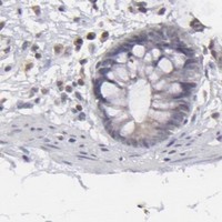


Comprehensive Human Protein Atlas IHC scoring per tissue (reliability: Enhanced — Medium consistency between antibody staining and RNA expression data. Presumed off target binding observed and disregarded.). Rows are taken directly from the HPA tissue chart — click any row's HPA link to view the source.
| Tissue | Cell type | Level | Evidence | Source |
|---|---|---|---|---|
| Gallbladder | Glandular cells | High | Protein (IHC) | HPA → |
| Kidney | Proximal tubules (cell body) | Medium | Protein (IHC) | HPA → |
| Tissue | Cell type | Level | Evidence | Source |
|---|---|---|---|---|
| Adipose tissue | Adipocytes | Not detected | Protein (IHC) | HPA → |
| Appendix | Glandular cells | Not detected | Protein (IHC) | HPA → |
| Bone marrow | Hematopoietic cells | Not detected | Protein (IHC) | HPA → |
| Bronchus | Respiratory epithelial cells | Not detected | Protein (IHC) | HPA → |
| Caudate | Glial cells | Not detected | Protein (IHC) | HPA → |
Troubleshoot CLDN2 staining in paraffin sections by checking retrieval, tissue preservation, junctional localisation and cell-specific scoring.
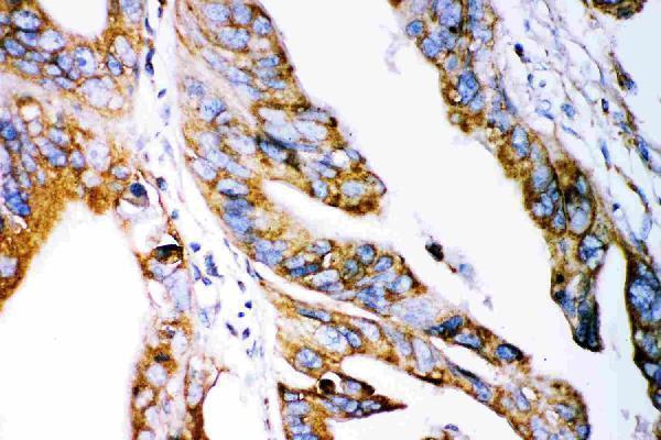
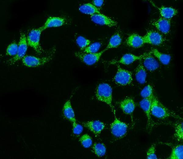
Anti-CLDN2 antibodies have image data for paraffin-section IHC in human intestinal cancer tissue (RP1042 IHC image caption) and IF/ICC in human Caco-2 cells (A03033-1 IF image caption).
RP1042 is listed for IHC and shows staining in a human intestinal cancer paraffin section (RP1042 applications; RP1042 IHC image caption). A03033-1 is listed for IF/ICC and shows staining in human Caco-2 cells (A03033-1 applications; A03033-1 IF image caption).
Which to pick: Choose RP1042 for tissue IHC: its image shows a paraffin section, its human IHC-P dilution is 0.5–1 μg/mL, and the fixative is unreported (RP1042 IHC image caption; RP1042 datasheet). Choose A03033-1 for IF/ICC in human cells; its pictured Caco-2 assay used 5 μg/mL (A03033-1 applications; A03033-1 IF image caption). For rat work, RP1042 is the only SKU listing rat reactivity, although its pictured IHC sample is human; clonality is unreported for both antibodies (catalog reactivity; RP1042 IHC image caption; catalog clone fields).