CLDN3 / Claudin-3 · IHC design guide

Design Immunohistochemistry for CLDN3

Plan CLDN3 paraffin-section IHC around membranous staining in gastrointestinal and glandular tissues (HPA tissue IHC). The catalog antibody has an IHC range of 2–5 μg/mL (datasheet A04393-5); breast glandular cells are a positive control and adipocytes a negative control (HPA tissue IHC).

Evidence assembled Oct 2026 · For research use; verify linked source records and product datasheet before use
Immunohistochemistry protocol sheet for CLDN3 (IHC for CLDN3): expected localisation Membranous tissue staining (HPA tissue IHC), antibody A04393-5, validated IHC image, and IHC protocol steps
Printable CLDN3 IHC protocol sheet — expected localisation Membranous tissue staining (HPA tissue IHC), antibody A04393-5, controls and protocol steps. Open the full CLDN3 IHC guide →

CLDN3 Immunohistochemistry Experimental Design Guide

Expected localisation, validated protocols, controls and antibodies — the at-a-glance facts below, then the full design guide.

Must know before staining
Expected localisation Membranous tissue staining (HPA tissue IHC)
Staining pattern Membranous staining in gastrointestinal and glandular tissues (HPA tissue IHC)
Antigen retrieval EDTA pH 8.0 HIER, heat-mediated (datasheet A04393-5)
Positive control ⓘ Appendix+4 more · see all
Negative control ⓘ Adipose tissue+4 more · see all
Important caveats
Reasons your staining may differ from the expected pattern.
Fixation Keep fixation consistent across paraffin sections (standard IHC practice; not target-specific)
Caveat Staining varies by cell type within tissues (HPA tissue IHC)
Regulation Expression regulation not annotated (UniProt)
Isoform / epitope No annotated isoforms; extracellular loops and cytoplasmic tail differ in epitope location (UniProt)
Section 1

Recommended CLDN3 IHC & IF Protocols

The catalog antibody protocol uses EDTA pH 8.0 retrieval (datasheet A04393-5). The following published IHC protocols cover breast, intestinal and lung tissue (PMC1064136; PMC13073207; PMC12790956; PMC6631579).

Recommended immunohistochemistry (IHC-P) protocol parameters
SampleParaffin-embedded human renal clear cell carcinoma tissue; fixative not specified (datasheet A04393-5)
FixationImage fixative and duration unreported (datasheet A04393-5); verify before use.
Sectioning4–5 µm sections on charged slides (standard)
DeparaffinisationXylene, graded ethanol series to water (standard)
Antigen retrievalHeat retrieval: EDTA pH 8.0 (datasheet A04393-5); 20 min, 95–100 °C (standard)
Peroxidase block3% H2O2, 10 min, room temperature (standard)
Blocking10% goat serum (datasheet A04393-5)
Primary antibodyRabbit anti-CLDN3, 2-5μg/ml (datasheet A04393-5)
Primary incubationOvernight at 4 °C (datasheet A04393-5)
DetectionStreptavidin-biotin complex (SABC), DAB chromogen (datasheet A04393-5)
CounterstainHematoxylin, blue, dehydrate and mount (standard)
Expected resultCLDN3-positive staining in endocrine cells of appendix (HPA tissue IHC: High). HPA tissue profile: Membranous expression in several tissues, including gastrointestinal tract and glandular tissues. No signal in the no-primary control.
💡Decision noteStart with heat-mediated EDTA pH 8.0 retrieval for the catalog antibody (datasheet A04393-5); the published protocols used other retrieval conditions with their respective antibodies (PMC1064136; PMC13073207; PMC12790956; PMC6631579).
Section 2

What Is the Expected CLDN3 Staining Pattern?

CLDN3 is a four-pass membrane protein of tight junctions, so expect junctional membrane staining in the cell types where tissue IHC detects it (UniProt O15551 topology; HPA tissue IHC). HPA reports high staining in selected intestinal endocrine cells and glandular cells, with an Enhanced tissue IHC reliability rating based on agreement between antibody staining and RNA expression (HPA tissue IHC).

What am I looking at on my slide?
Crisp membrane staining outlines contacts between the expected cells, such as breast glandular cells or colon endocrine cells (HPA tissue IHC).This fits CLDN3 localisation at tight junctions and the cell membrane (UniProt O15551 subcellular location). Score the named cell population and its junctional pattern together; an adjacent cell population need not share the same staining level (HPA tissue IHC).
Strong nuclear staining or broad cytoplasmic staining dominates while junctional borders are indistinct.That compartment pattern does not match the annotated tight-junction and membrane location (UniProt O15551 subcellular location). Treat it as suspect staining; compare with a no-primary control and reassess antibody and detection conditions before scoring it as CLDN3 (standard IHC practice).
Adipocytes, cardiomyocytes or esophageal squamous cells stain strongly (HPA: Not detected in those cell types).Those results conflict with the listed HPA tissue IHC observations, though they do not prove the cause. Check whether the signal persists without primary antibody; persistent color suggests endogenous detection activity, while primary-dependent staining calls for specificity review (standard IHC practice).
Color spreads across stroma, empty spaces or many cell types without clear membrane borders.A diffuse pattern is difficult to reconcile with CLDN3's junctional membrane localisation (UniProt O15551 subcellular location). Compare tissue morphology and a no-primary control, then review blocking, washes and detection background (standard IHC practice).
No junctional signal appears in breast glandular cells or colon endocrine cells on an otherwise interpretable section (HPA: High in those cells).A negative result in an HPA high-staining cell population warrants a run-level check, but HPA does not establish how fixation affects CLDN3. Verify the positive-control tissue, antibody validation, retrieval and detection workflow before interpreting the specimen as negative (HPA tissue IHC; standard IHC practice).
💡Expected CLDN3 appearanceCall positive when the expected cells show clear junctional membrane staining, potentially high in the cell populations listed as High by HPA; isolated nuclear color or diffuse staining without borders is a suspect result (HPA tissue IHC; UniProt O15551 subcellular location).
How each factor affects the staining
Membrane topologyCLDN3 has four transmembrane segments and both extracellular and cytoplasmic regions (UniProt O15551 topology). The antibody epitope is unspecified here, so topology alone cannot select a retrieval condition or predict access to that epitope.
Cell population and tissueHPA reports High staining in appendix, colon and duodenum endocrine cells and in several glandular populations; it reports Not detected in specified adipocytes and cardiomyocytes (HPA tissue IHC). Interpret staining against the named cells, not an entire organ.
IHC evidenceThe HPA tissue IHC reliability rating is Enhanced, reflecting consistency between antibody staining and RNA expression (HPA tissue IHC). HPA014361 has an Enhanced IHC validation status (HPA antibodies). These support the reported pattern but do not establish target-specific fixation sensitivity.
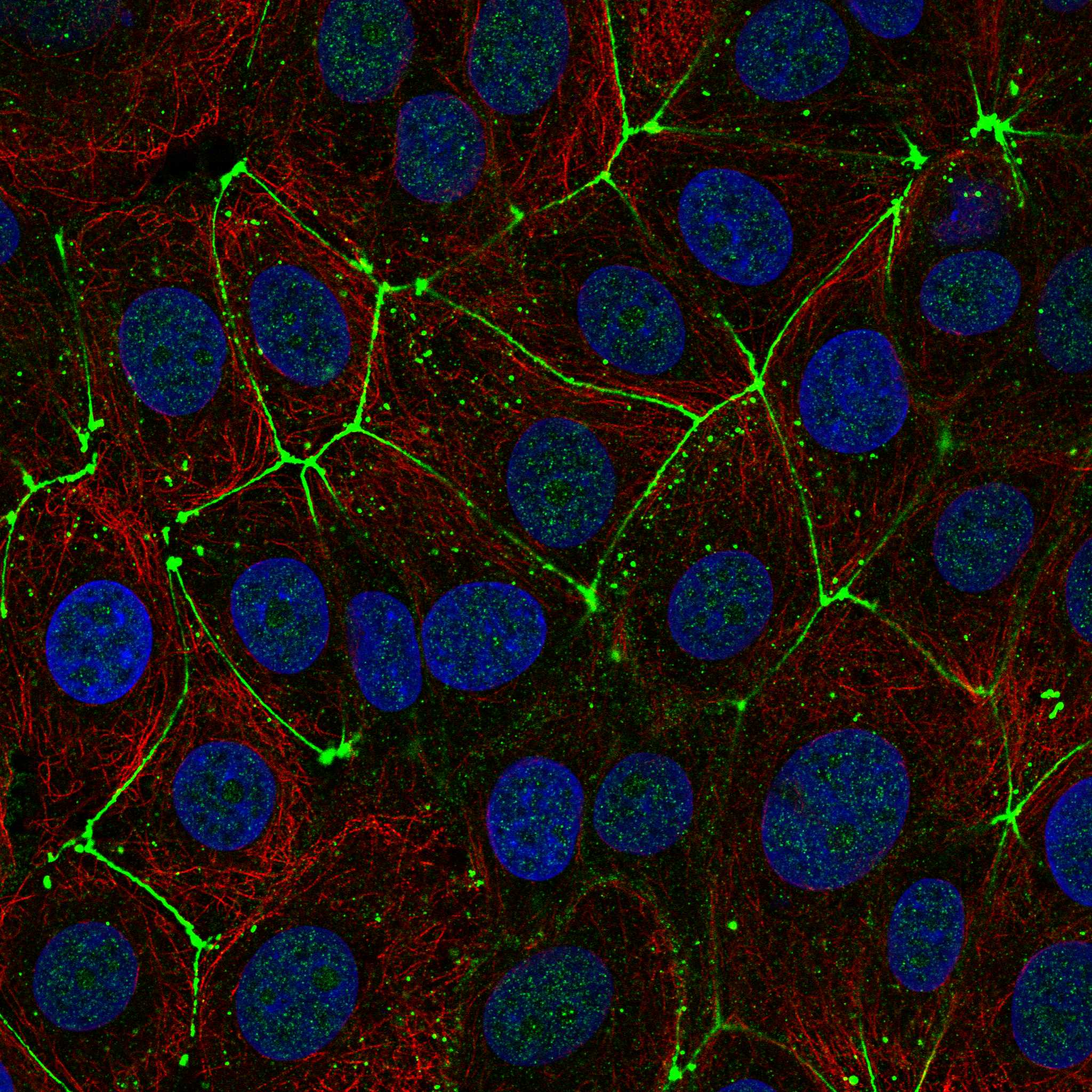
IF/ICC Q: Where should CLDN3 appear?A: At cell junctions; HPA calls that location supported and lists images from MCF-7 and U2OS (HPA subcellular ICC-IF). HPA014361 is ICC Approved (HPA antibodies). Use this as a localisation check; no IF/ICC protocol is supplied here.
Why is my staining missing, weak or wrong?
SituationLikely causeNext action
Expected positive cells show no signal.The run may have failed, or the specimen may differ from HPA's observed staining; the result alone cannot distinguish these explanations (HPA tissue IHC; standard IHC practice).Include a section containing an HPA High cell population, then verify antibody use, retrieval and chromogenic detection steps (HPA tissue IHC; standard IHC practice).
Nuclear or broad cytoplasmic color outweighs membrane borders.The location conflicts with CLDN3's annotated junctional membrane localisation; staining identity remains unresolved (UniProt O15551 subcellular location).Compare a no-primary control and tissue morphology; adjust background controls or reassess the primary antibody before assigning a CLDN3 score (standard IHC practice).
Unexpected color appears in HPA Not detected cells.For example, staining in adipocytes conflicts with HPA's reported result; cross-reactivity or endogenous detection activity is possible, not established (HPA: Not detected in adipocytes; standard IHC practice).Run a no-primary control. If using HRP detection, check endogenous-peroxidase blocking; investigate primary-dependent color with antibody controls (standard IHC practice).
The entire section has diffuse brown background.Widespread color without junctional boundaries is unlike the annotated location; nonspecific binding or detection background may contribute (UniProt O15551 subcellular location; standard IHC practice).Inspect the no-primary control, then review blocking, wash steps and detector exposure while retaining an HPA High positive cell population for comparison (standard IHC practice; HPA tissue IHC).
Only some cells in a tissue stain.HPA assigns levels to named cell types: colon endocrine cells are High, for example; that does not establish equal staining across every colonic cell (HPA tissue IHC).Identify the stained cells on the counterstained section and compare their compartment and intensity with the relevant HPA cell-type entry (HPA tissue IHC; standard IHC practice).
A result changes after changing retrieval conditions.Retrieval can alter IHC staining in general, but the supplied UniProt and HPA records do not establish a CLDN3-specific retrieval or fixation response (standard IHC practice; UniProt O15551; HPA tissue IHC).Compare conditions on the same HPA High positive cell population with matched controls; record which condition preserves interpretable junctional staining (HPA tissue IHC; standard IHC practice).

Sample controls for CLDN3 IHC & IF

🧪Run breast first: its glandular cells should show strong junctional staining (HPA: High in breast glandular cells; UniProt O15551: tight junction localization). Run adipose tissue as the negative: adipocytes should be unstained (HPA: Not detected in adipocytes). On the breast slide, use nonglandular stromal cells as internal negative comparators; they should lack the glandular junctional staining pattern (HPA: High in breast glandular cells; UniProt O15551: tight junction localization).
Positive control tissue: Appendix (Endocrine cells, HPA High)
Negative control tissue: Adipose tissue (HPA Not detected)
ICC-IF cell lines (HPA subcellular resource): HPA ICC-IF images show CLDN3 in MCF-7, U2OS, with annotated localisation: Cell Junctions (supported) (HPA subcellular).
Technical controls: Include a no-primary, secondary-only slide, a matched rabbit IgG isotype control, and a CLDN3 knockout biological negative (selected-SKU IHC caption: rabbit primary antibody; standard IHC practice). For the biotin-based DAB workflow, block endogenous peroxidase and check for endogenous biotin signal in breast tissue (selected-SKU IHC caption: biotinylated secondary, streptavidin–biotin complex and DAB; standard IHC practice).
⚠️Feasibility: A target-specific fixation window or fixation effect is unreported, and the exact selected-SKU tissue-IHC caption does not state the fixative (selected-SKU IHC caption). Paraffin-section staining is demonstrated after heat retrieval in EDTA at pH 8.0, but retrieval dependence has not been established by a comparison (selected-SKU IHC caption). Frozen sections have no supplied comparison showing they are easier; ICC-IF images support a cell-junction readout, while biotin-based detection can produce background in the breast IHC workflow (HPA subcellular: supported cell-junction localization and ICC-IF images; selected-SKU IHC caption: biotin-based detection; standard IHC practice).

HPA tissue IHC evidence for CLDN3

Comprehensive Human Protein Atlas IHC scoring per tissue (reliability: Enhanced — High consistency between antibody staining and RNA expression data.). Rows are taken directly from the HPA tissue chart — click any row's HPA link to view the source.

Positive expression · recommended positive controls

TissueCell typeLevelEvidenceSource
Appendix Endocrine cells High Protein (IHC) HPA →
Breast Glandular cells High Protein (IHC) HPA →
Colon Endocrine cells High Protein (IHC) HPA →
Duodenum Endocrine cells High Protein (IHC) HPA →
Endometrium Glandular cells High Protein (IHC) HPA →

Undetected expression · recommended negative controls

TissueCell typeLevelEvidenceSource
Adipose tissue Adipocytes Not detected Protein (IHC) HPA →
Adrenal gland Glandular cells Not detected Protein (IHC) HPA →
Bone marrow Hematopoietic cells Not detected Protein (IHC) HPA →
Caudate Glial cells Not detected Protein (IHC) HPA →
Cerebellum Cells in granular layer Not detected Protein (IHC) HPA →
Section 3

Advanced CLDN3 IHC Tips

Use membrane pattern, cell identity and matched controls to troubleshoot CLDN3 staining in paraffin sections.

Which retrieval conditions should I try when CLDN3 staining is weak?
Start with heat mediated retrieval in EDTA at pH 8.0 (datasheet A04393-5). The catalog antibody’s paraffin section image used this retrieval before incubation at 2 µg/mL overnight at 4°C (datasheet A04393-5). If staining remains weak, compare a citrate buffer at pH 6.0 as a fallback on adjacent sections, keeping detection and exposure to chromogen constant (standard IHC practice). Judge the result by clearer cell borders and preserved tissue morphology, because CLDN3 is a tight junction membrane protein and diffuse signal alone does not establish successful retrieval (UniProt O15551 localisation; standard IHC practice).
Could fixation explain weak or uneven CLDN3 staining in paraffin sections?
The catalog image identifies a paraffin embedded section but does not state its fixative, so target specific fixation sensitivity is unknown (datasheet A04393-5). Record the fixative, fixation duration and tissue processing for each specimen, and compare suspect sections with a section that stains reliably in the same run (standard IHC practice). Keep retrieval at EDTA pH 8.0 and the primary incubation at 2 µg/mL overnight at 4°C while assessing that difference (datasheet A04393-5). Patchy staining alongside poor morphology can flag a processing problem, but it cannot by itself establish a CLDN3 specific fixation effect (standard IHC practice).
What staining pattern supports CLDN3 at a tight junction?
Expect staining along cell borders, with emphasis where adjacent cells meet, because CLDN3 is assigned to the cell membrane and tight junctions (UniProt O15551 localisation). Supported subcellular evidence places it at cell junctions, while tissue IHC describes membranous expression in several gastrointestinal and glandular tissues (HPA subcellular; HPA tissue IHC). Examine sharply focused epithelial borders before interpreting diffuse cytoplasmic or nuclear chromogen as target signal, and compare the same compartments in a negative control (standard IHC practice). In kidney, Bowman's capsule is a reported high staining compartment; use that cellular context when assessing a kidney section (HPA tissue IHC).
How should epitope location affect CLDN3 staining decisions?
CLDN3 has 4 transmembrane segments, extracellular regions at residues 30–80 and 137–159, and a cytoplasmic tail at 181–220 (UniProt O15551 topology). Ask which region the catalog antibody recognizes before attributing weak paraffin staining to epitope masking; its position is not established by the supplied image caption (datasheet A04393-5). UniProt lists 0 annotated isoforms and phosphorylation sites at 198, 199 and 209, but these annotations do not show that this antibody’s binding changes with phosphorylation (UniProt O15551). Compare retrieval conditions on adjacent sections and retain the condition that improves junctional signal without degrading morphology (standard IHC practice).
How can I check a CLDN3 junctional pattern by immunofluorescence?
For a separate IF experiment, multiplex CLDN3 with an epithelial cell marker so that junctional signal can be assigned to the expected cells (HPA tissue IHC; UniProt O15551 localisation; standard IF practice). Choose fluorophores after imaging an unstained specimen, placing the weaker target signal in a channel with less tissue autofluorescence and checking single stain controls for bleed through (standard IF practice). Set permeabilisation according to the antibody’s mapped epitope: access to the cytoplasmic regions at 1–8, 102–115 or 181–220 may require it, whereas extracellular regions are accessible without crossing the membrane in intact cells (UniProt O15551 topology; standard IF practice). The paraffin IHC caption does not establish an IF fixation or permeabilisation condition (datasheet A04393-5).
What should I change when CLDN3 chromogen appears throughout the section?
First compare a no primary control with the stained section to identify signal from the detection system or endogenous tissue activity (standard IHC practice). The catalog image used biotinylated goat anti rabbit secondary antibody, a streptavidin biotin complex and DAB, so include controls appropriate to those reagents (datasheet A04393-5; standard IHC practice). Check the peroxidase block and shorten DAB development if broad brown staining persists in both sections; these are general chromogenic IHC adjustments (standard IHC practice). The catalog image used 10% goat serum blocking and 2 µg/mL primary antibody, useful reference points when assessing whether blocking or primary concentration drives background (datasheet A04393-5).
How should I score CLDN3 staining across paraffin sections? ⚠ ANSWER MARKED FOR VERIFICATION
Score the percentage of cells with junctional membrane staining and its intensity, then calculate an H score using 0–3 intensity categories if a single summary is needed (UniProt O15551 localisation; standard IHC scoring practice). Define the cell population and tissue area before scoring, excluding folds, necrosis and section edges where signal is unreliable (standard IHC practice). Normalise positive cell counts to all evaluable cells of the same type, or report positive junctional length relative to total evaluable cell border length when continuity matters (standard image analysis practice). Apply identical retrieval, chromogen development and scoring thresholds across sections; the catalog reference uses EDTA pH 8.0 retrieval (datasheet A04393-5; standard IHC practice).
How do I distinguish genuine CLDN3 staining from an artefact?
A convincing result follows contacts between appropriate cells, consistent with CLDN3 at cell membranes and tight junctions (UniProt O15551 localisation; HPA subcellular). Check whether staining matches the reported compartment: glandular cells in breast and endometrium and Bowman's capsule in kidney are high staining examples, whereas adipocytes are reported as undetected (HPA tissue IHC). Treat isolated nuclear signal, section edge staining and chromogen over necrotic areas cautiously, and compare them with the no primary control (UniProt O15551 localisation; standard IHC practice). If brown signal persists without primary antibody, investigate endogenous enzyme or detection system background before assigning it to CLDN3 (standard IHC practice).
Boster reagents

Best CLDN3 / Claudin-3 IHC Antibodies

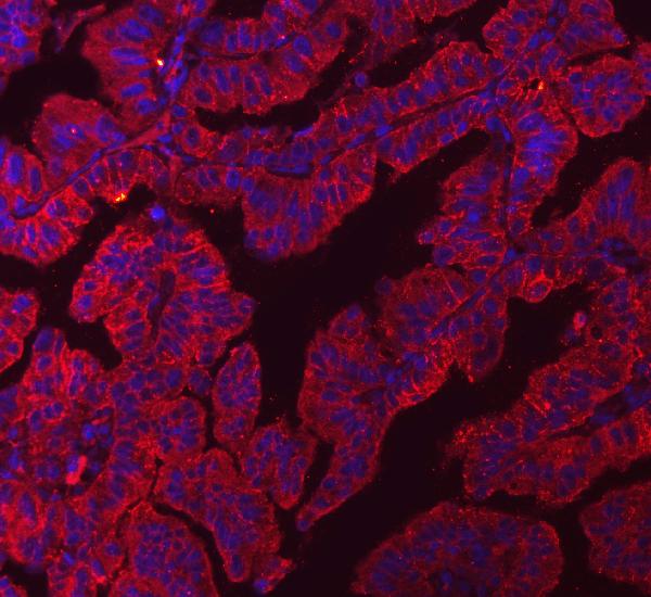
Human-reactive anti-CLDN3 antibodies have IHC images from paraffin-embedded tumor sections and IF images from MCF-7 cells or a paraffin-embedded lung tumor section (catalog reactivity and image captions).

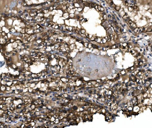
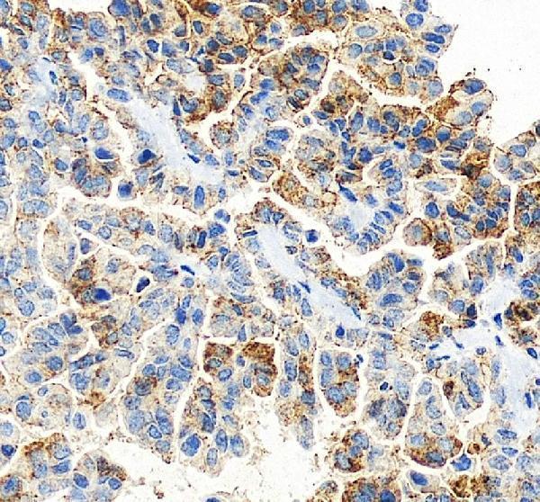
Real IHC data IHC analysis of Claudin 3/CLDN3 using anti-Claudin 3/CLDN3 antibody (A04393-5). Claudin 3/CLDN3 was detected in paraffin-embedded section of human renal clear cell carcinoma tissue. Heat mediated antigen retrieval was performed in EDTA buffer (pH8.0, epitope retrieval solution). The tissue section was blocked with 10% goat serum. The tissue section was then incubated with 2μg/ml rabbit anti-Claudin 3/CLDN3 Antibody (A04393-5) overnight at 4°C. Biotinylated goat anti-rabbit IgG was used as secondary antibody and incubated for 30 minutes at 37°C. The tissue section was developed using Strepavidin-Biotin-Complex (SABC) (Catalog # SA1022) with DAB as the chromogen.
Anti-Claudin 3/CLDN3 Antibody ®
Cat # A04393-5
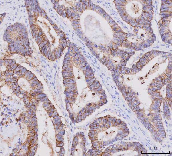
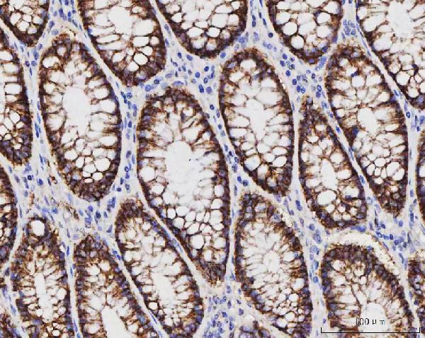
Real IHC data IHC analysis of Claudin 3/CLDN3 using anti-Claudin 3/CLDN3 antibody (M04393-2). Claudin 3/CLDN3 was detected in a paraffin-embedded section of human colorectal adenocarcinoma tissue. Heat mediated antigen retrieval was performed in EDTA buffer (pH 8.0, epitope retrieval solution). The tissue section was blocked with 10% goat serum. The tissue section was then incubated with 2 μg/ml mouse anti-Claudin 3/CLDN3 Antibody (M04393-2) overnight at 4°C. Peroxidase Conjugated Goat Anti-mouse IgG was used as secondary antibody and incubated for 30 minutes at 37°C. The tissue section was developed using HRP Conjugated Mouse IgG Super Vision Assay Kit (Catalog # SV0001) with DAB as the chromogen.
Anti-Claudin 3/CLDN3 Antibody ® (monoclonal, 4C7D2)
Cat # M04393-2
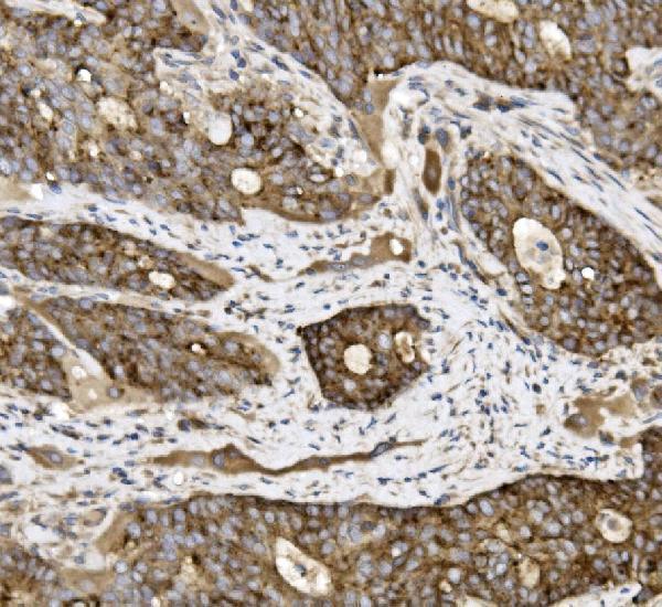
Real IHC data IHC analysis of Claudin 3/CLDN3 using anti-Claudin 3/CLDN3 antibody (A04393-3). Claudin 3/CLDN3 was detected in paraffin-embedded section of human pancreatic cancer tissue. Heat mediated antigen retrieval was performed in EDTA buffer (pH8.0, epitope retrieval solution). The tissue section was blocked with 10% goat serum. The tissue section was then incubated with 2μg/ml rabbit anti-Claudin 3/CLDN3 Antibody (A04393-3) overnight at 4°C. Biotinylated goat anti-rabbit IgG was used as secondary antibody and incubated for 30 minutes at 37°C. The tissue section was developed using Strepavidin-Biotin-Complex (SABC) (Catalog # SA1022) with DAB as the chromogen.
Anti-Claudin 3/CLDN3 Antibody ®
Cat # A04393-3

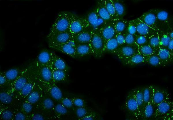
A04393-5 shows IHC in human renal clear cell carcinoma and IF/ICC in MCF-7 cells; M04393-2 shows IHC in human colorectal adenocarcinoma and IF in human lung adenocarcinoma (respective image captions). A04393-3 shows IHC in human pancreatic cancer and IF/ICC in MCF-7 cells (A04393-3 image captions).

Which to pick: For tissue IHC, choose the sample match: A04393-5 for human renal clear cell carcinoma (A04393-5 IHC caption), M04393-2 for human colorectal adenocarcinoma (M04393-2 IHC caption), or A04393-3 for human pancreatic cancer (A04393-3 IHC caption); all three captions describe paraffin sections with EDTA retrieval at pH 8.0, but do not report the fixative (respective IHC captions). For IF/ICC, A04393-5 and A04393-3 list both applications and show MCF-7 cell images; the mouse monoclonal M04393-2 lists IF and shows paraffin-section IF in human lung adenocarcinoma (catalog applications, host and clone; respective IF captions). None has documented nonhuman reactivity, so there is no supported cross-species pick (catalog reactivity: Human only).

Each figure is that product's own IHC / IF validation image from its datasheet.

References

  1. UniProt Consortium. UniProt entry O15551 (CLD3_HUMAN, Claudin-3).
  2. Human Protein Atlas. CLDN3 tissue IHC expression (reliability: Enhanced).
  3. Human Protein Atlas. CLDN3 subcellular location (ICC-IF): Localized to the cell junctions..
  4. Human Protein Atlas. CLDN3 antibody validation summary (1 antibodies).
  5. Claudin-1, -3 and -4 proteins and mRNA expression in benign and malignant breast lesions: a research study. Breast cancer research : BCR 2005 — PMC1064136.
  6. Investigation of the Effects of Postbiotics Obtained from Pediococcus acidilactici on Specific Biomarker Expressions in Intestinal Tissue. Foods (Basel, Switzerland) 2026 — PMC13073207.
  7. Reduced Claudin-3 Expression Is Linked to Unfavorable Tumor Features and Poor Prognosis in Non-Small Cell Lung Cancer. Lung Cancer (Auckland, N.Z.) 2026 — PMC12790956.
  8. Association of Cytokeratin 5 and Claudin 3 expression with BRCA1 and BRCA2 germline mutations in women with early breast cancer. BMC cancer 2019 — PMC6631579.
  9. PubMed PMID:9441748 — UniProt-cited evidence.
  10. PubMed PMID:9334247 — UniProt-cited evidence.
  11. PubMed PMID:15489334 — UniProt-cited evidence.