CLGN / Calmegin · IHC design guide

Design Immunohistochemistry for CLGN

Plan CLGN paraffin-section IHC with the catalog antibody at 2–5 μg/ml (datasheet A05261-1). Expect mainly cytoplasmic staining in pachytene spermatocytes and round spermatids (HPA tissue IHC).

Evidence assembled Oct 2026 · For research use; verify linked source records and product datasheet before use
Immunohistochemistry protocol sheet for CLGN (IHC for CLGN): expected localisation Cytoplasmic (HPA tissue IHC); ER membrane (UniProt), antibody A05261-1, validated IHC image, and IHC protocol steps
Printable CLGN IHC protocol sheet — expected localisation Cytoplasmic (HPA tissue IHC); ER membrane (UniProt), antibody A05261-1, controls and protocol steps. Open the full CLGN IHC guide →

CLGN Immunohistochemistry Experimental Design Guide

Expected localisation, validated protocols, controls and antibodies — the at-a-glance facts below, then the full design guide.

Must know before staining
Expected localisation Cytoplasmic (HPA tissue IHC); ER membrane (UniProt)
Staining pattern Mainly cytoplasmic in pachytene spermatocytes and round spermatids (HPA tissue IHC)
Antigen retrieval EDTA pH 8.0 HIER, heat-mediated (datasheet A05261-1)
Positive control ⓘ Endometrium+1 more · see all
Negative control ⓘ Adipose tissue+4 more · see all
Important caveats
Reasons your staining may differ from the expected pattern.
Fixation Keep fixation conditions consistent across sections (standard IHC practice; not target-specific)
Caveat Endometrial ciliated epithelial cells also stain strongly (HPA tissue IHC)
Regulation High in pachytene spermatocytes (HPA tissue IHC)
Isoform / epitope 2 isoforms; check whether the epitope is lumenal or cytoplasmic (UniProt)
Section 1

Recommended CLGN IHC & IF Protocols

Compare the catalog antibody’s IHC-P protocol (datasheet: A05261-1) with published CLGN IHC protocols from two articles (PMC9869145; PMC12698408).

Recommended immunohistochemistry (IHC-P) protocol parameters
SampleParaffin-embedded human mammary cancer tissue; fixative not specified (datasheet A05261-1)
FixationImage fixative and duration unreported (datasheet A05261-1); verify before use.
Sectioning4–5 µm sections on charged slides (standard)
DeparaffinisationXylene, graded ethanol series to water (standard)
Antigen retrievalHeat retrieval: EDTA pH 8.0 (datasheet A05261-1); 20 min, 95–100 °C (standard)
Peroxidase block3% H2O2, 10 min, room temperature (standard)
Blocking10% goat serum (datasheet A05261-1)
Primary antibodyRabbit anti-CLGN, 2-5 μg/ml (datasheet A05261-1)
Primary incubationOvernight at 4 °C (datasheet A05261-1)
DetectionHRP-conjugated secondary, DAB chromogen (datasheet A05261-1)
CounterstainHematoxylin, blue, dehydrate and mount (standard)
Expected resultCLGN-positive staining in ciliated epithelial cells of endometrium (HPA tissue IHC: High). HPA tissue profile: Cytoplasmic expression mainly in pachytene spermatocytes and round spermatids. No signal in the no-primary control.
💡Decision noteStart with heat-mediated EDTA retrieval at pH 8.0 (datasheet: A05261-1); citrate at pH 6.0 is a published alternative (PMC9869145 methods).
Section 2

What Is the Expected CLGN Staining Pattern?

CLGN is an endoplasmic reticulum membrane protein with a lumenal region at residues 20–471 and one transmembrane segment at 472–492 (UniProt O14967 topology). In tissue sections, expect mainly cytoplasmic staining in testicular pachytene spermatocytes and round spermatids (HPA tissue IHC: Enhanced reliability). HPA also reports high staining in endometrial ciliated epithelial cells (HPA tissue IHC: High).

What am I looking at on my slide?
Cytoplasmic stain in pachytene spermatocytes, with staining also seen in round spermatids.This matches the main testicular pattern (HPA tissue IHC). Compare cell types within the same section; a positive testis does not imply uniform staining across its cells (HPA tissue IHC: mainly pachytene spermatocytes and round spermatids).
Staining is predominantly nuclear, or forms a crisp plasma membrane rim in the expected testicular cells.That distribution conflicts with the reported cytoplasmic tissue pattern and endoplasmic reticulum location (HPA tissue IHC; UniProt O14967 subcellular location). Check staining specificity and detection controls before scoring it as CLGN (general IHC practice).
Strong stain appears in an unexpected cell type, while the expected cells are unstained.Investigate cross-reactivity or endogenous detection activity (general IHC practice). Cell identity matters: high staining in endometrial ciliated epithelial cells is reported, so it is a valid contextual positive (HPA tissue IHC: High).
Diffuse colour covers multiple compartments or tissue areas without a discernible cell pattern.Treat this as background until controls resolve it (general IHC practice). The reported signal is mainly cytoplasmic in defined testicular cells, rather than an indiscriminate tissue-wide stain (HPA tissue IHC).
No signal is visible in pachytene spermatocytes on a testis control section.The expected high-staining cell population is missing (HPA tissue IHC: High in pachytene spermatocytes). Check tissue preservation, retrieval, antibody and detection performance with appropriate controls before calling the sample negative (general IHC practice).
💡Expected CLGN appearanceCall a result positive when cytoplasmic stain is clear in pachytene spermatocytes, with the reported round-spermatid pattern; expect strong staining in pachytene spermatocytes, while nuclear-only or indiscriminate stain warrants review (HPA tissue IHC: High in pachytene spermatocytes; HPA tissue IHC profile; general IHC practice).
How each factor affects the staining
Cell type and tissue choiceTestis pachytene spermatocytes and endometrial ciliated epithelial cells are reported high-staining references (HPA tissue IHC: High). Adipocytes in adipose tissue are reported not detected; score those cells specifically when using them as a negative comparison (HPA tissue IHC: Not detected).
Compartment and epitope contextCLGN has a lumenal segment at 20–471, a transmembrane segment at 472–492 and a cytoplasmic segment at 493–610 (UniProt O14967 topology). The supplied evidence gives no antibody epitope, so topology cannot predict this antibody's retrieval needs.
Antibody validationHPA lists IHC support for HPA048761 and Enhanced IHC validation for HPA058627 and CAB020709 (HPA antibodies). Validation supports interpretation of the reported pattern, but each staining run still needs suitable positive and negative controls (general IHC practice).
Antigen retrieval in paraffin sectionsTarget-specific fixation effects are not established by the supplied assay evidence. Verify with a matched IHC source before attributing a result to fixation.
IF/ICC: where should signal appear?HPA reports a supported main location in the endoplasmic reticulum and approved additional locations in the sperm equatorial segment and mid piece (HPA subcellular ICC-IF). Interpret those sperm structures in their own context; they do not redefine the tissue IHC pattern (HPA tissue IHC).
Why is my staining missing, weak or wrong?
SituationLikely causeNext action
Testis control has no stain in pachytene spermatocytes.An expected high-staining population is absent (HPA tissue IHC: High); the failed step cannot be identified from appearance alone (general IHC practice).Check control section quality, retrieval, antibody incubation and detection reagents against the catalog antibody's IHC-P protocol (general IHC practice).
Colour appears throughout the section, including cell-free or implausible areas.Nonspecific reagent binding or detection background may obscure the cell pattern (general IHC practice).Review blocking, antibody dilution, washes and detection controls; score only a resolved cellular pattern (general IHC practice).
Strong signal is confined to nuclei in testis.Nuclear-only staining conflicts with the mainly cytoplasmic tissue pattern and endoplasmic reticulum assignment (HPA tissue IHC; UniProt O14967).Check antibody specificity and detection controls, then compare staining with pachytene spermatocytes on the same or a matched section (general IHC practice; HPA tissue IHC: High).
A supposed negative section stains strongly.The comparison may contain a documented positive cell type, or may show nonspecific staining (HPA tissue IHC; general IHC practice).Identify the stained cells first: endometrial ciliated epithelial cells are high, whereas adipose adipocytes are not detected (HPA tissue IHC). Then inspect detection controls (general IHC practice).
Pachytene spermatocytes stain, but every nearby cell is also called positive.Broad scoring may erase the cell-type distinction in the reported testis pattern (HPA tissue IHC: mainly pachytene spermatocytes and round spermatids).Score identified cell populations separately and use the counterstain to check morphology (general IHC practice); keep the reported cell distribution as the reference (HPA tissue IHC).
Signal changes substantially when retrieval conditions change.Retrieval can alter staining in paraffin IHC generally; no CLGN-specific fixation or retrieval response is established here (general IHC practice; UniProt O14967 and HPA source scope).Compare conditions on matched positive and negative control sections and follow the catalog antibody's IHC-P protocol when available (general IHC practice).

Sample controls for CLGN IHC & IF

🧪Run testis first and require staining in pachytene spermatocytes (HPA: High in pachytene spermatocytes; UniProt O14967: detected in testis). Use adipose tissue as the negative, with adipocytes unstained (HPA: Not detected in adipocytes); on the testis slide, cells outside the positive spermatocyte population should lack specific staining (HPA: High in pachytene spermatocytes).
Positive control tissue: Endometrium (Ciliated epithelial cells, HPA High)
Negative control tissue: Adipose tissue (HPA Not detected)
ICC-IF cell lines (HPA subcellular resource): HPA ICC-IF images show CLGN in PC-3, U-251MG, U2OS, Sperm, with annotated localisation: Endoplasmic reticulum (supported) (HPA subcellular).
Technical controls: Include no-primary (secondary-only) and matched rabbit IgG isotype controls, plus CLGN knockout tissue as a biological negative where available (caption: rabbit primary antibody; standard IHC practice). Block endogenous peroxidase and check for background before interpreting DAB staining in testis (caption: peroxidase detection with DAB; standard IHC practice).
⚠️Feasibility: A target-specific fixation window or fixation effect is unreported, and the selected A05261-1 paraffin-section caption does not state the fixative (selected-SKU tissue-IHC caption). That caption uses heat retrieval in EDTA at pH 8.0, but does not establish whether retrieval is required; the supplied evidence also does not establish whether frozen sections or IF are easier for CLGN (selected-SKU tissue-IHC caption). In testis, use cell morphology when scoring staining in the densely arranged germ-cell layers (HPA: High in pachytene spermatocytes; standard histology practice).

HPA tissue IHC evidence for CLGN

Comprehensive Human Protein Atlas IHC scoring per tissue (reliability: Enhanced — High consistency between antibody staining and RNA expression data.). Rows are taken directly from the HPA tissue chart — click any row's HPA link to view the source.

Positive expression · recommended positive controls

TissueCell typeLevelEvidenceSource
Endometrium Ciliated epithelial cells High Protein (IHC) HPA →
Testis Pachytene spermatocytes High Protein (IHC) HPA →

Undetected expression · recommended negative controls

TissueCell typeLevelEvidenceSource
Adipose tissue Adipocytes Not detected Protein (IHC) HPA →
Adrenal gland Glandular cells Not detected Protein (IHC) HPA →
Appendix Glandular cells Not detected Protein (IHC) HPA →
Bone marrow Hematopoietic cells Not detected Protein (IHC) HPA →
Breast Adipocytes Not detected Protein (IHC) HPA →
Section 3

Advanced CLGN IHC Tips

Troubleshoot CLGN staining in paraffin sections by checking retrieval, cell identity, and the expected endoplasmic reticulum pattern before comparing staining intensity.

Which retrieval condition should I start with for CLGN in paraffin sections?
Start with heat mediated retrieval in EDTA pH 8.0 for this catalog antibody (datasheet A05261-1). The selected tissue image used that condition before overnight incubation with 2 μg/ml primary antibody at 4°C (caption A05261-1). If staining is weak, first confirm that sections were fully deparaffinised, heated evenly, and cooled consistently; then compare a second retrieval condition on adjacent sections while holding detection constant (standard IHC practice). Evaluate whether the change improves staining in pachytene spermatocytes without increasing signal in nearby unstained cells (HPA tissue IHC: High in pachytene spermatocytes; standard IHC practice).
How should I troubleshoot fixation when CLGN staining is weak or uneven?
CLGN specific sensitivity to fixation is unknown from the supplied evidence; the selected paraffin section caption does not state a fixative (caption A05261-1). Record the actual fixative, fixation duration, specimen thickness, and processing history for each section before changing the staining protocol (standard IHC practice). Compare sections with documented, consistent handling using the same EDTA pH 8.0 retrieval and 2 μg/ml primary concentration (datasheet A05261-1). If signal varies across a section, inspect morphology and processing artefacts, then repeat with matched material rather than attributing the difference to a CLGN specific fixation effect (standard IHC practice).
What staining pattern supports a specific CLGN result in tissue?
Look for cytoplasmic staining in pachytene spermatocytes and round spermatids, the pattern reported for tissue IHC (HPA tissue IHC). CLGN is an endoplasmic reticulum membrane protein with a lumenal region at residues 20–471 and a cytoplasmic region at 493–610 (UniProt O14967 topology). On chromogenic sections, assess whether cellular staining is coherent with that distribution while allowing for the limited resolution of brightfield microscopy (UniProt O14967 subcellular location; standard IHC practice). Predominantly nuclear staining, isolated section edges, or diffuse signal across unrelated cells warrants comparison with a negative control and a cell type with documented CLGN expression (HPA tissue IHC; standard IHC practice).
Could epitope position or isoforms explain inconsistent CLGN staining?
CLGN has 2 annotated isoforms, so check the antibody's stated immunogen and isoform coverage before interpreting absent staining (UniProt O14967 isoforms; standard IHC practice). Its membrane span is at residues 472–492, separating the lumenal 20–471 region from the cytoplasmic 493–610 region (UniProt O14967 topology). The record also lists modified residues, including phosphoserines at 560, 576, 579, and 581, but supplies no evidence that they affect this antibody (UniProt O14967 modified residues). Compare retrieval conditions on adjacent sections and document the antibody epitope if available; do not assign an isoform or modification from chromogenic staining alone (standard IHC practice).
How can I follow up an IHC result with CLGN immunofluorescence?
Treat IF as a separate optimisation: the selected antibody evidence describes paraffin section chromogenic IHC, with no IF conditions supplied (caption A05261-1). For tissue multiplexing, pair CLGN with a validated marker of pachytene spermatocytes or round spermatids and check each channel separately (HPA tissue IHC: cytoplasmic expression in those cells; standard IF practice). Choose spectrally separated fluorophores after checking tissue autofluorescence with an unstained control, and include single stain controls (standard IF practice). If the epitope is lumenal at residues 20–471, optimise permeabilisation for access to the endoplasmic reticulum lumen; a cytoplasmic epitope at 493–610 may have different access requirements (UniProt O14967 topology; standard IF practice).
What should I check when CLGN DAB staining is diffuse?
First inspect a no primary control for staining from the detection system, including endogenous peroxidase activity (standard chromogenic IHC practice). The selected image used 10% goat serum blocking, 2 μg/ml primary antibody overnight at 4°C, and a peroxidase conjugated secondary for 30 minutes at 37°C (caption A05261-1). If background persists, verify peroxidase blocking, washing, and DAB development time, then adjust one variable at a time (standard chromogenic IHC practice). Compare background in adipocytes or glandular cells reported as undetected with staining in pachytene spermatocytes, while recognising that tissue processing and detection can also differ (HPA tissue IHC; standard IHC practice).
How should I quantify CLGN IHC across tissue sections? ⚠ ANSWER MARKED FOR VERIFICATION
Define the cell population and sampling regions before scoring, and keep acquisition and DAB development settings consistent across compared sections (standard IHC practice). In testis, score pachytene spermatocytes separately from round spermatids because both are reported as cytoplasmically positive (HPA tissue IHC). Record the percentage of positive cells and intensity to calculate an H-score, or report positive cells per mm² when cell density is the question (standard IHC practice). Normalise counts to the number of evaluable cells in the specified population, or area measurements to viable tissue area; exclude folds and necrotic regions using prespecified criteria (standard IHC practice).
How can I distinguish true CLGN signal from artefact?
Give the most weight to reproducible cellular staining in pachytene spermatocytes, which HPA reports as High, with a cytoplasmic pattern in spermatocytes and round spermatids (HPA tissue IHC). An endoplasmic reticulum associated pattern fits CLGN localisation, whereas dominant nuclear staining needs further investigation (UniProt O14967 subcellular location; standard IHC practice). Review section edges, folds, and necrotic areas for local DAB accumulation, and check a no primary control for endogenous enzyme or detection signal (standard chromogenic IHC practice). The selected antibody stained a human mammary cancer paraffin section, but that image alone does not establish which cells express CLGN or that every stained cell is specific (caption A05261-1; standard IHC interpretation).
Boster reagents

Best CLGN / Calmegin IHC Antibodies

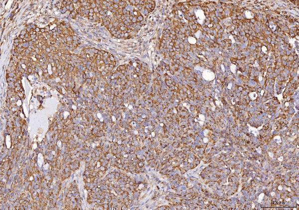
Real CLGN IHC images cover paraffin sections of human mammary and ovarian cancers and mouse and rat testes (catalog image captions). IF/ICC is listed for two monoclonals without image data (catalog applications; catalog image records).

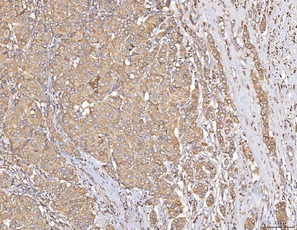
Real IHC data IHC analysis of Calmegin/CLGN using anti-Calmegin/CLGN antibody (A05261-1). Calmegin/CLGN was detected in a paraffin-embedded section of human mammary cancer tissue. Heat mediated antigen retrieval was performed in EDTA buffer (pH 8.0, epitope retrieval solution). The tissue section was blocked with 10% goat serum. The tissue section was then incubated with 2 μg/ml rabbit anti-Calmegin/CLGN Antibody (A05261-1) overnight at 4°C. Peroxidase Conjugated Goat Anti-rabbit IgG was used as secondary antibody and incubated for 30 minutes at 37°C. The tissue section was developed using HRP Conjugated Rabbit IgG Super Vision Assay Kit (Catalog # SV0002) with DAB as the chromogen.
Anti-Calmegin/CLGN Antibody ®
Cat # A05261-1

A05261-1 is the sole card with a rendered IHC figure, showing a human mammary cancer paraffin section (card figure caption). Its additional IHC captions show human ovarian cancer and mouse and rat testis paraffin sections (catalog image captions).

Which to pick: Choose A05261-1 for paraffin-section IHC: its own captions document EDTA retrieval at pH 8.0 and primary antibody at 2 μg/ml; the fixative is unreported (A05261-1 image captions). For IF/ICC, M05261 (clone 18C26) and M05261-1 (clone 23C69) list those applications, but neither has an IF image in the payload (catalog applications; catalog image records). For cross-species paraffin IHC, A05261-1 has captioned examples from human, mouse and rat tissue (A05261-1 image captions).

Each figure is that product's own IHC / IF validation image from its datasheet.

References

  1. UniProt Consortium. UniProt entry O14967 (CLGN_HUMAN, Calmegin).
  2. Human Protein Atlas. CLGN tissue IHC expression (reliability: Enhanced).
  3. Human Protein Atlas. CLGN subcellular location (ICC-IF): Mainly localized to the endoplasmic reticulum. In addition localized to the equatorial segment and mid piece..
  4. Human Protein Atlas. CLGN antibody validation summary (3 antibodies).
  5. The upregulation of CLGN in hepatocellular carcinoma is potentially regulated by hsa-miR-194-3p and associated with patient progression. Frontiers in oncology 2022 — PMC9869145.
  6. Targeting endoplasmic reticulum stress-induced CLGN resensitizes hepatocellular carcinoma to apoptosis: paeonol synergistically enhances efficacy by dual inhibition of CLGN and NF-κB. Frontiers in oncology 2025 — PMC12698408.
  7. Transcriptomics, Epigenetics, and Metabolomics of Primary Aldosteronism. Cancers 2021 — PMC8583505.
  8. PubMed PMID:9434179 — UniProt-cited evidence.
  9. PubMed PMID:14702039 — UniProt-cited evidence.
  10. PubMed PMID:15489334 — UniProt-cited evidence.