This website uses cookies to ensure you get the best experience on our website.


- Table of Contents
Plan CLN5 chromogenic IHC in paraffin sections using the cytoplasmic tissue pattern reported by HPA (HPA tissue IHC). Compare staining across consistently fixed samples, and interpret intensity with HPA’s low staining–RNA concordance in mind (HPA tissue IHC).

Expected localisation, validated protocols, controls and antibodies — the at-a-glance facts below, then the full design guide.
| Expected localisation | Cytoplasmic in tissue (HPA tissue IHC); lysosomal membrane expected (UniProt) | |
| Staining pattern | Cytoplasmic staining in most tissues (HPA tissue IHC) | |
| Antigen retrieval | Citrate pH 6.0 HIER, 95–98 °C, 20 min (rule: cytoplasmic / membrane antigen) | |
| Positive control | Caudate+4 more · see all | |
| Negative control | Lymph node+3 more · see all |
| Fixation | Matched tissue-IHC evidence does not establish the fixation claim. Validate the specimen-specific method before use. (selected-SKU IHC image A04893-2) | |
| Caveat | Low staining–RNA concordance complicates interpretation (HPA tissue IHC) | |
| Regulation | No specific staining regulator reported (UniProt) | |
| Isoform / epitope | No isoforms; cytoplasmic N-terminus absent from 44–358 form (UniProt) |
The catalog antibody provides the paraffin-section protocol (datasheet). A published sheep-brain CLN5 IHC method provides additional chromogenic staining conditions (PMC10628725).
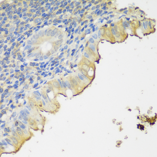
| Sample | Paraffin-embedded human appendix tissue; fixative not specified (datasheet A04893-2) |
| Fixation | Image fixative and duration unreported (datasheet A04893-2); verify before use. |
| Sectioning | 4–5 µm sections on charged slides (standard) |
| Deparaffinisation | Xylene, graded ethanol series to water (standard) |
| Antigen retrieval | Heat-induced epitope retrieval in citrate buffer, pH 6.0, 20 min at 95–98 °C (standard rule: cytoplasmic / membrane antigen) |
| Peroxidase block | 3% H2O2, 10 min, room temperature (standard) |
| Blocking | 10% normal serum of the secondary host, 30 min, room temperature (standard) |
| Primary antibody | Rabbit anti-CLN5, 1:50-1:200 (datasheet A04893-2) |
| Primary incubation | Overnight at 4 °C (standard) |
| Detection | HRP-polymer secondary, DAB chromogen 5–10 min (standard) |
| Counterstain | Hematoxylin, blue, dehydrate and mount (standard) |
| Expected result | CLN5-positive staining in glial cells of caudate (HPA tissue IHC: High). HPA tissue profile: Cytoplasmic expression in most tissues. No signal in the no-primary control. |
CLN5 is a lysosomal protein associated with membranes; residues 24–40 form a transmembrane segment, with residues 41–358 facing the lumen (UniProt O75503 topology). In paraffin sections, expect cytoplasmic staining, including strong signal in lung macrophages, cerebral cortex glial cells, and duodenal glandular cells (HPA: tissue IHC). HPA rates its IHC staining Approved but reports low consistency with RNA expression (HPA: reliability).
| Granular cytoplasmic staining in lung macrophages, with stronger signal than nearby unstained cells (HPA: High in lung macrophages). | This fits the reported cell pattern and is compatible with lysosomal localisation (HPA: tissue IHC; UniProt O75503 subcellular location). Granules alone do not prove lysosomal identity; compare cell morphology and controls (general IHC practice). |
| Predominantly nuclear staining, especially when cytoplasmic signal is absent (HPA: cytoplasmic expression in most tissues). | A nuclear-only result conflicts with the reported tissue pattern and lysosomal localisation (HPA: tissue IHC; UniProt O75503 subcellular location). Review counterstain, detection controls, and antibody conditions before assigning it to CLN5 (general IHC practice). |
| Strong staining in skeletal muscle myocytes or lymph node germinal center cells (HPA: Not detected in those cells). | That distribution conflicts with these HPA examples and may reflect cross-reactivity or endogenous detection activity (HPA: tissue IHC; general IHC practice). A negative cell population is a comparison, not proof that every positive cell is specific. |
| Broad, fairly even color over cells and extracellular areas, obscuring cellular boundaries (general IHC practice). | Interpret this as background until detection controls and slide processing are checked (general IHC practice). HPA describes cytoplasmic expression in most tissues, so cytoplasmic staining alone cannot distinguish true signal from widespread background (HPA: tissue IHC). |
| No signal in lung macrophages or duodenal glandular cells (HPA: High in both cell populations). | A blank result in a reported high-staining population warrants a technical check before a negative CLN5 call (HPA: tissue IHC; general IHC practice). HPA's Approved rating has low RNA–staining consistency, so corroborate unexpected biology independently (HPA: reliability). |
| Cell and tissue selection (HPA: tissue IHC). | HPA reports High staining in lung macrophages, cerebral cortex glial cells, hippocampal neuronal cells, and several glandular populations; it reports Not detected in skeletal muscle myocytes and selected lymph node cells (HPA: tissue IHC). Score the specified cell population within each tissue. |
| Antibody evidence (HPA: antibody validation; HPA: reliability). | The listed antibody, HPA041788, is Approved for IHC; HPA also notes low consistency between staining and RNA data (HPA: antibody validation; HPA: reliability). Treat an unexpected tissue pattern as provisional and inspect appropriate controls (general IHC practice). |
| Compartment and epitope position (UniProt O75503 topology). | CLN5 spans the membrane at residues 24–40, with residues 41–358 lumenal (UniProt O75503 topology). Epitope position is not supplied, so this record cannot predict which antibody conditions best expose its epitope. |
| Processing and glycosylation (UniProt O75503 processing and glycosylation). | UniProt lists a full-length 1–358 chain, a 44–358 secreted form, and eight glycosylation sites (UniProt O75503). Their effects on the supplied antibody's IHC staining are unreported; do not infer a tissue pattern or retrieval requirement from them. |
| Antigen retrieval (general IHC practice). | Use the IHC-validated antibody's documented retrieval conditions where available, then compare a reported high-staining tissue with detection controls (general IHC practice; HPA: tissue IHC). Neither source here establishes CLN5-specific retrieval or fixation sensitivity. |
| IF/ICC: what pattern can this guide confirm? (HPA: subcellular ICC-IF). | UniProt supports a lysosomal localisation, but HPA supplies no ICC-IF image set or main subcellular location for CLN5 (UniProt O75503 subcellular location; HPA: subcellular ICC-IF). An IF punctate pattern remains a hypothesis for separate validation. |
| Situation | Likely cause | Next action |
|---|---|---|
| A reported high-staining population is blank (HPA: High in lung macrophages or duodenal glandular cells). | Possible assay failure or inadequate signal development (general IHC practice); this result alone does not establish absent CLN5. | Check tissue identity, the detection control, antibody application, and the documented IHC retrieval conditions; repeat with a reported high-staining section (general IHC practice; HPA: tissue IHC). |
| All cells stain strongly, including HPA not-detected populations (HPA: tissue IHC). | Excess background, cross-reactivity, or endogenous detection activity is possible (general IHC practice). | Inspect a no-primary control and detection background; adjust blocking, antibody concentration, or development time as indicated by those controls (general IHC practice). |
| Signal is mainly nuclear (HPA: cytoplasmic expression in most tissues). | The compartment conflicts with reported cytoplasmic staining and lysosomal localisation (HPA: tissue IHC; UniProt O75503 subcellular location). | Confirm the counterstain and cell boundaries, then compare no-primary and reported high-staining tissue controls before scoring (general IHC practice; HPA: tissue IHC). |
| Only extracellular deposits stain (HPA: cytoplasmic expression in most tissues). | Deposits alone do not match HPA's usual cellular pattern; UniProt's secreted form does not establish an extracellular IHC pattern (HPA: tissue IHC; UniProt O75503 processing). | Examine morphology and detection controls; require a supported cellular pattern before interpreting the deposits as CLN5 (general IHC practice; HPA: tissue IHC). |
| Staining differs from an RNA-based expectation (HPA: low RNA–staining consistency). | HPA explicitly reports low consistency between antibody staining and RNA expression (HPA: reliability). | Score the observed cell population and compartment; use independent evidence before claiming that the discrepancy is biological (HPA: tissue IHC and reliability; general IHC practice). |
| An IF image seems to show lysosomal puncta (UniProt O75503 subcellular location). | The appearance is plausible, but HPA provides no CLN5 ICC-IF images or main location to verify it (HPA: subcellular ICC-IF). | Treat the IF assignment as unconfirmed and validate localisation with appropriate controls in the separate IF/ICC workflow (general IF practice). |


Comprehensive Human Protein Atlas IHC scoring per tissue (reliability: Approved — Low consistency between antibody staining and RNA expression data.). Rows are taken directly from the HPA tissue chart — click any row's HPA link to view the source.
| Tissue | Cell type | Level | Evidence | Source |
|---|---|---|---|---|
| Caudate | Glial cells | High | Protein (IHC) | HPA → |
| Cerebral cortex | Glial cells | High | Protein (IHC) | HPA → |
| Duodenum | Glandular cells | High | Protein (IHC) | HPA → |
| Gallbladder | Glandular cells | High | Protein (IHC) | HPA → |
| Hippocampus | Neuronal cells | High | Protein (IHC) | HPA → |
| Tissue | Cell type | Level | Evidence | Source |
|---|---|---|---|---|
| Lymph node | Germinal center cells | Not detected | Protein (IHC) | HPA → |
| Parathyroid gland | Glandular cells | Not detected | Protein (IHC) | HPA → |
| Skeletal muscle | Myocytes | Not detected | Protein (IHC) | HPA → |
| Spleen | Cells in red pulp | Not detected | Protein (IHC) | HPA → |
CLN5 IHC troubleshooting centers on retrieval, compartment-aware controls, and cautious interpretation of cytoplasmic staining (UniProt O75503 localisation; HPA tissue IHC).
A04893-2 has real IHC data from a human paraffin section (A04893-2 image caption). Human, Mouse, and Rat reactivity is listed; no IF data are shown (catalog reactivity; IF image alts).
A04893-2 will render with an IHC figure of paraffin-embedded human appendix stained at 1:150 after microwave retrieval in 10 mM PBS, pH 7.2 (A04893-2 image caption). IHC is listed for Human, Mouse, and Rat, but the figure documents human tissue only (catalog applications/reactivity; A04893-2 image caption).
Which to pick: For tissue IHC, choose A04893-2 for paraffin sections; its own caption supports that preparation, but does not report the fixative or establish tissue use (A04893-2 image caption). For IF/ICC, there is no listed IF application or IF figure, and clonality is unreported (catalog applications; IF image alts; catalog clone field). For cross-species IHC, A04893-2 lists Human, Mouse, and Rat reactivity, with pictured IHC evidence limited to human appendix (catalog reactivity; A04893-2 image caption). The selected A04893-2 tissue-IHC caption documents paraffin sections, but does not specify the fixative (selected-SKU IHC image A04893-2).