CLN6 / Ceroid-lipofuscinosis neuronal protein 6 · IHC design guide

Design Immunohistochemistry for CLN6

Plan CLN6 paraffin IHC around cytoplasmic staining in glandular and kidney tubular cells (HPA tissue IHC). The protein resides in the ER membrane (UniProt); start the IHC-validated antibody at 1:50–1:200 (datasheet: IHC) and assess nuclear signal seen in a stomach carcinoma example (datasheet: IHC).

Evidence assembled Oct 2026 · For research use; verify linked source records and product datasheet before use
Immunohistochemistry protocol sheet for CLN6 (IHC for CLN6): expected localisation Cytoplasmic in tissue (HPA tissue IHC); ER membrane (UniProt), antibody A03538, validated IHC image, and IHC protocol steps
Printable CLN6 IHC protocol sheet — expected localisation Cytoplasmic in tissue (HPA tissue IHC); ER membrane (UniProt), antibody A03538, controls and protocol steps. Open the full CLN6 IHC guide →

CLN6 Immunohistochemistry Experimental Design Guide

Expected localisation, validated protocols, controls and antibodies — the at-a-glance facts below, then the full design guide.

Must know before staining
Expected localisation Cytoplasmic in tissue (HPA tissue IHC); ER membrane (UniProt)
Staining pattern Cytoplasmic staining in glandular and kidney tubular cells (HPA tissue IHC)
Antigen retrieval Citrate pH 6.0 HIER, 95–98 °C, 20 min (rule: cytoplasmic / membrane antigen)
Positive control ⓘ Colon+4 more · see all
Negative control ⓘ Adipose tissue+4 more · see all
Important caveats
Reasons your staining may differ from the expected pattern.
Fixation Matched tissue-IHC evidence does not establish the fixation claim. Validate the specimen-specific method before use. (selected-SKU IHC image A03538)
Caveat Carcinoma example includes nuclear staining (datasheet IHC)
Regulation No expression regulator annotated (UniProt)
Isoform / epitope 2 isoforms; epitope side and coverage are unknown (UniProt)
Section 1

Recommended CLN6 IHC & IF Protocols

The catalog antibody protocol is followed by a published CLN6 protocol for dog paraffin sections (PMC11203140).

Recommended immunohistochemistry (IHC-P) protocol parameters
SampleParaffin-embedded human stomach carcinoma tissue; fixative not specified (datasheet A03538)
FixationImage fixative and duration unreported (datasheet A03538); verify before use.
Sectioning4–5 µm sections on charged slides (standard)
DeparaffinisationXylene, graded ethanol series to water (standard)
Antigen retrievalHeat-induced epitope retrieval in citrate buffer, pH 6.0, 20 min at 95–98 °C (standard rule: cytoplasmic / membrane antigen)
Peroxidase block3% H2O2, 10 min, room temperature (standard)
Blocking10% normal serum of the secondary host, 30 min, room temperature (standard)
Primary antibodyRabbit anti-CLN6, 1:50-1:200 (datasheet A03538)
Primary incubationOvernight at 4 °C (standard)
DetectionHRP-polymer secondary, DAB chromogen 5–10 min (standard)
CounterstainHematoxylin, blue, dehydrate and mount (standard)
Expected resultCLN6-positive staining in glandular cells of colon (HPA tissue IHC: High). HPA tissue profile: Cytoplasmic expression in several tissues. No signal in the no-primary control.
💡Decision noteTry citrate pH 6.0 retrieval at 95–98 °C for 20 min (page antigen-retrieval setting); CLN6 is an ER membrane protein (UniProt Q9NWW5).
Section 2

What Is the Expected CLN6 Staining Pattern?

CLN6 is a seven-pass endoplasmic reticulum (ER) membrane protein (UniProt Q9NWW5 topology and subcellular location). In paraffin-section IHC, expect cytoplasmic staining in several tissues, especially colon and duodenum glandular cells and kidney tubular cells, where HPA reports high staining (HPA tissue IHC). HPA rates the tissue pattern Approved, with medium consistency between antibody staining and RNA expression (HPA tissue IHC reliability).

What am I looking at on my slide?
Cytoplasmic staining in colon or duodenum glandular cells, or kidney tubular cells (HPA tissue IHC).This matches HPA's high-staining cell populations and CLN6's ER location (HPA tissue IHC; UniProt Q9NWW5 subcellular location). Assess staining within the named cells; an HPA level describes those cells rather than every cell in the section (HPA tissue IHC).
Predominantly nuclear, nucleolar, or cell-surface staining in paraffin sections.Recheck specificity and detection controls: UniProt places CLN6 at the ER membrane (UniProt Q9NWW5 subcellular location). HPA reports nucleolar staining only as an uncertain additional ICC-IF location, so it does not establish a nucleolar IHC pattern (HPA subcellular).
Strong staining in adipocytes or esophageal squamous epithelial cells.These named cell populations were not detected in HPA tissue IHC (HPA: adipocytes and esophageal squamous epithelial cells, Not detected). Consider cross-reactivity or endogenous detection activity; inspect the no-primary control and verify the cell identity before interpreting the signal (general IHC practice).
Diffuse colour across cells and surrounding tissue, with little cell-specific contrast.Treat this as background until controls support a cellular pattern (general IHC practice). HPA describes cytoplasmic expression in several tissues, with high staining assigned to specific cell populations; uniform haze does not reproduce that distribution (HPA tissue IHC).
No staining in colon glandular cells or kidney tubular cells.A negative result in these HPA high-staining populations needs a technical check before a biological conclusion (HPA tissue IHC; general IHC practice). Review tissue integrity, primary-antibody use, retrieval and detection controls; HPA's Approved rating has medium consistency with RNA data, so the pattern is a guide rather than a guarantee for every section (HPA tissue IHC reliability).
💡Expected CLN6 appearanceCall a convincing positive result cell-associated cytoplasmic staining in HPA high-staining glandular or kidney tubular cells, with ER localization as the biological reference; diffuse haze or dominant nuclear staining warrants review (HPA tissue IHC; UniProt Q9NWW5 subcellular location; general IHC practice).
How each factor affects the staining
Membrane topologyCLN6 has seven annotated transmembrane segments and an ER membrane location (UniProt Q9NWW5 topology and subcellular location). Use cytoplasmic distribution as the paraffin-IHC readout; routine chromogenic IHC does not by itself resolve individual ER membranes (general IHC practice).
Isoforms and processingUniProt lists two isoforms, one chain spanning residues 1–311, and no annotated signal peptide, propeptide or glycosylation sites (UniProt Q9NWW5). The payload gives no antibody epitope, so it cannot establish isoform coverage or an epitope-specific processing effect.
Strength of tissue evidenceHPA rates its tissue IHC pattern Approved but reports medium consistency with RNA expression (HPA tissue IHC reliability). Use the named high-staining cells as reference observations, and investigate disagreements with controls rather than treating a single stain as definitive.
Antigen retrievalRetrieval is a routine variable in paraffin-section IHC (general IHC practice). No CLN6-specific retrieval condition or fixation-sensitivity result is supplied by UniProt or HPA; assess retrieval with the antibody's validated IHC procedure and an appropriate control section.
Why is my staining missing, weak or wrong?
SituationLikely causeNext action
Known HPA high-staining cells show no colour.The assay may have failed, or the sampled cells may not match the HPA reference population (HPA tissue IHC; general IHC practice).Confirm glandular or tubular cell identity, inspect section quality, and check primary-antibody, retrieval and detection steps with a positive control (general IHC practice).
Adipocytes stain strongly.HPA reports adipocytes as Not detected; nonspecific binding or endogenous detection activity is possible (HPA tissue IHC; general IHC practice).Compare a no-primary control, review blocking and detection chemistry, then reassess signal restricted to adipocytes (general IHC practice).
Colour forms a diffuse film over the section.Background from antibody or detection reagents can obscure cell-associated staining (general IHC practice).Check the no-primary control, washes and blocking; adjust primary concentration only within the antibody's validated IHC conditions (general IHC practice).
Nuclei dominate the chromogenic signal.Dominant nuclear IHC conflicts with the annotated ER location; HPA's additional nucleolar ICC-IF location is uncertain (UniProt Q9NWW5; HPA subcellular).Verify staining with an appropriate control and review whether cytoplasmic signal remains after separating chromogen from counterstain (general IHC practice).
The stain appears weaker than expected in one tissue.HPA levels differ by cell population: adrenal glandular cells are Medium, while several glandular and tubular populations are High (HPA tissue IHC).Compare the same named cell population and the same staining run before judging a weak result; retain a HPA high-staining tissue as a reference (HPA tissue IHC; general IHC practice).
An IF image shows puncta or nucleolar signal alongside ER staining.HPA supports the ER as the main ICC-IF location but marks vesicles and nucleoli as uncertain additional locations (HPA subcellular).Interpret those additional signals cautiously using IF controls; use the separate IF/ICC guide for assay setup, and do not substitute its localization image for paraffin-IHC validation (HPA subcellular; general IHC practice).

Sample controls for CLN6 IHC & IF

🧪Run colon first and require staining in glandular cells (HPA: High in colon glandular cells). Use adipose tissue adipocytes as the negative tissue (HPA: Not detected in adipocytes); on the colon slide, assess adjacent nonglandular cells as internal comparators only if they remain unstained, since their CLN6 status is unreported (HPA: colon glandular-cell row).
Positive control tissue: Colon (Glandular cells, HPA High)
Negative control tissue: Adipose tissue (HPA Not detected)
ICC-IF cell lines (HPA subcellular resource): HPA ICC-IF images show CLN6 in A-549, MCF-7, U2OS, with annotated localisation: Endoplasmic reticulum (supported) (HPA subcellular).
Technical controls: Include a no-primary, secondary-only control and a host- and immunoglobulin-class-matched control IgG for the polyclonal primary; confirm specificity with CLN6 knockout material or a validated peptide-blocking control (selected A03538 tissue-IHC caption: pAb; standard IHC practice). For chromogenic colon sections, block endogenous peroxidase and assess endogenous biotin background when using the caption’s avidin–biotin detection (selected A03538 tissue-IHC caption; standard IHC practice).
⚠️Feasibility: A CLN6-specific fixation window and retrieval effect are unreported, and the selected paraffin-section caption does not state the fixative (selected A03538 tissue-IHC caption). Pilot antigen retrieval for IHC and assess cytoplasmic staining in light of CLN6’s ER localization; the supplied evidence does not establish whether frozen sections or IF are easier (UniProt Q9NWW5 subcellular; HPA: supported ER localization in ICC-IF). Endogenous biotin in colon glandular epithelium may complicate interpretation with avidin–biotin detection (selected A03538 tissue-IHC caption; standard IHC practice).

HPA tissue IHC evidence for CLN6

Comprehensive Human Protein Atlas IHC scoring per tissue (reliability: Approved — Medium consistency between antibody staining and RNA expression data.). Rows are taken directly from the HPA tissue chart — click any row's HPA link to view the source.

Positive expression · recommended positive controls

TissueCell typeLevelEvidenceSource
Colon Glandular cells High Protein (IHC) HPA →
Duodenum Glandular cells High Protein (IHC) HPA →
Kidney Cells in tubules High Protein (IHC) HPA →
Ovary Follicle cells High Protein (IHC) HPA →
Parathyroid gland Glandular cells High Protein (IHC) HPA →

Undetected expression · recommended negative controls

TissueCell typeLevelEvidenceSource
Adipose tissue Adipocytes Not detected Protein (IHC) HPA →
Bone marrow Hematopoietic cells Not detected Protein (IHC) HPA →
Caudate Glial cells Not detected Protein (IHC) HPA →
Cerebral cortex Endothelial cells Not detected Protein (IHC) HPA →
Endometrium Cells in endometrial stroma Not detected Protein (IHC) HPA →
Section 3

Advanced CLN6 IHC Tips

Troubleshoot CLN6 staining in paraffin sections by checking retrieval, compartment, controls and cell-specific scoring before interpreting chromogenic signal.

What retrieval should I try first when CLN6 staining is weak?
Start with citrate pH 6.0 HIER at 95–98 °C for 20 min (page retrieval specification). CLN6 has 7 transmembrane segments and is assigned to the endoplasmic reticulum membrane, so assess retrieval by staining in intact cell cytoplasm (UniProt Q9NWW5 topology and subcellular location). Compare serial sections processed together, including a section without primary antibody, before increasing retrieval intensity (standard IHC practice). Excessive heating can damage tissue morphology or increase background, making an apparent gain in signal difficult to interpret (standard IHC practice). Record the heating device, measured temperature and cooling conditions so the comparison is reproducible (standard IHC practice).
Can I infer the best fixation conditions from the CLN6 tissue image?
Target-specific fixation sensitivity is unknown from the supplied evidence; the A03538 image caption reports paraffin-embedded human stomach carcinoma tissue but does not state a fixative (A03538 tissue-IHC caption). Its reported 1:50 antibody dilution therefore cannot establish how CLN6 staining responds to different fixatives (A03538 tissue-IHC caption). Document fixative, fixation duration and processing history for each specimen, then compare similarly processed sections using the same citrate pH 6.0 retrieval (standard IHC practice; page retrieval specification). Include a primary-omission control and assess morphology alongside signal, since damaged or unevenly processed regions can mislead chromogenic interpretation (standard IHC practice).
Should I score nuclear staining as convincing CLN6 expression?
Give greatest weight to cytoplasmic staining compatible with an endoplasmic reticulum membrane protein (UniProt Q9NWW5 subcellular location; HPA tissue IHC: cytoplasmic expression). The A03538 stomach carcinoma caption reports nuclear and strong cytoplasmic staining, but the nucleolar location reported by HPA is uncertain (A03538 tissue-IHC caption; HPA subcellular location). Record nuclear staining separately from cytoplasmic staining rather than combining them into one positive score (standard IHC practice). Compare both compartments with a primary-omission section and inspect whether nuclear color tracks damaged tissue or section edges (standard IHC practice). Nuclear staining alone needs further specificity evidence before assignment to CLN6 (UniProt Q9NWW5 subcellular location; HPA subcellular location).
Could isoforms or epitope access explain inconsistent CLN6 staining?
CLN6 has 2 annotated isoforms and 7 transmembrane segments, so an antibody’s recognized sequence and its accessibility matter when comparing specimens (UniProt Q9NWW5 isoforms and topology). The supplied A03538 caption does not define the recognized epitope or establish which isoform it detects (A03538 tissue-IHC caption). Check the antibody’s documented immunogen against both isoform sequences before attributing a staining difference to alternative splicing (standard antibody-validation practice; UniProt Q9NWW5 isoforms). UniProt annotates 0 glycosylation sites and 0 modified residues here, which provides no basis for assigning a staining change to either modification (UniProt Q9NWW5 record). Keep retrieval and scoring conditions matched during comparisons (standard IHC practice).
How could IF help resolve ambiguous chromogenic CLN6 staining?
Use IF as an orthogonal localization check for ambiguous IHC, with an endoplasmic reticulum marker and a marker identifying the expected glandular or tubular cell population (UniProt Q9NWW5 subcellular location; HPA tissue IHC: glandular and tubular cells). HPA supports a main endoplasmic reticulum location in ICC/IF, while its nucleolar and vesicular locations are uncertain (HPA subcellular location). Choose spectrally separated fluorophores and assess tissue autofluorescence in an unstained section before interpreting overlap (standard IF practice). The recognized epitope’s membrane-facing side is unspecified here; select and verify permeabilisation for that side, including access to the ER lumen if required (UniProt Q9NWW5 topology; standard IF practice).
What should I check when CLN6 chromogenic staining looks diffuse?
First compare the section with a primary-omission control processed through the same secondary and chromogenic steps; the A03538 image used PBS instead of primary antibody for its negative control (A03538 tissue-IHC caption). Check endogenous peroxidase blocking, secondary-antibody background and chromogen development across serial sections before assigning diffuse color to CLN6 (standard chromogenic IHC practice). CLN6 is an endoplasmic reticulum membrane protein, and HPA describes cytoplasmic tissue staining, making uniform extracellular color a poor localization match (UniProt Q9NWW5 subcellular location; HPA tissue IHC). Compare intact cells with section edges, folds and damaged areas, and adjust one processing variable at a time (standard IHC practice).
How should I quantify CLN6 staining across tissue sections? ⚠ ANSWER MARKED FOR VERIFICATION
Define the cell population and cytoplasmic scoring compartment before reading slides, because HPA reports high staining in specific glandular cells and kidney tubule cells (HPA tissue IHC). For chromogenic IHC, record the percentage of positive target cells and an intensity-based H-score from 0–300; retain nuclear staining as a separate observation (standard IHC scoring practice; UniProt Q9NWW5 subcellular location). Normalize cell counts to the number of evaluable target cells, or positive-cell density to measured tissue area in mm² (standard IHC quantification practice). Apply the same thresholds, tissue-region rules and imaging settings to every section, excluding necrotic or damaged regions consistently (standard IHC practice).
When is a positive CLN6 stain likely to be artefactual?
A credible result should occur in intact cells with predominantly cytoplasmic staining compatible with CLN6’s endoplasmic reticulum location (UniProt Q9NWW5 subcellular location; HPA tissue IHC). HPA reports high staining in colon and stomach glandular cells and kidney tubule cells, providing cell-specific comparisons for those tissues (HPA tissue IHC). Treat isolated nuclear color cautiously: the A03538 caption reports nuclear staining, while HPA calls the additional nucleolar location uncertain (A03538 tissue-IHC caption; HPA subcellular location). Color concentrated at edges or necrotic regions, or persisting without primary antibody, warrants investigation of processing and endogenous enzyme activity before scoring (standard chromogenic IHC practice).
Boster reagents

Best CLN6 / Ceroid-lipofuscinosis neuronal protein 6 IHC Antibodies

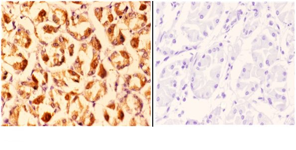
A03538 has real paraffin-section IHC data in human stomach carcinoma (catalog image caption); the catalog lists Human, Mouse and Rat reactivity (catalog: reactivity) but no IF image (catalog: IF images).

Real IHC data Immunohistochemistry (IHC) analyzes of CLN6 (N258) pAb in paraffin-embedded human stomach carcinoma tissue at 1:50.showing nuclear and strong cytoplasmic staining. Negative control (the right)Using PBS instead of primary antibody, secondary antibody is Goat Anti-Rabbit IgG-biotin followed by avidin-peroxidase.
Anti-CLN6 (N258) Antibody
Cat # A03538

A03538 is listed for IHC and WB in Human, Mouse and Rat (catalog: applications and reactivity). Its IHC image shows paraffin-embedded human stomach carcinoma at 1:50, with nuclear and strong cytoplasmic staining (catalog image caption).

Which to pick: Choose A03538 for paraffin-section tissue IHC: its image documents human stomach carcinoma staining, and the listed IHC range is 1:50–1:200 (catalog image caption; datasheet: IHC dilution). The caption does not report the fixative (catalog image caption). For IF/ICC, A03538 has no listed IF application, dilution or image; for Mouse or Rat IHC, reactivity is listed but the supplied IHC image shows Human tissue only (catalog: applications, IF dilution, IF images and reactivity; catalog image caption).

Each figure is that product's own IHC / IF validation image from its datasheet.

References

  1. UniProt Consortium. UniProt entry Q9NWW5 (CLN6_HUMAN, Ceroid-lipofuscinosis neuronal protein 6).
  2. Human Protein Atlas. CLN6 tissue IHC expression (reliability: Approved).
  3. Human Protein Atlas. CLN6 subcellular location (ICC-IF): Mainly localized to the endoplasmic reticulum. In addition localized to the nucleoli and vesicles..
  4. Human Protein Atlas. CLN6 antibody validation summary (2 antibodies).
  5. Neuronal Ceroid Lipofuscinosis in a Mixed-Breed Dog with a Splice Site Variant in CLN6. Genes 2024 — PMC11203140.
  6. Intracranial delivery of AAV9 gene therapy partially prevents retinal degeneration and visual deficits in CLN6-Batten disease mice. Molecular therapy. Methods & clinical development 2021 — PMC7887332.
  7. A murine model of variant late infantile ceroid lipofuscinosis recapitulates behavioral and pathological phenotypes of human disease. PloS one 2013 — PMC3815212.
  8. Progressive retinal degeneration and glial activation in the CLN6 (nclf) mouse model of neuronal ceroid lipofuscinosis: a beneficial effect of DHA and curcumin supplementation. PloS one 2013 — PMC3790850.
  9. PubMed PMID:14702039 — UniProt-cited evidence.
  10. PubMed PMID:16572171 — UniProt-cited evidence.
  11. PubMed PMID:15489334 — UniProt-cited evidence.