CLOCK / Circadian locomoter output cycles protein kaput · IHC design guide

Design Immunohistochemistry for CLOCK

Plan chromogenic CLOCK IHC in paraffin sections around the nuclear tissue staining reported by HPA (HPA tissue IHC). Consider collection time when comparing samples, because CLOCK shuttles between nucleus and cytoplasm under circadian and BMAL1-dependent control (UniProt).

Evidence assembled Oct 2026 · For research use; verify linked source records and product datasheet before use
Immunohistochemistry protocol sheet for CLOCK (IHC for CLOCK): expected localisation Predominantly nuclear tissue staining (HPA tissue IHC); cytoplasmic shuttling is possible (UniProt), antibody M00064, validated IHC image, and IHC protocol steps
Printable CLOCK IHC protocol sheet — expected localisation Predominantly nuclear tissue staining (HPA tissue IHC); cytoplasmic shuttling is possible (UniProt), antibody M00064, controls and protocol steps. Open the full CLOCK IHC guide →

CLOCK Immunohistochemistry Experimental Design Guide

Expected localisation, validated protocols, controls and antibodies — the at-a-glance facts below, then the full design guide.

Must know before staining
Expected localisation Predominantly nuclear tissue staining (HPA tissue IHC); cytoplasmic shuttling is possible (UniProt)
Staining pattern Nuclear staining across tissues; high in testis Leydig cells (HPA tissue IHC)
Antigen retrieval Tris-EDTA pH 9.0 HIER, 95–98 °C, 20 min (rule: nuclear antigen)
Positive control ⓘ Testis+4 more · see all
Negative control ⓘ None in HPA (detected in all 45 tissues); use no-primary + isotype controls
Important caveats
Reasons your staining may differ from the expected pattern.
Fixation Keep fixation consistent across samples. (standard IHC practice; not target-specific)
Caveat Circadian phase may shift nuclear versus cytoplasmic signal (UniProt)
Regulation BMAL1-dependent circadian shuttling (UniProt)
Isoform / epitope No annotated isoforms; one chain spans residues 1–846 (UniProt)
Section 1

Recommended CLOCK IHC & IF Protocols

The catalog antibody’s IHC-P protocol is followed by four published CLOCK IHC methods, covering frozen vascular sections and paraffin-embedded bone and liver sections (PMC6085090; PMC5765856; PMC9098081; PMC5985434).

Recommended immunohistochemistry (IHC-P) protocol parameters
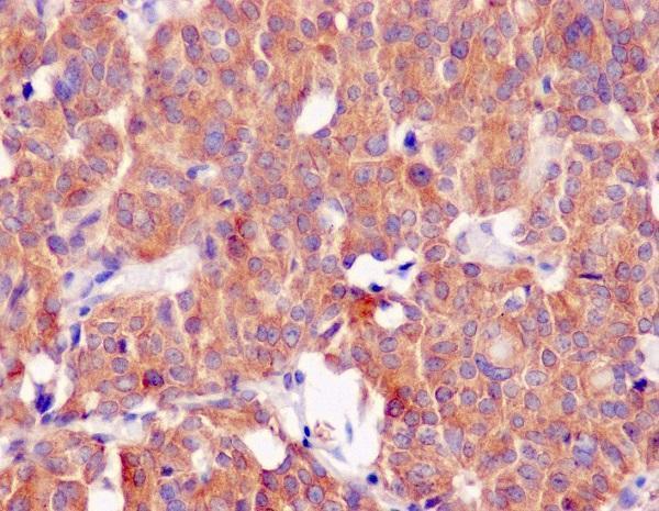
SampleParaffin-embedded human thyroid carcinoma tissue; fixative not specified (datasheet M00064)
FixationImage fixative and duration unreported (datasheet M00064); verify before use.
Sectioning4–5 µm sections on charged slides (standard)
DeparaffinisationXylene, graded ethanol series to water (standard)
Antigen retrievalHeat-induced epitope retrieval in Tris-EDTA buffer, pH 9.0, 20 min at 95–98 °C (standard rule: nuclear antigen)
Peroxidase block3% H2O2, 10 min, room temperature (standard)
Blocking10% normal serum of the secondary host, 30 min, room temperature (standard)
Primary antibodyRabbit monoclonal (clone ACCH-3) anti-CLOCK, 1:50 (datasheet M00064)
Primary incubationOvernight at 4 °C (standard)
DetectionHRP-polymer secondary, DAB chromogen 5–10 min (standard)
CounterstainHematoxylin, blue, dehydrate and mount (standard)
Expected resultCLOCK-positive staining in leydig cells of testis (HPA tissue IHC: High). HPA tissue profile: Ubiquitous nuclear expression. No signal in the no-primary control.
💡Decision noteStart with Tris-EDTA pH 9.0 HIER at 95–98 °C for 20 min (page retrieval rule); the bone and liver papers report different retrieval methods (PMC9098081; PMC5985434).
Section 2

What Is the Expected CLOCK Staining Pattern?

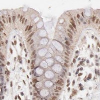
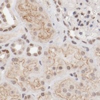
CLOCK is chiefly nuclear in tissue IHC, with a ubiquitous nuclear pattern rated Supported by HPA; that rating reflects medium consistency between staining and RNA data (HPA: tissue IHC). Expect especially strong staining in testicular Leydig cells, with staining also reported in several other cell types (HPA: High in Leydig cells; Medium in listed tissues). Cytoplasmic staining can occur because CLOCK shuttles between cytoplasm and nucleus (UniProt O15516: subcellular location); CLOCK has no transmembrane segment (UniProt O15516: topology).

What am I looking at on my slide?
Distinct nuclear chromogen in Leydig cells, with weaker nuclear staining in other tissues (HPA: tissue IHC).This fits the reported ubiquitous nuclear pattern and High Leydig cell staining. Compare intensity within the same run; HPA levels are observational categories, not a quantitative calibration (HPA: tissue IHC).
Predominantly cytoplasmic staining with little nuclear signal in an otherwise positive section (HPA: ubiquitous nuclear IHC pattern).Treat a persistent compartment mismatch as a possible staining artefact and check controls. Do not reject every cytoplasmic signal: CLOCK shuttles, and its nonphosphorylated form is reported in the cytoplasm (UniProt O15516: subcellular location).
Strong staining in cells outside the reported HPA pattern, especially when expected nuclei are unstained (HPA: tissue IHC).Consider antibody cross-reactivity or endogenous chromogenic activity. Compare morphology and a no-primary control before assigning the signal to CLOCK (general IHC practice). HPA's listed cells do not define an exhaustive set of positive cell types (HPA: tissue IHC).
Diffuse chromogen covers tissue or obscures nuclear borders, including low-staining areas (HPA: Low in listed cells).The distribution cannot be scored reliably against HPA's nuclear pattern (HPA: tissue IHC). Background may reflect nonspecific binding, incomplete blocking, or detection chemistry; assess it with no-primary and reagent controls (general IHC practice).
No nuclear signal in testicular Leydig cells (HPA: High in Leydig cells).First suspect an uninformative run, then check section identity, tissue preservation, retrieval, antibody and detection controls (general IHC practice). One negative section cannot overturn a pattern HPA rates Supported with medium RNA consistency (HPA: tissue IHC).
💡Expected CLOCK appearanceCall a section positive when clearly bounded nuclei stain in the expected cells, with strong Leydig cell staining as a useful reference (HPA: ubiquitous nuclear pattern; High in Leydig cells); diffuse chromogen or isolated signal in unexpected cells warrants control checks (general IHC practice).
How each factor affects the staining
Cell type and tissue selectionLeydig cells offer a reported High IHC signal; adipocytes and several listed glandular or neuronal cells are Medium, while listed low-staining cells are weak comparators, not true negatives (HPA: tissue IHC).
Cellular distribution and sampling timeCLOCK shuttling is circadian regulated and BMAL1 dependent; nuclear and cytoplasmic proportions may differ across samples (UniProt O15516: subcellular location). Record collection time when comparing groups (general experimental practice).
Protein form and epitope claimsUniProt lists one chain spanning residues 1–846, no signal peptide or propeptide, and several phosphorylation sites (UniProt O15516: processing and modified residues). These facts do not identify this antibody's epitope or establish a retrieval condition.
Antibody evidenceTwo listed antibodies, HPA001867 and HPA027565, have Supported IHC and ICC status (HPA: antibody validation). HPA calls the tissue pattern Supported with medium RNA consistency; neither statement establishes performance for every specimen (HPA: tissue IHC).
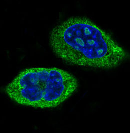
IF/ICC Q&A: what location should fluorescence show?Mainly nucleoplasmic signal, with additional vesicular localization reported in ICC-IF (HPA: subcellular, supported nucleoplasm; approved vesicles). UniProt also reports cytoplasmic localization and regulated shuttling (UniProt O15516: subcellular location).
Why is my staining missing, weak or wrong?
SituationLikely causeNext action
Leydig cell nuclei are blank in the testis reference section (HPA: High in Leydig cells).The run may have failed at retrieval, primary incubation, or detection; the HPA observation alone cannot identify which step (HPA: tissue IHC; general IHC practice).Verify section identity and morphology, then review the IHC-validated antibody's instructions and run a known-positive control beside the sample (general IHC practice).
Chromogen is mainly cytoplasmic throughout a section expected to show nuclear staining (HPA: ubiquitous nuclear pattern).Background or a staining artefact is possible, though cytoplasmic CLOCK is biologically plausible (UniProt O15516: subcellular location).Check nuclear counterstain and no-primary control; assess whether cytoplasmic signal is cell bounded and reproducible before interpreting localization (general IHC practice).
Strong color appears in an unexpected cell population (HPA: tissue IHC).Cross-reactivity or endogenous detection activity may mimic staining; HPA's listed observations cannot settle the identity of every stained cell (HPA: tissue IHC; general IHC practice).Compare morphology, no-primary and detection-only controls, and the expected nuclear distribution; investigate persistent signal with an independently validated antibody if available (general IHC practice).
Background obscures nuclei across the section (HPA: nuclear tissue pattern).Nonspecific primary or secondary binding, insufficient blocking, or chromogen development may contribute (general IHC practice).Check no-primary background, blocking and wash steps; adjust detection or primary conditions using the IHC-validated antibody's instructions (general IHC practice).
A low-staining comparison tissue shows no visible CLOCK signal (HPA: Low in listed cells).A Low HPA category can fall below the run's visual threshold; it is not a documented negative control (HPA: tissue IHC).Judge assay performance using a reported High reference such as Leydig cells, while retaining the low-staining section as a distribution comparison (HPA: High in Leydig cells; Low in listed cells).
Nuclear intensity differs between otherwise comparable specimens (HPA: ubiquitous nuclear pattern).Regulated cytoplasm-to-nucleus shuttling is one biological possibility; technical variation is another (UniProt O15516: subcellular location; general IHC practice).Record collection times, stain comparison sections together, and score nuclear signal with the same criteria and controls (general experimental and IHC practice).

Sample controls for CLOCK IHC & IF

🧪Run testis first and confirm CLOCK staining in Leydig cells (HPA: High in Leydig cells). HPA detects CLOCK in all 45 scored tissues, so use no-primary and isotype controls for negative reference; other cells on the testis slide should show only background where they lack specific staining, without being treated as proven CLOCK-negative cells (HPA: no negative tissue listed; standard IHC practice).
Positive control tissue: Testis (Leydig cells, HPA High)
Negative control tissue: None in HPA: CLOCK is detected in all 45 scored tissues. Use a no-primary (secondary-only) and an isotype control instead.
ICC-IF cell lines (HPA subcellular resource): HPA ICC-IF images show CLOCK in A-431, U-251MG, U2OS, with annotated localisation: Nucleoplasm (supported) (HPA subcellular).
Technical controls: Include a no-primary, secondary-only slide and an isotype control matched to the primary antibody’s host species, immunoglobulin class and clonality; confirm specificity with CLOCK-knockout material if available (standard IHC practice). For chromogenic testis IHC, block endogenous peroxidase and check background in the negative controls (standard IHC practice).
⚠️Feasibility: A target-specific fixation window and retrieval dependency are unreported in the supplied evidence; optimize antigen retrieval empirically for paraffin sections (standard IHC practice). The selected M00064 paraffin-section caption does not report a fixative, so it does not establish fixation (selected-SKU tissue-IHC caption: fixative unreported). The evidence does not establish that frozen sections or IF are easier; in IF, assess nucleoplasmic signal against vesicular signal and tissue autofluorescence (HPA subcellular: nucleoplasm supported, vesicles approved; standard IF practice). The selected M00064 tissue-IHC caption documents paraffin sections, but does not specify the fixative (selected-SKU IHC image M00064).

HPA tissue IHC evidence for CLOCK

Comprehensive Human Protein Atlas IHC scoring per tissue (reliability: Supported — Medium consistency between antibody staining and RNA expression data.). Rows are taken directly from the HPA tissue chart — click any row's HPA link to view the source.

Positive expression · recommended positive controls

TissueCell typeLevelEvidenceSource
Testis Leydig cells High Protein (IHC) HPA →
Adipose tissue Adipocytes Medium Protein (IHC) HPA →
Adrenal gland Glandular cells Medium Protein (IHC) HPA →
Appendix Glandular cells Medium Protein (IHC) HPA →
Bone marrow Hematopoietic cells Medium Protein (IHC) HPA →

Undetected expression · recommended negative controls

TissueCell typeLevelEvidenceSource
None in HPA: CLOCK is detected in all 45 scored tissues. Use a no-primary (secondary-only) and an isotype control instead.
Section 3

Advanced CLOCK IHC Tips

Troubleshoot CLOCK staining in paraffin sections by checking retrieval, nuclear localisation, controls and scoring before interpreting biological differences.

How should I retrieve CLOCK in paraffin sections with weak nuclear staining?
Start with Tris-EDTA at pH 9.0, heated to 95–98 °C for 20 min (page retrieval setting). Let slides cool in retrieval buffer, then compare the same tissue across a small retrieval-time series while holding antibody concentration, detection and imaging conditions constant (standard IHC practice). Check whether staining becomes sharper in nuclei of morphologically intact cells, since CLOCK is mainly nucleoplasmic in the subcellular reference and shuttles between nucleus and cytoplasm (HPA subcellular; UniProt O15516 localisation). If staining remains weak, trial a milder citrate retrieval as a fallback, recording its pH and time and checking for tissue damage alongside signal (standard IHC practice).
Could fixation explain weak CLOCK staining in my paraffin sections?
Target-specific fixation sensitivity is unknown from the supplied evidence, so document the actual fixative and fixation duration before changing the staining workflow (selected M00064 caption: fixative not stated). For a controlled comparison, stain sections from differently processed blocks together using the page retrieval condition of Tris-EDTA pH 9.0 at 95–98 °C for 20 min (page retrieval setting; standard IHC practice). Review morphology and nuclear counterstain before attributing a faint result to antigen loss, and include a no-primary control for detection background (standard IHC practice). The paraffin-section thyroid carcinoma caption establishes a stained specimen for M00064 but does not identify its fixative (selected M00064 caption).
Should CLOCK staining appear only in nuclei?
Assess nuclear and cytoplasmic staining separately: the tissue profile describes ubiquitous nuclear expression, while the subcellular reference places CLOCK mainly in the nucleoplasm (HPA tissue IHC; HPA subcellular). Cytoplasmic signal can still be biologically plausible because CLOCK shuttles under circadian regulation, with BMAL1-dependent nuclear entry and different reported locations for phosphorylated and nonphosphorylated forms (UniProt O15516 localisation). Use the nuclear counterstain to define compartments, then compare intact cells within the same staining run before calling diffuse colour positive (standard IHC practice). Record collection time and relevant experimental timing, since a compartment shift could reflect circadian regulation rather than a change in total CLOCK abundance (UniProt O15516 function and localisation).
How can epitope position change my interpretation of CLOCK staining?
First obtain the catalog antibody's immunogen or epitope information; the supplied record does not locate its binding site, and it lists 0 isoforms for CLOCK (UniProt O15516 isoforms; supplied catalog evidence). Map any disclosed epitope against the bHLH region at residues 34–84, PAS regions at 107–177 and 262–332, and PAC region at 336–379 (UniProt O15516 domains). CLOCK also has reported phosphoserines, so an epitope overlapping a modified region warrants comparison with an independently validated antibody when staining varies (UniProt O15516 modified residues; standard IHC practice). Do not assign a staining difference to a particular isoform or modification without direct epitope and specificity evidence (UniProt O15516 isoforms; standard IHC practice).
How should I check CLOCK localisation in a multiplex IF experiment?
On the separate IF/ICC workflow, pair CLOCK with a marker for the expected cell type in the specimen and a nuclear dye, keeping channels distinct during acquisition (standard IF practice). Choose fluorophores after inspecting unstained tissue for autofluorescence, and assign the weaker signal to a channel with lower background where the available optics permit (standard IF practice). Permeabilise for an intracellular epitope when needed, since CLOCK lacks a transmembrane segment and is reported in nuclear and cytoplasmic compartments; optimise detergent exposure to preserve morphology (UniProt O15516 topology and localisation; standard IF practice). Compare nuclear and cytoplasmic signals by cell, because CLOCK shuttling is circadian regulated and BMAL1 dependent (UniProt O15516 localisation).
What should I check when CLOCK IHC shows diffuse brown background?
Run a no-primary control through the same chromogenic detection steps to reveal background from detection reagents or endogenous tissue activity (standard IHC practice). Check the peroxidase block and examine whether colour follows tissue folds, section edges, pigment or damaged areas rather than intact cell nuclei (standard IHC practice). Reduce nonspecific staining by optimising blocking, primary antibody concentration and washes one variable at a time, while retaining a known stained section in the run (standard IHC practice; selected M00064 caption). CLOCK is mainly nucleoplasmic in the subcellular reference, so widespread extracellular colour should prompt a background review before biological interpretation (HPA subcellular; standard IHC practice).
How should I score CLOCK staining across paraffin sections? ⚠ ANSWER MARKED FOR VERIFICATION
Define the cell population and nuclear compartment before scoring, using morphology and a consistent counterstain to segment evaluable cells (standard IHC practice; HPA tissue IHC: ubiquitous nuclear expression). Report the percentage of positive nuclei and staining intensity, or calculate a nuclear H-score from the percentages at intensity grades 0–3 (standard IHC practice). Normalise counts to the number of evaluable cells, or report positive-cell density per mm² when tissue area is the relevant denominator (standard IHC practice). Keep thresholds and image settings fixed across batches, and record sampling time because CLOCK localisation is under circadian regulation (standard IHC practice; UniProt O15516 localisation).
How can I distinguish true CLOCK staining from artefact?
Prioritise reproducible staining in intact nuclei: the HPA tissue profile reports ubiquitous nuclear expression, and its subcellular reference supports a mainly nucleoplasmic location (HPA tissue IHC; HPA subcellular). Evaluate staining within identified cell types rather than treating all brown material as CLOCK; Leydig cells are reported high, whereas several listed cell populations have low staining (HPA tissue IHC). Exclude section-edge concentration, necrotic areas and colour reproduced in the no-primary control, and review endogenous peroxidase blocking when DAB appears outside cells (standard IHC practice). Interpret genuine cytoplasmic staining cautiously because CLOCK can shuttle between compartments, and record collection time when comparing groups (UniProt O15516 localisation; standard IHC practice).
Boster reagents

Best CLOCK / Circadian locomoter output cycles protein kaput IHC Antibodies

Anti-CLOCK IHC images show paraffin sections of human thyroid carcinoma, human cancer tissue, and mouse and rat brain (M00064 and RP1082 IHC captions); IF images show HeLa and U2OS cells (M00064 and RP1082 IF captions).

Real IHC data Immunohistochemical analysis of paraffin-embedded human thyroid carcinoma, using CLOCK Antibody.
Anti-CLOCK Monoclonal Antibody
Cat # M00064

M00064 will render with an IHC image of a paraffin-embedded human thyroid carcinoma section (M00064 IHC caption). Its catalog also lists IF/ICC, with an IF image of HeLa cells (M00064 applications and IF caption).

Which to pick: For tissue IHC, M00064 has a human paraffin-section image; RP1082 adds documented mouse and rat brain paraffin-section images and a 2 μg/ml staining example (M00064 IHC caption; RP1082 IHC captions). For IF/ICC, both list the application and show cell images: HeLa for M00064 and U2OS for RP1082 (M00064 and RP1082 applications and IF captions). For cross-species tissue IHC, RP1082 has the stronger image evidence; M00064 lists human, mouse and rat reactivity but shows only human tissue, and neither SKU’s IHC caption reports a fixative (RP1082 IHC captions; M00064 reactivity and IHC caption).

Each figure is that product's own IHC / IF validation image from its datasheet.

References

  1. UniProt Consortium. UniProt entry O15516 (CLOCK_HUMAN, Circadian locomoter output cycles protein kaput).
  2. Human Protein Atlas. CLOCK tissue IHC expression (reliability: Supported).
  3. Human Protein Atlas. CLOCK subcellular location (ICC-IF): Mainly localized to the nucleoplasm. In addition localized to vesicles..
  4. Human Protein Atlas. CLOCK antibody validation summary (2 antibodies).
  5. Immunohistochemistry of the circadian clock in mouse and human vascular tissues. Vessel plus 2018 — PMC6085090.
  6. Differential Regulation of BMAL1, CLOCK, and Endothelial Signaling in the Aortic Arch and Ligated Common Carotid Artery. Journal of vascular research 2016 — PMC5765856.
  7. A comprehensive analysis of the circRNA-miRNA-mRNA network in osteocyte-like cell associated with Mycobacterium leprae infection. PLoS neglected tropical diseases 2022 — PMC9098081.
  8. Melatonin Attenuates Dysregulation of the Circadian Clock Pathway in Mice With CCl(4)-Induced Fibrosis and Human Hepatic Stellate Cells. Frontiers in pharmacology 2018 — PMC5985434.
  9. PubMed PMID:10198158 — UniProt-cited evidence.
  10. PubMed PMID:9205841 — UniProt-cited evidence.
  11. PubMed PMID:14702039 — UniProt-cited evidence.