This website uses cookies to ensure you get the best experience on our website.


- Table of Contents
Plan CLPB paraffin IHC around granular cytoplasmic staining in kidney tubules and seminal vesicle glands (HPA tissue IHC). This guide covers fixation consistency, the catalog antibody’s 0.5–1 μg/mL IHC range (datasheet A02912-2), and interpretation of the expected mitochondrial location (UniProt).

Expected localisation, validated protocols, controls and antibodies — the at-a-glance facts below, then the full design guide.
| Expected localisation | Granular cytoplasm (HPA tissue IHC); mitochondrial intermembrane space (UniProt) | |
| Staining pattern | Granular cytoplasmic staining in kidney tubule cells (HPA tissue IHC) | |
| Antigen retrieval | EDTA pH 8.0 HIER, heat-mediated (datasheet A02912-2) | |
| Positive control | Kidney+4 more · see all | |
| Negative control | None in HPA (detected in all 45 tissues); use no-primary + isotype controls |
| Fixation | Keep fixation conditions consistent across sections. (standard IHC practice; not target-specific) | |
| Caveat | Staining–RNA discordance; external verification pending (HPA tissue IHC) | |
| Regulation | Widely expressed; low in granulocytes (UniProt) | |
| Isoform / epitope | 5 isoforms; processed chains start at residues 37 or 127; epitope unknown (UniProt) |
The catalog antibody protocol uses EDTA pH 8.0 retrieval (datasheet A02912-2). One published CLPB IHC protocol uses citrate pH 6.0 retrieval (PMC11641810).
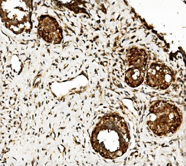
| Sample | Paraffin-embedded human mammary cancer tissue; fixative not specified (datasheet A02912-2) |
| Fixation | Image fixative and duration unreported (datasheet A02912-2); verify before use. |
| Sectioning | 4–5 µm sections on charged slides (standard) |
| Deparaffinisation | Xylene, graded ethanol series to water (standard) |
| Antigen retrieval | Heat retrieval: EDTA pH 8.0 (datasheet A02912-2); 20 min, 95–100 °C (standard) |
| Peroxidase block | 3% H2O2, 10 min, room temperature (standard) |
| Blocking | 10% goat serum (datasheet A02912-2) |
| Primary antibody | Rabbit anti-CLPB, 0.5-1μg/ml (datasheet A02912-2) |
| Primary incubation | Overnight at 4 °C (datasheet A02912-2) |
| Detection | Streptavidin-biotin complex (SABC), DAB chromogen (datasheet A02912-2) |
| Counterstain | Hematoxylin, blue, dehydrate and mount (standard) |
| Expected result | CLPB-positive staining in cells in tubules of kidney (HPA tissue IHC: High). HPA tissue profile: General cytoplasmic expression, with a granular pattern. No signal in the no-primary control. |
CLPB resides in the mitochondrial intermembrane space and has no transmembrane segment (UniProt Q9H078). In paraffin sections, expect granular cytoplasmic staining, especially in kidney tubule cells and seminal vesicle glandular cells (HPA: general granular cytoplasmic pattern; High in both cell types). HPA rates tissue IHC reliability Enhanced, while reporting very low agreement with RNA data and pending external verification (HPA: tissue reliability).
| Strong granular cytoplasmic staining in kidney tubule cells or seminal vesicle glandular cells. | This matches the reported High staining in both cell types (HPA: tissue IHC). The granules are consistent with mitochondrial localisation, although chromogenic IHC cannot resolve the intermembrane space (UniProt Q9H078; general IHC practice). |
| Predominantly nuclear, surface, or extracellular signal with little granular cytoplasmic staining. | Treat this as a localisation mismatch and investigate artefact or nonspecific binding: CLPB is assigned to the mitochondrial intermembrane space without a transmembrane segment (UniProt Q9H078). Check the negative reagent control and whether the apparent signal follows tissue edges or pigment (general IHC practice). |
| Strong staining in cells expected to be low, while adjacent expected positive cells remain weak. | Consider cross-reactivity or endogenous chromogen activity before calling this CLPB. For example, HPA reports Low staining in hippocampal glia and Medium staining in cortical neurons (HPA: tissue IHC). Cell identity and the control pattern matter more than colour alone (general IHC practice). |
| Diffuse colour across stroma, nuclei, and multiple cell types without a granular pattern. | This does not match the reported general granular cytoplasmic profile (HPA: tissue IHC). Uneven blocking, excess antibody or detection reagent, and incomplete washes can produce broad background; assess these as general IHC workflow causes, not established CLPB-specific effects (general IHC practice). |
| No specific signal in kidney tubules or seminal vesicle glands. | These are reported High staining sites, so a blank result warrants a technical check (HPA: tissue IHC). Confirm that tissue morphology is intact, a run control developed, and primary antibody and detection reagents were applied correctly (general IHC practice). A blank slide alone does not establish absent CLPB. |
| Compartment and image resolution | CLPB is in the mitochondrial intermembrane space (UniProt Q9H078). HPA describes granular cytoplasm in tissue IHC; a chromogenic section supports that pattern but cannot identify the mitochondrial subcompartment by itself (HPA: tissue IHC; general IHC practice). |
| Antibody evidence | HPA039005 and HPA039006 are IHC Enhanced; HPA064571 is IHC Approved (HPA: antibody validation). Enhanced tissue reliability has an explicit caveat: antibody staining and RNA expression show very low consistency, and external verification is pending (HPA: tissue reliability). |
| Sequence forms and epitope | UniProt lists 5 isoforms and chains beginning at residues 37 and 127 (UniProt Q9H078). Without the antibody epitope, coverage of each isoform or processed form cannot be inferred; compare the catalog antibody's stated immunogen or epitope before interpreting a missing signal. |
| IF/ICC evidence | HPA supplies no main ICC-IF location or cell line images, and lists no ICC status for these three antibodies (HPA: subcellular record; antibody validation). The tissue IHC pattern therefore does not establish an IF staining pattern or an IF protocol. |
| Retrieval and fixation evidence | No CLPB-specific retrieval condition or fixation sensitivity is supplied (UniProt Q9H078; HPA: tissue IHC). Record the retrieval conditions used and evaluate them with run controls as general IHC practice; do not attribute a change in signal to a known CLPB fixation effect. |
| Situation | Likely cause | Next action |
|---|---|---|
| Granular signal is weak in an expected positive tissue. | The assay may have low sensitivity, or the sampled cells may differ from the reported positive population (HPA: High in kidney tubules and seminal vesicle glands). | Verify the cell type and a successful run control, then review the catalog antibody's IHC-P instructions for retrieval and dilution; make no CLPB-specific retrieval assumption (general IHC practice). |
| Colour appears mainly in nuclei or at tissue edges. | That distribution conflicts with mitochondrial localisation and the reported granular cytoplasmic tissue pattern (UniProt Q9H078; HPA: tissue IHC). | Compare matched negative reagent controls and inspect section edges and pigment; repeat staining if the distribution tracks an artefact (general IHC practice). |
| All tissue compartments show similar diffuse colour. | Broad background may reflect nonspecific binding, inadequate blocking, or incomplete washing (general IHC practice). | Review blocking, reagent concentration, wash steps, and the negative reagent control; score only cell-associated granular cytoplasm against that background (general IHC practice; HPA: tissue IHC pattern). |
| Signal persists when primary antibody is omitted. | Detection chemistry or endogenous tissue activity can create colour independently of the primary antibody (general IHC practice). | Check the primary-omission control and the detection system's endogenous-activity controls before assigning that colour to CLPB (general IHC practice). |
| A low-staining cell population appears stronger than the expected positive population. | Cross-reactivity, background, or cell misidentification is possible; HPA reports Low hippocampal glia and High kidney tubule cells (HPA: tissue IHC). | Recheck cell identity and localisation, compare controls, and seek agreement with another IHC-validated antibody where available (HPA: antibody validation; general IHC practice). |
| An IF result disagrees with the paraffin-section IHC pattern. | HPA provides no ICC-IF image-based location or ICC validation status for the listed antibodies (HPA: subcellular record; antibody validation). | Interpret the IF experiment on its own controls and documented antibody validation; do not use the IHC result as an IF protocol or subcellular confirmation (general IF practice). |
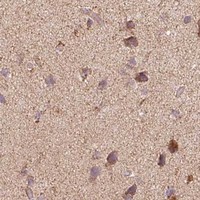


Comprehensive Human Protein Atlas IHC scoring per tissue (reliability: Enhanced — Very low consistency between antibody staining and RNA expression data. External characterization data supports antibody staining. Pending external verification.). Rows are taken directly from the HPA tissue chart — click any row's HPA link to view the source.
| Tissue | Cell type | Level | Evidence | Source |
|---|---|---|---|---|
| Kidney | Cells in tubules | High | Protein (IHC) | HPA → |
| Seminal vesicle | Glandular cells | High | Protein (IHC) | HPA → |
| Adrenal gland | Glandular cells | Medium | Protein (IHC) | HPA → |
| Appendix | Glandular cells | Medium | Protein (IHC) | HPA → |
| Breast | Glandular cells | Medium | Protein (IHC) | HPA → |
| Tissue | Cell type | Level | Evidence | Source |
|---|---|---|---|---|
| None in HPA: CLPB is detected in all 45 scored tissues. Use a no-primary (secondary-only) and an isotype control instead. | ||||
Use the paraffin-section CLPB image as a starting point, then assess staining against mitochondrial localisation and cell-specific controls.
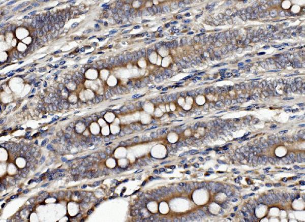
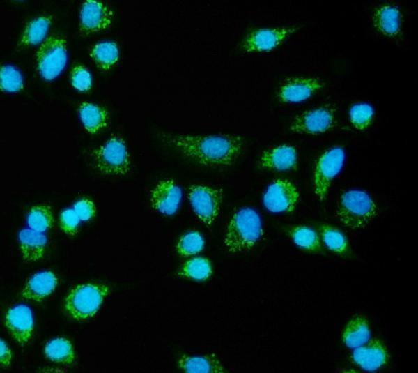
Anti-CLPB A02912-2 has IHC data from human paraffin sections of mammary and rectal cancer (catalog IHC captions) and IF data from A549 cells (catalog IF caption).
A02912-2 will render with an IHC figure from a human mammary cancer paraffin section (card image alt); another catalog caption documents IHC in a human rectal cancer paraffin section (catalog IHC caption). The same SKU lists IHC, ICC and IF applications (catalog applications) and shows IF staining in A549 cells (catalog IF caption).
Which to pick: Choose A02912-2 for tissue IHC in paraffin sections: its own figure caption reports EDTA retrieval at pH 8.0 and a 1 μg/ml primary antibody concentration in human mammary cancer tissue (A02912-2 IHC caption); the fixative is unreported (A02912-2 IHC caption). For IF/ICC, A02912-2 lists a 4 μg/ml IF concentration and shows staining in A549 cells (catalog dilution; catalog IF caption). For cross-species planning, A02912-2 lists Human, Monkey, Mouse and Rat reactivity (catalog reactivity), while its supplied IHC captions show human sections only (catalog IHC captions); clonality is unreported (catalog clone field).