CMPK1 / UMP-CMP kinase · IHC design guide

Design Immunohistochemistry for CMPK1

Plan CMPK1 staining in paraffin sections using its observed cytoplasmic and nuclear tissue pattern (HPA tissue IHC). The guide covers fixation, controls, detection and scoring around the IHC-validated antibody’s 1:50–1:200 dilution range (datasheet).

Evidence assembled Oct 2026 · For research use; verify linked source records and product datasheet before use
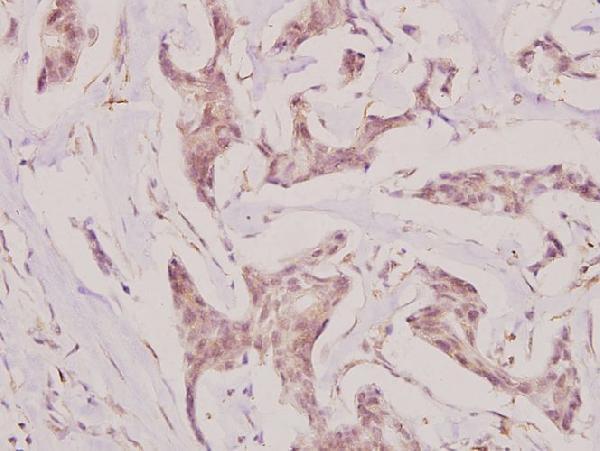
Immunohistochemistry protocol sheet for CMPK1 (IHC for CMPK1): expected localisation Cytoplasmic and nuclear staining in most tissues (HPA tissue IHC), antibody A07962-1, validated IHC image, and IHC protocol steps
Printable CMPK1 IHC protocol sheet — expected localisation Cytoplasmic and nuclear staining in most tissues (HPA tissue IHC), antibody A07962-1, controls and protocol steps. Open the full CMPK1 IHC guide →

CMPK1 Immunohistochemistry Experimental Design Guide

Expected localisation, validated protocols, controls and antibodies — the at-a-glance facts below, then the full design guide.

Must know before staining
Expected localisation Cytoplasmic and nuclear staining in most tissues (HPA tissue IHC)
Staining pattern Most tissues show cytoplasmic and nuclear cell staining (HPA tissue IHC)
Antigen retrieval Tris-EDTA pH 9.0 HIER, 95–98 °C, 20 min (rule: nuclear antigen)
Positive control ⓘ Appendix+4 more · see all
Negative control ⓘ Adipose tissue+1 more · see all
Important caveats
Reasons your staining may differ from the expected pattern.
Fixation Matched tissue-IHC evidence does not establish the fixation claim. Validate the specimen-specific method before use. (selected-SKU IHC image A07962-1)
Caveat Staining has medium consistency with RNA expression (HPA tissue IHC)
Regulation Specific expression regulation unreported (UniProt)
Isoform / epitope 3 isoforms; epitope differences unknown (UniProt)
Section 1

Recommended CMPK1 IHC & IF Protocols

The catalog antibody’s IHC-P protocol (datasheet) is accompanied by published CMPK1 IHC methods for liver samples (PMC11299646) and tissue sections (PMC8013733).

Recommended immunohistochemistry (IHC-P) protocol parameters
SampleParaffin-embedded human breast carcinoma tissue; fixative not specified (datasheet A07962-1)
FixationImage fixative and duration unreported (datasheet A07962-1); verify before use.
Sectioning4–5 µm sections on charged slides (standard)
DeparaffinisationXylene, graded ethanol series to water (standard)
Antigen retrievalHeat-induced epitope retrieval in Tris-EDTA buffer, pH 9.0, 20 min at 95–98 °C (standard rule: nuclear antigen)
Peroxidase block3% H2O2, 10 min, room temperature (standard)
Blocking10% normal serum of the secondary host, 30 min, room temperature (standard)
Primary antibodyRabbit anti-CMPK1, 1:50-1:200 (datasheet A07962-1)
Primary incubationOvernight at 4 °C (standard)
DetectionHRP-polymer secondary, DAB chromogen 5–10 min (standard)
CounterstainHematoxylin, blue, dehydrate and mount (standard)
Expected resultCMPK1-positive staining in glandular cells of appendix (HPA tissue IHC: High). HPA tissue profile: Cytoplasmic and nuclear expression in most tissues. No signal in the no-primary control.
💡Decision noteStart with Tris-EDTA pH 9.0 retrieval at 95–98 °C for 20 min (page retrieval setting); assess cytoplasmic and nuclear staining (UniProt P30085 localisation).
Section 2

What Is the Expected CMPK1 Staining Pattern?

CMPK1 staining is expected in the cytoplasm and nuclei of many tissue cell types (HPA tissue IHC: cytoplasmic and nuclear expression in most tissues). UniProt describes CMPK1 as predominantly cytoplasmic, with less than 15% nuclear, and annotates no transmembrane segment (UniProt P30085: subcellular location and topology). HPA rates its tissue IHC evidence Approved, with medium consistency between staining and RNA data; external verification is pending (HPA tissue IHC: reliability).

What am I looking at on my slide?
Glandular cells in appendix or duodenum show strong cytoplasmic and nuclear staining.This fits the reported compartment pattern and High staining in those cells (HPA tissue IHC: profile; High in appendix and duodenum glandular cells). Compare signal within identified cells: HPA's tissue summary supports both compartments, while UniProt describes a predominantly cytoplasmic distribution (UniProt P30085: subcellular location).
A crisp plasma-membrane rim or extracellular deposit dominates the signal.Treat this as a compartment mismatch: CMPK1 has no annotated transmembrane segment, and its reported locations are cytoplasm and nucleus (UniProt P30085: topology and subcellular location; HPA tissue IHC: profile). Check whether the pattern follows cell boundaries or section deposits before scoring it as CMPK1 staining (general IHC practice).
Adipocytes or caudate glial cells stain strongly while nearby expected-positive cells do not.Those cell types are reported as Not detected by HPA, so strong selective staining warrants a specificity check (HPA tissue IHC: adipocytes and caudate glial cells Not detected). Consider antibody cross-reactivity or endogenous chromogenic activity; neither is established by the HPA record for this slide (general IHC practice).
Brown signal spreads across stroma, section edges, and cells without clear boundaries.A diffuse distribution makes compartment and cell-type scoring unreliable (general IHC practice). The expected tissue pattern is cellular cytoplasmic and nuclear expression (HPA tissue IHC: profile). Review detection controls and background before interpreting the haze as widespread CMPK1 expression (general IHC practice).
No staining appears in an appendix glandular-cell reference section.HPA reports High staining in appendix glandular cells, making an absent signal a reason to check the assay and section (HPA tissue IHC: appendix glandular cells High; general IHC practice). It does not alone prove CMPK1 is absent from the specimen; HPA's IHC evidence is Approved but awaits external verification (HPA tissue IHC: reliability).
💡Expected CMPK1 appearanceCall a positive result when identified cells show cytoplasmic and nuclear staining, with strong signal plausible in HPA High cell types; a dominant membrane rim or cell-free deposit is suspect (HPA tissue IHC: profile and High entries; UniProt P30085: topology).
How each factor affects the staining
Compartment evidenceHPA tissue IHC reports cytoplasmic and nuclear expression, whereas UniProt describes CMPK1 as predominantly cytoplasmic with less than 15% nuclear (HPA tissue IHC: profile; UniProt P30085: subcellular location). Record both compartments rather than requiring equal intensity (general IHC practice).
Cell-type distributionHPA reports High staining in several glandular, respiratory epithelial, endothelial, and cardiomyocyte entries, but Not detected in adipocytes and caudate glial cells (HPA tissue IHC: positive and negative entries). Select and score reference cells by their named cell type, not the tissue label alone (general IHC practice).
Evidence and antibody validationThe HPA tissue profile is Approved with medium staining–RNA consistency and pending external verification; both listed antibodies are IHC Approved, while HPA053730 is ICC Supported (HPA tissue IHC: reliability; HPA antibodies: HPA053730, HPA058604). Interpret agreement as support, without treating it as independent external confirmation.
Isoform coverageUniProt lists three CMPK1 isoforms, but the supplied antibody records give no epitope or isoform coverage (UniProt P30085: isoforms; HPA antibodies: supplied fields). A change in staining cannot be assigned to a particular isoform from these data.
Chromogenic backgroundEndogenous tissue activity or nonspecific detection can produce color independent of the intended antigen (general IHC practice). Interpret weak widespread color against detection controls and the expected cellular distribution (general IHC practice; HPA tissue IHC: profile).
Why is my staining missing, weak or wrong?
SituationLikely causeNext action
Expected-positive glandular cells remain blank.Possible assay or section failure; HPA reports High appendix and duodenum glandular-cell staining (HPA tissue IHC: positive entries).Confirm that the named cells are present, then review the IHC-validated antibody dilution, antigen retrieval, detection reagents, and positive control under the assay's documented conditions (general IHC practice). No CMPK1-specific retrieval condition or dilution is supplied here.
A membrane-like outline dominates staining.The distribution conflicts with the reported cytoplasmic and nuclear locations and lack of a transmembrane segment (HPA tissue IHC: profile; UniProt P30085: topology).Inspect the pattern at higher magnification and compare a negative reagent control; score CMPK1 only where signal resolves within cells in supported compartments (general IHC practice; HPA tissue IHC: profile).
Adipocytes or caudate glial cells show strong selective color.These cell types are listed as Not detected; cross-reactivity or endogenous detection activity is possible, not demonstrated (HPA tissue IHC: negative entries; general IHC practice).Verify cell identity and compare the detection control with an HPA High reference cell type before accepting the signal (general IHC practice; HPA tissue IHC: positive entries).
Diffuse brown background obscures cell boundaries.Nonspecific binding or endogenous chromogenic activity can obscure a cellular pattern (general IHC practice).Review blocking, washes, detection controls, and reagent concentration using the assay instructions; then reassess whether cytoplasmic and nuclear signal is distinguishable (general IHC practice; HPA tissue IHC: profile).
Nuclear staining seems stronger than expected in tissue IHC.UniProt describes predominantly cytoplasmic CMPK1, but HPA tissue IHC includes nuclear expression; intensity differences alone do not identify an artefact (UniProt P30085: subcellular location; HPA tissue IHC: profile).Record nuclear and cytoplasmic staining separately, identify the stained cell type, and compare controls before judging specificity (general IHC practice).
Q: Does nucleoplasmic IF/ICC staining invalidate the tissue IHC result?A: HPA reports the nucleoplasm as the supported main ICC-IF location, while its tissue IHC profile includes cytoplasm and nuclei (HPA subcellular: main location; HPA tissue IHC: profile).Interpret each application against its own HPA observation and cell context; do not require identical compartment emphasis across ICC-IF and paraffin IHC (HPA subcellular: main location; HPA tissue IHC: profile).

Sample controls for CMPK1 IHC & IF

🧪Run appendix first: its glandular cells should stain (HPA: High in appendix glandular cells). Use adipose tissue as the negative tissue, assessing adipocytes (HPA: Not detected in adipocytes); on the appendix slide, cells outside the glandular compartment should show only background where they are demonstrably CMPK1-negative, since CMPK1 is broadly expressed (UniProt P30085: ubiquitously expressed).
Positive control tissue: Appendix (Glandular cells, HPA High)
Negative control tissue: Adipose tissue (HPA Not detected)
ICC-IF cell lines (HPA subcellular resource): HPA ICC-IF images show CMPK1 in MCF-7, PC-3, U2OS, NIH 3T3, with annotated localisation: Nucleoplasm (supported) (HPA subcellular).
Technical controls: Include a no-primary, secondary-only control; an isotype control matched to the primary antibody’s host species and clonality; and CMPK1 knockout material, if available, as a biological negative (standard IHC practice). Check appendix sections for endogenous peroxidase signal in blood or inflammatory cells before interpreting chromogenic staining (standard IHC practice).
⚠️Feasibility: A CMPK1-specific fixation window and antigen-retrieval dependency are unreported in the supplied evidence; the selected A07962-1 caption shows staining in a paraffin section at 1:100 but does not report the fixative (selected SKU tissue-IHC caption). The evidence does not establish whether frozen sections or IF would be easier: HPA ICC-IF reports mainly nucleoplasmic localization, while UniProt describes CMPK1 as predominantly cytoplasmic (HPA: subcellular summary; UniProt P30085: subcellular location). In appendix, inflammatory-cell peroxidase can complicate chromogenic interpretation (standard IHC practice).

HPA tissue IHC evidence for CMPK1

Comprehensive Human Protein Atlas IHC scoring per tissue (reliability: Approved — Medium consistency between antibody staining and RNA expression data. Pending external verification.). Rows are taken directly from the HPA tissue chart — click any row's HPA link to view the source.

Positive expression · recommended positive controls

TissueCell typeLevelEvidenceSource
Appendix Glandular cells High Protein (IHC) HPA →
Bronchus Respiratory epithelial cells High Protein (IHC) HPA →
Colon Endothelial cells High Protein (IHC) HPA →
Duodenum Glandular cells High Protein (IHC) HPA →
Endometrium Glandular cells Medium Protein (IHC) HPA →

Undetected expression · recommended negative controls

TissueCell typeLevelEvidenceSource
Adipose tissue Adipocytes Not detected Protein (IHC) HPA →
Caudate Glial cells Not detected Protein (IHC) HPA →
Section 3

Advanced CMPK1 IHC Tips

Troubleshoot CMPK1 staining in paraffin sections by checking retrieval, compartment patterns, controls and cell specific scoring.

What retrieval conditions should I try first for weak CMPK1 staining?
Use Tris-EDTA at pH 9.0 for heat induced epitope retrieval at 95–98 °C for 20 minutes (page retrieval specification). Deparaffinize sections completely, keep them covered by buffer during heating, and cool them consistently before antibody incubation (standard IHC practice). Compare a retrieved section with a matched section processed without retrieval to see whether heating improves specific cellular signal (standard IHC practice). If staining remains weak, optimize heating time on matched sections while checking for tissue damage and increased background (standard IHC practice). Score cytoplasmic and nuclear staining separately because both occur in tissue IHC (HPA: tissue profile).
Could fixation explain variable CMPK1 staining between paraffin sections?
The selected A07962-1 paraffin tissue caption does not report a fixative, so CMPK1 sensitivity to fixation remains unknown for this antibody (selected tissue IHC caption). Record the fixative, fixation duration, processing schedule and section age for every specimen before comparing stain intensity (standard IHC practice). If these variables differ, run matched sections together with the same retrieval, antibody incubation and chromogenic development conditions (standard IHC practice). Inspect morphology alongside signal, since poorly preserved tissue can make cellular boundaries and compartment scoring unreliable (standard IHC practice). Do not assign a CMPK1 specific fixation effect without a controlled comparison using the same antibody (selected tissue IHC caption).
How should I evaluate nuclear versus cytoplasmic CMPK1 staining?
Score nuclear and cytoplasmic staining separately: tissue IHC reports both compartments in most tissues (HPA: tissue profile). UniProt describes CMPK1 as predominantly cytoplasmic, with less than 15% nuclear, whereas the subcellular imaging summary identifies supported nucleoplasmic localisation (UniProt P30085: subcellular location; HPA: subcellular summary). Use a nuclear counterstain to define boundaries, then compare intensity within the same cell type and staining run (standard IHC practice). Report the observed compartment pattern rather than resolving this source difference by assumption (UniProt P30085: subcellular location; HPA: tissue and subcellular profiles). Diffuse staining beyond identifiable cells warrants review of background and morphology (standard IHC practice).
Can epitope differences account for inconsistent CMPK1 staining?
CMPK1 has 3 annotated isoforms, but the supplied antibody evidence does not identify the epitope or establish equal recognition of each isoform (UniProt P30085: isoforms; selected tissue IHC caption). Its 196 residue annotated chain has no transmembrane segment, so a membrane restricted stain would need independent validation (UniProt P30085: chain and topology). Annotated modifications include phosphoserine 33 and 180 and acetyllysine 43 and 55; their effect on this antibody is unknown (UniProt P30085: modified residues; selected tissue IHC caption). Compare staining on serial sections under matched processing before attributing a difference to isoforms or modifications (standard IHC practice).
How can I use IF to investigate an ambiguous CMPK1 IHC pattern?
Use IF as a compartment check alongside IHC, while treating performance of the catalog antibody in IF as unestablished by its tissue IHC caption (selected tissue IHC caption). Multiplex with an epithelial marker when examining bronchial respiratory epithelial cells, a reported high expression population, and include single color controls (HPA: bronchus respiratory epithelial cells, High; standard IF practice). Choose a far red fluorophore after checking tissue autofluorescence and spectral overlap (standard IF practice). Because CMPK1 is reported in cytoplasm and nucleus and lacks a transmembrane segment, test permeabilisation that permits access to cytosolic and nuclear epitopes (UniProt P30085: location and topology; standard IF practice). Compare compartment patterns with the paired chromogenic section (standard IF and IHC practice).
How do I separate CMPK1 signal from chromogenic background?
Start with a no primary control and a matched section stained with the catalog antibody; the selected paraffin tissue image used 1:100, which is a documented example rather than an established optimum for every sample (selected tissue IHC caption; standard IHC practice). Apply a peroxidase block before enzyme based detection and interpret DAB deposition in the control as possible detection background (standard chromogenic IHC practice). Check blocking, wash consistency and chromogen development when diffuse stain obscures cell boundaries (standard IHC practice). Compare staining within identifiable glandular or respiratory epithelial cells, where high expression is reported (HPA: appendix glandular cells, High; bronchus respiratory epithelial cells, High).
What is a defensible way to quantify CMPK1 IHC? ⚠ ANSWER MARKED FOR VERIFICATION
Define the cell population and compartment before scoring, because tissue IHC reports cytoplasmic and nuclear expression (HPA: tissue profile). An H-score can combine the percentage of cells at each intensity from 0 to 3, yielding 0–300; alternatively, report percentage positive cells using a prespecified threshold (standard IHC scoring practice). Normalize counts to viable cells of the same type, or report positive cell density per mm² of viable tissue when cell counts are impractical (standard pathology quantification practice). Keep staining, imaging and threshold settings consistent across the comparison set (standard IHC practice). Record nuclear and cytoplasmic results separately to preserve the observed compartment pattern (HPA: tissue profile).
When is an apparent CMPK1 positive cell likely to be artefactual?
A plausible positive pattern is cellular cytoplasmic or nuclear staining, with reported high signal in appendix glandular cells and bronchial respiratory epithelial cells (HPA: tissue profile and positive populations). Treat exclusively membrane outlined staining cautiously because CMPK1 has no transmembrane segment (UniProt P30085: topology). Check whether unexpected staining concentrates at section edges, in necrotic regions or in the no primary control, where processing or detection artefacts can occur (standard IHC practice). Peroxidase activity can contribute chromogenic signal, so review the peroxidase blocked control and cellular morphology before calling such deposits positive (standard chromogenic IHC practice). Report discordance explicitly rather than treating a single stained cell as proof of CMPK1 expression (standard IHC interpretation practice).
Boster reagents

Best CMPK1 / UMP-CMP kinase IHC Antibodies

A07962-1 has real IHC data from paraffin-embedded human breast carcinoma tissue (catalog: IHC image caption). Listed reactivity covers human, mouse, and rat (catalog: reactivity); no IF data are supplied (catalog: IF images).

Real IHC data Immunohistochemistry (IHC) analyzes of UMP-CMP Kinase (E36) pAb in paraffin-embedded human breast carcinoma tissue at 1:100.
Anti-UMP-CMP Kinase (E36) CMPK1 Antibody
Cat # A07962-1

A07962-1 is listed for IHC and has an image of paraffin-embedded human breast carcinoma tissue stained at 1:100 (catalog: applications; IHC image caption). It is listed as reactive with human, mouse, and rat (catalog: reactivity), but the supplied image documents human tissue only (catalog: IHC image caption).

Which to pick: Choose A07962-1 for paraffin-section IHC: its rabbit polyclonal antibody is IHC-listed, with a pictured human breast carcinoma section and a listed IHC dilution of 1:50–1:200 (catalog: host; IHC image caption; applications; IHC dilution). The caption does not report the fixative (catalog: IHC image caption). No SKU here has documented IF/ICC use (catalog: applications; IF images); A07962-1 lists mouse and rat reactivity, but the supplied IHC image demonstrates human tissue only (catalog: reactivity; IHC image caption).

Each figure is that product's own IHC / IF validation image from its datasheet.

References

  1. UniProt Consortium. UniProt entry P30085 (KCY_HUMAN, UMP-CMP kinase).
  2. Human Protein Atlas. CMPK1 tissue IHC expression (reliability: Approved).
  3. Human Protein Atlas. CMPK1 subcellular location (ICC-IF): Mainly localized to the nucleoplasm..
  4. Human Protein Atlas. CMPK1 antibody validation summary (2 antibodies).
  5. Integrated Transcriptomics and Proteomics Identified CMPK1 as a Potential Biomarker for Type 2 Diabetes Mellitus. Diabetes, metabolic syndrome and obesity : targets and therapy 2024 — PMC11299646.
  6. CMPK1 Regulated by miR-130b Attenuates Response to 5-FU Treatment in Gastric Cancer. Frontiers in oncology 2021 — PMC8013733.
  7. 4EBP1/eIF4E and p70S6K/RPS6 axes play critical and distinct roles in hepatocarcinogenesis driven by AKT and N-Ras proto-oncogenes in mice. Hepatology (Baltimore, Md.) 2015 — PMC4280310.
  8. Oncogenic KSHV-encoded interferon regulatory factor upregulates HMGB2 and CMPK1 expression to promote cell invasion by disrupting a complex lncRNA-OIP5-AS1/miR-218-5p network. PLoS pathogens 2019 — PMC6370251.
  9. PubMed PMID:10462544 — UniProt-cited evidence.
  10. PubMed PMID:11681623 — UniProt-cited evidence.
  11. PubMed PMID:11912132 — UniProt-cited evidence.