This website uses cookies to ensure you get the best experience on our website.


- Table of Contents
Plan chromogenic CMTM6 IHC in paraffin sections using the predominantly cytoplasmic tissue pattern (HPA tissue IHC). Start with the catalog antibody at 1:100–1:300 (datasheet), and compare high-staining endometrial glandular cells with undetected staining in skeletal myocytes (HPA tissue IHC).

Expected localisation, validated protocols, controls and antibodies — the at-a-glance facts below, then the full design guide.
| Expected localisation | Predominantly cytoplasmic tissue staining (HPA tissue IHC) | |
| Staining pattern | Cytoplasmic staining in most tissues (HPA tissue IHC) | |
| Antigen retrieval | Citrate pH 6.0 HIER, 95–98 °C, 20 min (rule: cytoplasmic / membrane antigen) | |
| Positive control | Cerebellum+4 more · see all | |
| Negative control | Hippocampus+1 more · see all |
| Fixation | Keep paraffin-section fixation consistent across samples (standard IHC practice; not target-specific) | |
| Caveat | Staining has medium consistency with RNA expression (HPA tissue IHC) | |
| Regulation | Expression regulation unspecified (UniProt) | |
| Isoform / epitope | No isoforms reported; epitope side matters across 4 transmembrane spans (UniProt) |
The catalog antibody’s IHC-P protocol is accompanied by published CMTM6 protocols for gastric cancer, hepatocellular carcinoma, and renal cell carcinoma (PMC7842018; PMC8353897; PMC13039633).
| Sample | Paraffin-embedded human oophoroma tissue; fixative not specified (datasheet A14107) |
| Fixation | Image fixative and duration unreported (datasheet A14107); verify before use. |
| Sectioning | 4–5 µm sections on charged slides (standard) |
| Deparaffinisation | Xylene, graded ethanol series to water (standard) |
| Antigen retrieval | Heat-induced epitope retrieval in citrate buffer, pH 6.0, 20 min at 95–98 °C (standard rule: cytoplasmic / membrane antigen) |
| Peroxidase block | 3% H2O2, 10 min, room temperature (standard) |
| Blocking | 10% normal serum of the secondary host, 30 min, room temperature (standard) |
| Primary antibody | Rabbit anti-CMTM6, 1:100-1:300 (datasheet A14107) |
| Primary incubation | Overnight at 4 °C (standard) |
| Detection | HRP-polymer secondary, DAB chromogen 5–10 min (standard) |
| Counterstain | Hematoxylin, blue, dehydrate and mount (standard) |
| Expected result | CMTM6-positive staining in purkinje cells of cerebellum (HPA tissue IHC: High). HPA tissue profile: Cytoplasmic expression in most tissues. No signal in the no-primary control. |
CMTM6 is a four-pass membrane protein found at the cell membrane and in early and recycling endosomes (UniProt Q9NX76 topology and subcellular location). In tissue IHC, expect chiefly cytoplasmic staining, with signal in glandular cells, urothelial cells and Purkinje cells among the HPA high-staining examples (HPA tissue IHC). HPA rates its tissue staining reliability Enhanced, while reporting medium consistency between antibody staining and RNA expression (HPA tissue IHC).
| Cytoplasmic staining in endometrial, fallopian tube or rectal glandular cells. | This matches HPA's chiefly cytoplasmic tissue profile and High staining in these cell types (HPA tissue IHC). A cytoplasmic appearance can be compatible with endosomal membrane localization; IHC alone does not identify the stained organelle (UniProt Q9NX76 subcellular location; general IHC practice). |
| Membrane-associated signal, possibly alongside cytoplasmic signal, in cells with expected staining. | A membrane component is compatible with CMTM6 at the cell membrane and on early and recycling endosome membranes (UniProt Q9NX76 subcellular location). Judge it alongside HPA's observed cytoplasmic tissue pattern; a membrane-only result is not the sole required appearance (HPA tissue IHC). |
| Predominantly nuclear staining, with little cytoplasmic or membrane signal. | A nuclear-dominant pattern does not match the reported cell membrane, endosomal or tissue IHC locations (UniProt Q9NX76 subcellular location; HPA tissue IHC). Treat it as suspect and assess background and antibody specificity before scoring cells as CMTM6 positive (general IHC practice). |
| Strong staining in hippocampal glial cells or skeletal myocytes. | HPA reports CMTM6 as Not detected in those respective cells, so strong signal is discordant with these reference observations (HPA tissue IHC). Cross-reactivity or endogenous detection activity are possibilities to investigate, although one discordant slide does not establish either cause (general IHC practice). |
| No signal in a tissue containing a documented high-staining cell population. | Absence in, for example, endometrial glandular cells or urinary bladder urothelial cells conflicts with HPA's High observations (HPA tissue IHC). First check that the relevant cells are present and assess the staining run; HPA's Enhanced reliability does not guarantee every specimen will stain (HPA tissue IHC; general IHC practice). |
| Cell population and reference intensity | HPA reports High staining in Purkinje cells, several glandular populations and urinary bladder urothelial cells, but Not detected staining in hippocampal glial cells and skeletal myocytes (HPA tissue IHC). Compare like cell types when choosing and reading controls; whole-tissue labels can obscure which cells matter (general IHC practice). |
| Compartment and topology | CMTM6 has four transmembrane segments and reported cell membrane and endosomal membrane locations (UniProt Q9NX76 topology and subcellular location). HPA describes tissue staining as cytoplasmic, so interpret visible cytoplasmic signal in that context without claiming that chromogenic IHC resolves a specific endosome (HPA tissue IHC; general IHC practice). |
| Evidence and antibody validation | HPA calls tissue IHC reliability Enhanced and notes medium agreement between staining and RNA data; HPA026980 has Enhanced IHC validation (HPA tissue IHC; HPA antibodies). These support a reference pattern, while unexpected compartments or cell types still warrant controls and review (general IHC practice). |
| Detection background | Endogenous detection activity and nonspecific reagent staining can produce chromogen unrelated to primary-antibody binding (general IHC practice). A no-primary control helps assess detection-system background; it cannot by itself establish that remaining signal is specific to CMTM6 (general IHC practice). |
| IF/ICC question | Q: What localization should an IF/ICC image be compared with? A: HPA reports mainly vesicular CMTM6 signal and an Approved ICC result for HPA063491; UniProt also reports cell membrane and recycling endosome localization (HPA subcellular; HPA antibodies; UniProt Q9NX76 subcellular location). Use the separate IF/ICC guide for its workflow. |
| Situation | Likely cause | Next action |
|---|---|---|
| Expected high-staining cells are blank. | The target cells may be absent from the section, or a staining-run step may have failed; the image alone cannot distinguish these explanations (general IHC practice). | Confirm the relevant cell population and run a documented positive tissue alongside the sample; review the IHC-validated antibody's specified workflow and the run's detection controls (HPA tissue IHC; general IHC practice). |
| Most tissue compartments show a diffuse brown haze. | Broad signal without cell or compartment definition is consistent with nonspecific staining or detection background (general IHC practice). | Compare with a no-primary control, inspect wash and blocking steps, and reassess the signal only after background is separated from the cellular pattern (general IHC practice). |
| Strong signal is concentrated in nuclei. | This location conflicts with reported membrane and endosomal localization and the HPA cytoplasmic tissue profile (UniProt Q9NX76 subcellular location; HPA tissue IHC). | Check whether the nuclear signal persists in controls, then evaluate primary-antibody specificity before including it in a positive score (general IHC practice). |
| Hippocampal glia or skeletal myocytes stain strongly. | Those cell types are listed as Not detected by HPA; cross-reactivity or endogenous detection activity may account for discordant signal (HPA tissue IHC; general IHC practice). | Verify the cell identity, compare a no-primary control and review staining in a documented High cell population within the same run (HPA tissue IHC; general IHC practice). |
| Only an apparent membrane rim is visible. | Cell membrane localization is supported, but a rim alone does not reproduce HPA's chiefly cytoplasmic tissue profile (UniProt Q9NX76 subcellular location; HPA tissue IHC). | Inspect the expected cell populations at suitable magnification and compare the complete cellular pattern with a positive tissue control before assigning a compartment-based score (HPA tissue IHC; general IHC practice). |
| Signal varies sharply between otherwise comparable sections. | Target-specific fixation effects are not established by the supplied assay evidence. Verify with a matched IHC source before attributing a result to fixation. | Compare matching cell populations and concurrent controls, then review the recorded IHC workflow for run differences without attributing the variation to a target-specific fixation effect (general IHC practice). |


Comprehensive Human Protein Atlas IHC scoring per tissue (reliability: Enhanced — Medium consistency between antibody staining and RNA expression data.). Rows are taken directly from the HPA tissue chart — click any row's HPA link to view the source.
| Tissue | Cell type | Level | Evidence | Source |
|---|---|---|---|---|
| Cerebellum | Purkinje cells | High | Protein (IHC) | HPA → |
| Endometrium | Glandular cells | High | Protein (IHC) | HPA → |
| Fallopian tube | Glandular cells | High | Protein (IHC) | HPA → |
| Rectum | Glandular cells | High | Protein (IHC) | HPA → |
| Urinary bladder | Urothelial cells | High | Protein (IHC) | HPA → |
Troubleshoot CMTM6 staining in paraffin sections by checking retrieval, tissue context, subcellular pattern and controls before scoring chromogenic signal.
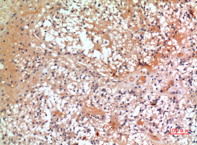
A14107 is a human- and mouse-reactive anti-CMTM6 antibody listed for IHC and IF (catalog: applications and reactivity). Its IHC image shows a paraffin-embedded human oophoroma section (A14107 IHC image caption).
A14107 will render with IHC data from a paraffin-embedded human oophoroma section at 1:100 (A14107 IHC image caption). The catalog lists A14107 for human and mouse IHC and IF, but supplies no IF image (catalog: applications, reactivity, and image captions).
Which to pick: Choose A14107 for paraffin-section IHC because its own image caption documents that preparation; the fixative is unreported (A14107 IHC image caption). For IF/ICC planning, A14107 lists IF at 1:50, but ICC validation and an IF image are unreported (catalog: applications, IF dilution, and image captions). A14107 is the listed cross-species option because the catalog reports human and mouse reactivity; it is a rabbit polyclonal antibody (catalog: reactivity, host, and dilution_raw).