This website uses cookies to ensure you get the best experience on our website.


- Table of Contents
Plan CNN2 staining in paraffin sections using the catalog antibody’s documented tonsil IHC conditions (datasheet A08680-2). Compare the result with cytoplasmic immune-cell staining and medium staining in tonsil germinal center cells (HPA tissue IHC).

Expected localisation, validated protocols, controls and antibodies — the at-a-glance facts below, then the full design guide.
| Expected localisation | Cytoplasmic staining in immune cells (HPA tissue IHC) | |
| Staining pattern | Cytoplasmic immune cells, including germinal center cells (HPA tissue IHC) | |
| Antigen retrieval | EDTA pH 8.0 HIER, heat-mediated (datasheet A08680-2) | |
| Positive control | Lung+2 more · see all | |
| Negative control | Adipose tissue+4 more · see all |
| Fixation | Keep paraffin-section fixation consistent. (standard IHC practice; not target-specific) | |
| Caveat | Immune-cell staining may complicate interpretation (HPA tissue IHC) | |
| Regulation | No expression regulator specified (UniProt) | |
| Isoform / epitope | 2 isoforms; antibody epitope coverage is unspecified (UniProt) |
The catalog antibody uses EDTA retrieval (datasheet A08680-2); one published CNN2 IHC protocol uses citrate retrieval (PMC10298237).
| Sample | Paraffin-embedded human tonsil tissue; fixative not specified (datasheet A08680-2) |
| Fixation | Image fixative and duration unreported (datasheet A08680-2); verify before use. |
| Sectioning | 4–5 µm sections on charged slides (standard) |
| Deparaffinisation | Xylene, graded ethanol series to water (standard) |
| Antigen retrieval | Heat retrieval: EDTA pH 8.0 (datasheet A08680-2); 20 min, 95–100 °C (standard) |
| Peroxidase block | 3% H2O2, 10 min, room temperature (standard) |
| Blocking | 10% goat serum (datasheet A08680-2) |
| Primary antibody | Rabbit anti-CNN2, 2-5 μg/ml (datasheet A08680-2) |
| Primary incubation | Overnight at 4 °C (datasheet A08680-2) |
| Detection | Streptavidin-biotin complex (SABC), DAB chromogen (datasheet A08680-2) |
| Counterstain | Hematoxylin, blue, dehydrate and mount (standard) |
| Expected result | CNN2-positive staining in alveolar cells of lung (HPA tissue IHC: Medium). HPA tissue profile: Cytoplasmic expression in immune cells. No signal in the no-primary control. |
CNN2 is reported as cytoplasmic in immune cells by tissue IHC, with medium staining in lung alveolar cells and germinal center cells of lymph node and tonsil (HPA tissue IHC). ICC-IF places it mainly on actin filaments (HPA subcellular). It has no annotated subcellular location or transmembrane segment in UniProt (UniProt Q99439). The tissue IHC profile is Approved but pending external verification (HPA tissue IHC).
| Cytoplasmic staining in germinal center cells, or in the reported lung alveolar cells, at about medium intensity (HPA tissue IHC). | This matches the reported cell types and intensity (HPA tissue IHC). Score the named cells separately from surrounding tissue; a positive cell population does not make every cell in that section positive. |
| Predominantly nuclear, surface-restricted, or extracellular signal in place of cytoplasmic signal (HPA tissue IHC: cytoplasmic profile). | Treat this as a localization mismatch requiring review, rather than confirmed CNN2. Check morphology, the negative control, and whether detection signal follows tissue edges or deposits (general IHC practice). |
| Signal in adipocytes or bronchial respiratory epithelial cells, both reported as not detected (HPA tissue IHC). | Consider cross-reactivity or endogenous detection activity, especially if a control also stains (general IHC practice). These HPA calls concern the specified cell types; they do not establish that the whole tissue is negative. |
| Weak color spread across many structures, with little separation between reported positive and negative cells (HPA tissue IHC). | This is difficult to score as cell-specific CNN2. Review blocking, washing, detection reagent, and counterstain balance using matched controls (general IHC practice); HPA does not identify a CNN2-specific cause. |
| No signal in the reported medium germinal center cells of lymph node or tonsil (HPA tissue IHC). | The run lacks its expected reference pattern. Check the section and staining controls, antibody dilution, retrieval and detection steps (general IHC practice) before interpreting other cells as negative. |
| Cell and compartment reference | Use the HPA cell-level calls: medium in the named lung and germinal center cells, not detected in listed adipocytes and bronchial respiratory epithelial cells (HPA tissue IHC). Do not transfer a cell-level call to an entire organ. |
| Localization and topology | The cytoplasmic tissue profile agrees with the actin-filament ICC-IF location (HPA tissue IHC; HPA subcellular). CNN2 has no transmembrane segment or signal peptide (UniProt Q99439); tissue IHC need not resolve individual filaments. |
| Strength of antibody evidence | The tissue profile is Approved and pending external verification; HPA049095 is Approved for IHC, with no Enhanced IHC status supplied (HPA tissue IHC; HPA antibodies). Compare unexpected staining with the controls before treating it as a new pattern. |
| Isoforms and epitope uncertainty | UniProt lists 2 CNN2 isoforms (UniProt Q99439). No antibody epitope or isoform-specific IHC result is supplied, so a staining difference cannot be assigned to an isoform from this evidence. |
| IF/ICC Q: where should CNN2 appear? | A: Mainly on actin filaments (HPA subcellular, ICC-IF). That observation supports an intracellular interpretation but does not establish that filament detail will be visible in chromogenic paraffin-section IHC (HPA subcellular; general IHC practice). |
| Situation | Likely cause | Next action |
|---|---|---|
| The reported positive germinal center cells are unstained (HPA tissue IHC). | An unsuccessful staining run or an unsuitable section is possible; HPA supplies no CNN2-specific retrieval or fixation sensitivity. | Confirm tissue morphology and run controls, then review antibody dilution, retrieval and detection records (general IHC practice). Avoid calling test sections negative until a reference stains. |
| Signal is chiefly nuclear, despite the cytoplasmic tissue profile (HPA tissue IHC). | Localization mismatch may reflect nonspecific detection or misleading counterstain (general IHC practice). | Compare the negative control and counterstain, then assess whether cytoplasmic signal is actually present in reported positive cells (HPA tissue IHC; general IHC practice). |
| Adipocytes or bronchial respiratory epithelial cells stain strongly (HPA tissue IHC: not detected). | Cross-reactivity or endogenous detection activity is possible; the HPA call is specific to these cells (HPA tissue IHC; general IHC practice). | Inspect a matched negative control and check endogenous-activity blocking where relevant to the detection system (general IHC practice). Recheck cell identity before scoring. |
| Color covers the section diffusely and obscures cell boundaries. | Background from blocking, washing, or detection conditions can limit interpretation (general IHC practice); no CNN2-specific background mechanism is supplied. | Review matched controls and the blocking, wash, detection and counterstain steps (general IHC practice). Score only cells with distinguishable signal and morphology. |
| Extracellular deposits or edge staining dominate the slide. | This distribution conflicts with the cytoplasmic tissue profile (HPA tissue IHC) and may reflect a staining artifact (general IHC practice). | Check the negative control and section quality, and separate deposited color from intracellular signal in the named positive cells (HPA tissue IHC; general IHC practice). |
| Low-level thyroid glandular staining is called a strong positive (HPA tissue IHC: Low). | The intensity call may have been overinterpreted; HPA reports low staining for these cells (HPA tissue IHC). | Rescore intensity against matched controls and the reported medium reference cells, recording cell type and compartment as well as color strength (HPA tissue IHC; general IHC practice). |


Comprehensive Human Protein Atlas IHC scoring per tissue (reliability: Approved — Pending external verification. Antibody staining in cells/structures not annotated, view images.). Rows are taken directly from the HPA tissue chart — click any row's HPA link to view the source.
| Tissue | Cell type | Level | Evidence | Source |
|---|---|---|---|---|
| Lung | Alveolar cells | Medium | Protein (IHC) | HPA → |
| Lymph node | Germinal center cells | Medium | Protein (IHC) | HPA → |
| Tonsil | Germinal center cells | Medium | Protein (IHC) | HPA → |
| Tissue | Cell type | Level | Evidence | Source |
|---|---|---|---|---|
| Adipose tissue | Adipocytes | Not detected | Protein (IHC) | HPA → |
| Adrenal gland | Glandular cells | Not detected | Protein (IHC) | HPA → |
| Bone marrow | Hematopoietic cells | Not detected | Protein (IHC) | HPA → |
| Bronchus | Respiratory epithelial cells | Not detected | Protein (IHC) | HPA → |
| Caudate | Glial cells | Not detected | Protein (IHC) | HPA → |
Troubleshoot CNN2 staining in paraffin sections by checking retrieval, cell identity, cytoplasmic pattern, and controls before comparing chromogenic signal across samples.
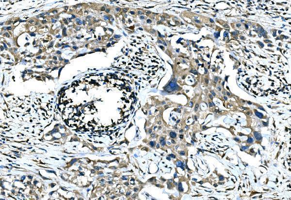
A08680-2 has real IHC data from a paraffin-embedded human tonsil section; no IF figure is supplied (catalog: A08680-2 image captions).
A08680-2 is listed for human IHC (catalog: A08680-2 applications and reactivity). Its IHC figure shows a paraffin-embedded human tonsil section stained with 2 μg/ml primary antibody after EDTA pH 8.0 retrieval (A08680-2 image caption).
Which to pick: Choose A08680-2 for human paraffin-section IHC: it is a rabbit antibody listed for human IHC at 2–5 μg/ml, and its own figure documents staining in human tonsil (catalog: A08680-2; A08680-2 image caption). No SKU in the payload is listed for IF/ICC or non-human reactivity, and clonality is not reliably specified (catalog: A08680-2 applications, reactivity, and dilution_raw). The IHC caption does not report the fixative, so fixation cannot be inferred (A08680-2 image caption). The selected A08680-2 tissue-IHC caption documents paraffin sections, but does not specify the fixative (selected-SKU IHC image A08680-2).