This website uses cookies to ensure you get the best experience on our website.


- Table of Contents
Plan CNPY2 chromogenic IHC around the widespread cytoplasmic tissue pattern (HPA tissue IHC) and its ER localization (UniProt). Use the catalog antibody’s 1:50–1:200 IHC dilution range as a starting point (datasheet A10020), and interpret staining with the reported multi-gene targeting caution (HPA tissue IHC).

Expected localisation, validated protocols, controls and antibodies — the at-a-glance facts below, then the full design guide.
| Expected localisation | Cytoplasmic staining (HPA tissue IHC) | |
| Staining pattern | Widespread cytoplasmic staining across cell types (HPA tissue IHC) | |
| Antigen retrieval | Citrate pH 6.0 HIER, heat-mediated (datasheet A10020) | |
| Positive control | Cervix+4 more · see all | |
| Negative control | Oral mucosa |
| Fixation | Keep paraffin-section fixation consistent (standard IHC practice; not target-specific); Matched tissue-IHC evidence does not establish the fixation claim. Validate the specimen-specific method before use. (selected-SKU IHC image A10020) | |
| Caveat | Staining may reflect protein from more than one gene (HPA tissue IHC) | |
| Regulation | Staining intensity varies by tissue (HPA tissue IHC) | |
| Isoform / epitope | 2 isoforms; signal peptide 1–20 is cleaved (UniProt) |
The catalog antibody protocol is followed by three published chromogenic IHC protocols for CNPY2 in liver, myocardium, and tissue sections (PMC8306219; PMC6443342; PMC4230931).
| Sample | Paraffin-embedded mouse kidney tissue; fixative not specified (datasheet A10020) |
| Fixation | Image fixative and duration unreported (datasheet A10020); verify before use. |
| Sectioning | 4–5 µm sections on charged slides (standard) |
| Deparaffinisation | Xylene, graded ethanol series to water (standard) |
| Antigen retrieval | Heat retrieval: Citrate pH 6.0 (datasheet A10020); 20 min, 95–100 °C (standard) |
| Peroxidase block | 3% H2O2, 10 min, room temperature (standard) |
| Blocking | 10% normal serum of the secondary host, 30 min, room temperature (standard) |
| Primary antibody | Rabbit anti-CNPY2, 1:50-1:200 (datasheet A10020) |
| Primary incubation | Overnight at 4 °C (standard) |
| Detection | HRP-polymer secondary, DAB chromogen 5–10 min (standard) |
| Counterstain | Hematoxylin, blue, dehydrate and mount (standard) |
| Expected result | CNPY2-positive staining in glandular cells of cervix (HPA tissue IHC: High). HPA tissue profile: Ubiquitous cytoplasmic expression. No signal in the no-primary control. |
CNPY2 should appear predominantly cytoplasmic in tissue IHC (HPA: ubiquitous cytoplasmic expression), consistent with an endoplasmic reticulum annotation and no transmembrane segment (UniProt Q9Y2B0: location and topology). Expect strong staining in placental trophoblastic cells and in glandular cells of the cervix, epididymis, fallopian tube and thyroid (HPA: High). HPA rates its tissue pattern Supported, while cautioning that the staining may represent proteins from more than one gene (HPA: reliability).
| Cytoplasmic staining in placental trophoblastic cells or the listed glandular cells. | This matches the reported High tissue IHC pattern (HPA: tissue IHC). Compare cell types and compartments before calling a section positive; intensity alone cannot resolve HPA's multi-gene staining caveat (HPA: reliability). |
| Predominantly nuclear staining, with little cytoplasmic signal. | That compartment does not match the reported cytoplasmic tissue pattern or the endoplasmic reticulum annotation (HPA: tissue IHC; UniProt Q9Y2B0: location). Check staining and counterstain interpretation before assigning it to CNPY2 (general IHC practice). |
| Strong signal in oral mucosal squamous epithelium. | HPA reports CNPY2 as Not detected in these cells (HPA: oral mucosa). Unexpected staining warrants a specificity check; cross-reactivity or endogenous chromogen activity are possibilities, not diagnoses from appearance alone (general IHC practice). |
| Diffuse colour across cells and surrounding tissue, without a clear cellular pattern. | The pattern is hard to reconcile with HPA's cytoplasmic cell staining (HPA: tissue IHC). Review background in a no-primary control and inspect blocking, washing and detection steps (general IHC practice). |
| No staining in placental trophoblastic cells despite an apparently intact section. | This conflicts with HPA's High report for that cell type (HPA: placenta). Check section identity, antibody and detection performance, and controls before interpreting other unstained tissues as negative (general IHC practice). |
| Tissue and cell choice | Placental trophoblastic cells and the listed glandular cells offer reported High staining; oral mucosal squamous cells are reported Not detected (HPA: tissue IHC). These are cell-specific comparisons, not guarantees for every section (general IHC practice). |
| Antibody evidence | HPA038465 and HPA038466 each have Supported IHC status (HPA: antibodies). HPA also cautions that tissue staining may target proteins from more than one gene, so a matching pattern alone does not establish unique CNPY2 detection (HPA: reliability). |
| Protein location and processing | UniProt places CNPY2 in the endoplasmic reticulum, finds no transmembrane segment, and records a signal peptide at residues 1–20 followed by a 21–182 chain (UniProt Q9Y2B0). The antibody epitope is unspecified; these annotations cannot predict its staining performance. |
| Isoforms | UniProt records 2 CNPY2 isoforms (UniProt Q9Y2B0: isoforms). The payload does not map an antibody epitope to either isoform, so staining intensity cannot be assigned to a particular isoform. |
| IF/ICC Q&A: should its pattern match tissue IHC? | HPA reports approved cytosolic ICC-IF localisation and images from A-431, U-251MG and U2OS (HPA: subcellular; HPA038465: ICC Approved). Tissue IHC reports a cytoplasmic profile (HPA: tissue IHC); compare compartment, while evaluating IF/ICC on its own guide page. |
| Situation | Likely cause | Next action |
|---|---|---|
| Placental trophoblastic cells show no visible chromogenic signal (HPA: placenta, High). | A failed staining or detection step is possible; the image alone cannot identify which step failed (general IHC practice). | Confirm the cell type, inspect a positive control, then review antibody application, retrieval and detection records (general IHC practice). |
| Most colour is nuclear, with little cytoplasmic staining. | This conflicts with the reported cytoplasmic IHC profile and endoplasmic reticulum annotation (HPA: tissue IHC; UniProt Q9Y2B0: location). | Review counterstain and chromogen channels, then repeat with appropriate staining controls if the compartment remains unclear (general IHC practice). |
| Oral mucosal squamous epithelium stains strongly (HPA: Not detected). | Unexpected signal could reflect nonspecific antibody binding or endogenous detection activity; HPA's multi-gene caution limits attribution (HPA: reliability; general IHC practice). | Compare a no-primary control and the expected positive cell types in the same run; reassess specificity before scoring (general IHC practice). |
| Colour is diffuse across cells and tissue spaces. | Background from detection or washing is possible; diffuse colour does not show HPA's cell-associated cytoplasmic profile (HPA: tissue IHC; general IHC practice). | Check the no-primary control, blocking and wash steps, then reassess whether cellular boundaries and cytoplasm can be scored (general IHC practice). |
| Positive cells stain, but their intensity varies among sections. | Different cell populations may have different reported levels; HPA records High, Medium and Low categories across specified tissues and cells (HPA: tissue IHC). | Compare like cell types under the same scoring conditions and check run controls before interpreting a weaker section (general IHC practice). |
| An apparently plausible cytoplasmic pattern is being called CNPY2-specific. | HPA calls its IHC evidence Supported but cautions that staining may represent proteins from more than one gene (HPA: reliability). | Report the observed cell type, compartment and intensity separately from target attribution; use an independent specificity check if the conclusion requires unique CNPY2 detection (general IHC practice). |


Comprehensive Human Protein Atlas IHC scoring per tissue (reliability: Supported — High consistency between antibody staining and RNA expression data. Caution, targets protein from more than one gene.). Rows are taken directly from the HPA tissue chart — click any row's HPA link to view the source.
| Tissue | Cell type | Level | Evidence | Source |
|---|---|---|---|---|
| Cervix | Glandular cells | High | Protein (IHC) | HPA → |
| Epididymis | Glandular cells | High | Protein (IHC) | HPA → |
| Fallopian tube | Glandular cells | High | Protein (IHC) | HPA → |
| Placenta | Trophoblastic cells | High | Protein (IHC) | HPA → |
| Thyroid gland | Glandular cells | High | Protein (IHC) | HPA → |
| Tissue | Cell type | Level | Evidence | Source |
|---|---|---|---|---|
| Oral mucosa | Squamous epithelial cells | Not detected | Protein (IHC) | HPA → |
Troubleshoot CNPY2 staining in paraffin sections by checking retrieval, compartment, cell identity, and controls before comparing staining intensity.
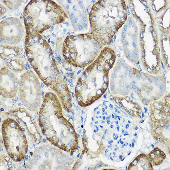
A10020 has a real IHC image of paraffin-embedded mouse kidney (IHC caption). Its listed reactivity covers human, mouse and rat; no IF/ICC data are provided (catalog: A10020).
A10020 is shown staining paraffin-embedded mouse kidney at 1:100 after high-pressure retrieval in 10 mM citrate, pH 6.0 (IHC caption). Its listed applications include IHC, and its listed reactivity covers human, mouse and rat; the payload provides no IF image (catalog: A10020).
Which to pick: Choose A10020 for paraffin-section IHC because its own image documents staining in mouse kidney, while the fixative is unreported (IHC caption). No listed SKU has IF/ICC validation, so there is no supported IF/ICC pick here (catalog: A10020). A10020 is a rabbit polyclonal antibody with listed human, mouse and rat reactivity; its pictured IHC evidence is limited to mouse kidney (catalog: A10020; IHC caption).