CNPY3 / Protein canopy homolog 3 · IHC design guide

Design Immunohistochemistry for CNPY3

For paraffin CNPY3 IHC, start the catalog antibody at 1:50–1:200 (datasheet) and assess granular cytoplasmic staining (HPA tissue IHC). Compare high-staining bone marrow hematopoietic cells with adipocytes reported as undetected, while accounting for the tissue profile’s uncertain reliability (HPA tissue IHC).

Evidence assembled Oct 2026 · For research use; verify linked source records and product datasheet before use
Immunohistochemistry protocol sheet for CNPY3 (IHC for CNPY3): expected localisation Granular cytoplasm in tissue; ER at molecular level (HPA tissue IHC; UniProt), antibody A10971, validated IHC image, and IHC protocol steps
Printable CNPY3 IHC protocol sheet — expected localisation Granular cytoplasm in tissue; ER at molecular level (HPA tissue IHC; UniProt), antibody A10971, controls and protocol steps. Open the full CNPY3 IHC guide →

CNPY3 Immunohistochemistry Experimental Design Guide

Expected localisation, validated protocols, controls and antibodies — the at-a-glance facts below, then the full design guide.

Must know before staining
Expected localisation Granular cytoplasm in tissue; ER at molecular level (HPA tissue IHC; UniProt)
Staining pattern Granular cytoplasm in most tissues; high in marrow cells (HPA tissue IHC)
Antigen retrieval Citrate pH 6.0 HIER, 95–98 °C, 20 min (rule: cytoplasmic / membrane antigen)
Positive control ⓘ Bone marrow+4 more · see all
Negative control ⓘ Adipose tissue+4 more · see all
Important caveats
Reasons your staining may differ from the expected pattern.
Fixation Keep fixation consistent across sections (standard IHC practice; not target-specific)
Caveat Tissue staining has uncertain reliability (HPA tissue IHC)
Regulation Expression regulation unannotated (UniProt)
Isoform / epitope 2 isoforms; signal peptide 1–30 is cleaved; check epitope (UniProt)
Section 1

Recommended CNPY3 IHC & IF Protocols

The catalog antibody’s IHC-P protocol is accompanied by published CNPY3 protocols for colon and breast tissue microarrays (PMC11887163; PMC11271795; PMC12651230).

Recommended immunohistochemistry (IHC-P) protocol parameters
SampleParaffin-embedded mouse kidney tissue; fixative not specified (datasheet A10971)
FixationImage fixative and duration unreported (datasheet A10971); verify before use.
Sectioning4–5 µm sections on charged slides (standard)
DeparaffinisationXylene, graded ethanol series to water (standard)
Antigen retrievalHeat-induced epitope retrieval in citrate buffer, pH 6.0, 20 min at 95–98 °C (standard rule: cytoplasmic / membrane antigen)
Peroxidase block3% H2O2, 10 min, room temperature (standard)
Blocking10% normal serum of the secondary host, 30 min, room temperature (standard)
Primary antibodyRabbit anti-CNPY3, 1:50-1:200 (datasheet A10971)
Primary incubationOvernight at 4 °C (standard)
DetectionHRP-polymer secondary, DAB chromogen 5–10 min (standard)
CounterstainHematoxylin, blue, dehydrate and mount (standard)
Expected resultCNPY3-positive staining in hematopoietic cells of bone marrow (HPA tissue IHC: High). HPA tissue profile: Granular cytoplasmic expression in most tissues. No signal in the no-primary control.
💡Decision noteStart with citrate pH 6.0 retrieval at 95–98 °C for 20 min (page antigen retrieval rule); adjust for the tissue and antibody.
Section 2

What Is the Expected CNPY3 Staining Pattern?

CNPY3 is an endoplasmic reticulum protein with no transmembrane segment (UniProt Q9BT09 topology). In paraffin-section IHC, expect granular cytoplasmic staining in most tissues, with high staining reported in bone marrow hematopoietic cells and fallopian tube glandular cells (HPA tissue IHC). Treat the pattern as provisional: HPA rates tissue staining Uncertain because antibody staining and RNA expression show only medium consistency, pending external verification (HPA tissue IHC).

What am I looking at on my slide?
Granular cytoplasmic staining in bone marrow hematopoietic cells or fallopian tube glandular cells.This matches the reported compartment and High staining in those cells (HPA tissue IHC). Compare intensity within the same run; an HPA staining level is not a universal intensity threshold (general IHC practice).
Predominantly nuclear staining, with little granular cytoplasmic signal.Nuclear dominance conflicts with the reported IHC pattern (HPA tissue IHC) and ER localisation (UniProt Q9BT09). Review the staining and counterstain before calling it CNPY3; compartment mismatch suggests artefact or nonspecific detection (general IHC practice).
Strong staining in adipocytes or squamous epithelial cells of cervix, esophagus, oral mucosa or vagina.Those cells are reported as Not detected (HPA tissue IHC). Check for cross-reactivity or endogenous detection activity using appropriate controls (general IHC practice); one unexpected section cannot establish a new CNPY3 distribution.
Diffuse colour across many cells and the surrounding tissue, without a granular cell pattern.This obscures the reported granular cytoplasmic profile (HPA tissue IHC). Review blocking, washing and detection background with control sections (general IHC practice); diffuse colour alone is insufficient evidence of CNPY3.
No staining in bone marrow hematopoietic cells or fallopian tube glandular cells.These are reported High groups (HPA tissue IHC), so a blank result warrants a run-control check. Review retrieval and detection steps (general IHC practice), while retaining HPA's Uncertain tissue-IHC rating (HPA tissue IHC).
💡Expected CNPY3 appearanceCall a positive when granular cytoplasmic staining is clear in reported High cells, such as bone marrow hematopoietic cells (HPA tissue IHC); dominant nuclear colour or strong staining in HPA-negative adipocytes is suspect (HPA tissue IHC; UniProt Q9BT09).
How each factor affects the staining
Compartment and topologyUniProt places CNPY3 in the ER and reports no transmembrane segment (UniProt Q9BT09 topology). Interpret paraffin IHC against HPA's granular cytoplasmic profile; an isolated membrane outline does not reproduce that tissue-IHC profile (HPA tissue IHC).
Tissue and cell contextHPA reports low RNA tissue specificity and granular cytoplasmic staining in most tissues, but intensity varies by cell group (HPA tissue IHC). Use the named cells for comparisons; absence from one group does not make an entire organ negative.
Protein processing and isoformsThe annotated signal peptide spans residues 1–30; the mature chain spans 31–278, and two isoforms are listed (UniProt Q9BT09). The supplied record does not locate the IHC antibody epitope, so it cannot establish which forms the stain detects.
GlycosylationUniProt lists one glycosylation site at residue 153 (UniProt Q9BT09). Its effect on this antibody's IHC staining is unreported; the site alone cannot explain a weak or absent result.
Validation strengthThe listed antibody, HPA016560, is Uncertain for IHC and Approved for ICC (HPA antibodies). Tissue IHC remains Uncertain, pending external verification (HPA tissue IHC); interpret unexpected cell or compartment staining with controls.
IF/ICC Q&A: Is plasma membrane staining plausible?Yes. HPA reports approved ER and plasma membrane locations in ICC-IF (HPA subcellular). That cell-imaging observation does not establish a membrane-only pattern as the expected result in paraffin-section chromogenic IHC.
Why is my staining missing, weak or wrong?
SituationLikely causeNext action
The reported high-staining cells are blank.Retrieval, primary-antibody or detection failure is possible (general IHC practice); the cells are reported High, subject to Uncertain tissue-IHC reliability (HPA tissue IHC).Check a reported high-staining control section in the same run and review retrieval, reagent application and detection steps (general IHC practice).
Signal is mainly nuclear.The compartment conflicts with granular cytoplasmic tissue staining (HPA tissue IHC) and ER localisation (UniProt Q9BT09); nonspecific colour is possible.Compare with a no-primary control and inspect whether counterstain or detection colour is being mistaken for specific signal (general IHC practice).
Adipocytes or the listed squamous epithelial cells stain strongly.These cells are reported Not detected (HPA tissue IHC); cross-reactivity or endogenous detection activity may contribute (general IHC practice).Compare the cell pattern with no-primary and detection controls before assigning the colour to CNPY3 (general IHC practice).
Colour is diffuse across the section.General background can obscure granular cytoplasmic staining (general IHC practice; HPA tissue IHC profile).Review blocking, wash steps and detection exposure; judge cell-specific staining only after background is controlled (general IHC practice).
Only a sharp membrane outline appears in tissue IHC.Plasma membrane localisation is reported for ICC-IF (HPA subcellular), while tissue IHC is described as granular cytoplasmic (HPA tissue IHC).Check whether reported high-staining cells also show granular cytoplasmic colour; document membrane-only tissue staining as a discrepancy.
A weak cell group is scored as negative.HPA distinguishes Low skin fibroblasts from Not detected cell groups (HPA tissue IHC); thresholds can collapse that distinction (general IHC practice).Score cell type and intensity separately, using the same criteria across sections and a reported high-staining control for context (general IHC practice).

Sample controls for CNPY3 IHC & IF

🧪Run bone marrow first; its hematopoietic cells should stain (HPA: High in bone marrow hematopoietic cells). Use adipose tissue adipocytes as the negative comparator (HPA: Not detected in adipose adipocytes); on the bone marrow slide, independently confirmed CNPY3-negative cells should show only background chromogen, but the supplied HPA row does not identify an internal negative cell population (HPA: bone marrow hematopoietic cells High).
Positive control tissue: Bone marrow (Hematopoietic cells, HPA High)
Negative control tissue: Adipose tissue (HPA Not detected)
ICC-IF cell lines (HPA subcellular resource): HPA ICC-IF images show CNPY3 in GAMG, OE19, U2OS, with annotated localisation: Endoplasmic reticulum (approved), Plasma membrane (approved) (HPA subcellular).
Technical controls: Include a no-primary, secondary-only control; an isotype control matched to the primary antibody’s host species and immunoglobulin class; and CNPY3 knockout material, if available, as a biological negative (standard IHC practice). Block endogenous peroxidase when staining bone marrow hematopoietic cells with chromogenic detection (standard IHC practice).
⚠️Feasibility: A target-specific fixation window or fixation effect is unreported, and the exact A10971 mouse-kidney paraffin-section caption does not state a fixative (A10971 caption). That caption uses microwave antigen retrieval in 10 mM PBS, pH 7.2, with antibody at 1:100; retrieval should be checked for the chosen specimen (A10971 caption). Whether frozen sections or IF/ICC is easier is unreported; endogenous peroxidase may cause background in bone marrow chromogenic IHC (standard IHC practice), while HPA ICC-IF images show approved ER and plasma-membrane localization in GAMG, OE19 and U2OS cells (HPA subcellular).

HPA tissue IHC evidence for CNPY3

Comprehensive Human Protein Atlas IHC scoring per tissue (reliability: Uncertain — Medium consistency between antibody staining and RNA expression data. Pending external verification.). Rows are taken directly from the HPA tissue chart — click any row's HPA link to view the source.

Positive expression · recommended positive controls

TissueCell typeLevelEvidenceSource
Bone marrow Hematopoietic cells High Protein (IHC) HPA →
Cerebellum Cells in molecular layer High Protein (IHC) HPA →
Fallopian tube Glandular cells High Protein (IHC) HPA →
Adrenal gland Glandular cells Medium Protein (IHC) HPA →
Appendix Glandular cells Medium Protein (IHC) HPA →

Undetected expression · recommended negative controls

TissueCell typeLevelEvidenceSource
Adipose tissue Adipocytes Not detected Protein (IHC) HPA →
Cervix Squamous epithelial cells Not detected Protein (IHC) HPA →
Esophagus Squamous epithelial cells Not detected Protein (IHC) HPA →
Oral mucosa Squamous epithelial cells Not detected Protein (IHC) HPA →
Ovary Ovarian stroma cells Not detected Protein (IHC) HPA →
Section 3

Advanced CNPY3 IHC Tips

Troubleshoot CNPY3 staining in paraffin sections by checking retrieval, controls, compartment, and scoring before interpreting chromogenic signal.

Which retrieval conditions should I start with for CNPY3 paraffin IHC?
Start with citrate buffer at pH 6.0, heated to 95–98 °C for 20 min (page retrieval setting: CNPY3). Let sections cool in buffer, then compare signal and tissue preservation against an otherwise identical section; cooling and matched comparisons are standard IHC practice (standard IHC practice). If staining is weak, test microwave retrieval in 10 mM PBS, pH 7.2, the method reported for paraffin-embedded mouse kidney stained with SKU A10971 at 1:100 (A10971 tissue-IHC caption). Change one retrieval variable at a time, because excess heat can damage sections and make granular cytoplasmic staining difficult to assess (standard IHC practice; HPA tissue IHC: granular cytoplasmic profile).
Could fixation explain weak or uneven CNPY3 staining?
Target-specific sensitivity to fixation is unknown: the selected mouse-kidney paraffin caption does not state a fixative, and the supplied record gives no CNPY3 fixation comparison (A10971 tissue-IHC caption; supplied CNPY3 record). Document fixative, fixation interval, section thickness, and time in storage for every specimen before comparing staining (standard IHC practice). On serial sections, keep retrieval at citrate pH 6.0 for 20 min while assessing whether staining varies with documented handling (page retrieval setting: CNPY3; standard IHC practice). Compare intact tissue regions with matched controls, since patchy processing or damaged edges can mimic biological differences in chromogenic IHC (standard IHC practice).
Where should convincing CNPY3 signal appear in a paraffin section?
Assess fine granular cytoplasmic staining first: CNPY3 is annotated in the endoplasmic reticulum, while HPA reports a granular cytoplasmic tissue profile (UniProt Q9BT09: subcellular location; HPA tissue IHC: profile). HPA also reports approved endoplasmic reticulum and plasma-membrane locations from subcellular imaging, so a membrane accent can be assessed alongside cytoplasmic signal rather than scored alone (HPA subcellular: approved locations). The protein has no transmembrane segment and has a 1–30 signal sequence, which makes antibody epitope placement relevant to compartment interpretation (UniProt Q9BT09: topology and processing). Require consistent staining within intact cells and compare controls before accepting diffuse nuclear, luminal, or edge-only color as CNPY3 (standard IHC practice).
How can epitope choice change the CNPY3 IHC pattern?
Check the antibody’s stated immunogen against both annotated isoforms before assigning a negative section to absent CNPY3 (UniProt Q9BT09: 2 isoforms; standard IHC practice). The precursor has a 1–30 signal sequence and a mature 31–278 chain; an epitope confined to the removed signal sequence may not report mature protein (UniProt Q9BT09: processing). A glycosylation site at residue 153 and a Saposin B-type domain spanning 47–271 are additional features to consider when interpreting epitope accessibility (UniProt Q9BT09: glycosylation and domain). If epitope information is unavailable, compare retrieval conditions and appropriate controls without claiming that a particular isoform or modification caused weak staining (standard IHC practice).
How should I use IF to investigate an ambiguous CNPY3 IHC result?
Use IF/ICC as a separate corroborating assay and select a marker for the cell population being assessed in the IHC section; HPA reports high tissue staining in bone-marrow hematopoietic cells and fallopian-tube glandular cells (HPA tissue IHC: positive cells; standard IF practice). Choose fluorophores after checking tissue autofluorescence and include single-label controls to distinguish bleed-through from colocalisation (standard IF practice). Match permeabilisation to the antibody epitope’s side of the membrane: access to an intracellular epitope generally requires it, while a surface-accessible epitope may not (standard IF practice; UniProt Q9BT09: no transmembrane segment). Interpret any overlap in light of HPA’s approved endoplasmic-reticulum and plasma-membrane locations, without treating IF staining as validation of chromogenic IHC specificity (HPA subcellular: approved locations; standard assay-validation practice).
What should I check when CNPY3 chromogenic staining looks widespread?
First compare a primary-antibody omission control with the stained section, keeping peroxidase block, secondary detection, and DAB development matched (standard chromogenic IHC practice). Endogenous peroxidase, inadequate blocking, and overdevelopment can create color unrelated to antibody binding, especially where tissue is damaged or blood rich (standard chromogenic IHC practice). Titrate around the reported 1:100 use of SKU A10971 only as an assay starting point; that caption concerns paraffin-embedded mouse kidney and does not establish an optimum for other specimens (A10971 tissue-IHC caption; standard IHC practice). Accept a pattern only when intact-cell signal exceeds control background and remains compatible with CNPY3’s granular cytoplasmic tissue profile (HPA tissue IHC: profile; standard IHC practice).
How should I score CNPY3 IHC across heterogeneous tissue regions? ⚠ ANSWER MARKED FOR VERIFICATION
Define the cell population and viable tissue area before scoring, then apply identical staining and image settings to compared sections (standard quantitative IHC practice). For cellular staining, report the percentage of positive cells and an H-score using intensity categories 0–3; document the threshold and score cytoplasmic and membrane signal separately if both occur (standard quantitative IHC practice; HPA subcellular: approved locations). For dispersed positive cells, density per mm² can be useful when normalized to the sampled viable tissue area and, where relevant, the count of the intended cell population (standard quantitative IHC practice). Keep necrosis and edge artefacts outside the analysis mask, and report HPA’s tissue-IHC reliability as uncertain when using its pattern for comparison (standard quantitative IHC practice; HPA tissue IHC: reliability).
How do I distinguish genuine CNPY3 staining from artefact?
Favor reproducible granular cytoplasmic staining in intact cells, with any membrane component evaluated against CNPY3’s reported locations (HPA tissue IHC: profile; HPA subcellular: approved locations; standard IHC practice). Check whether the stained cells match the population being scored: HPA reports high staining in bone-marrow hematopoietic cells, but its tissue-IHC reliability is uncertain and pending external verification (HPA tissue IHC: positive cells and reliability). Treat edge-restricted color, necrotic regions, and signal reproduced by primary-antibody omission as artefact until resolved (standard chromogenic IHC practice). Because endogenous enzyme activity can produce DAB color, confirm adequate peroxidase blocking and compare controls before attributing a 1:100 stain to CNPY3 (standard chromogenic IHC practice; A10971 tissue-IHC caption: dilution).
Boster reagents

Best CNPY3 / Protein canopy homolog 3 IHC Antibodies

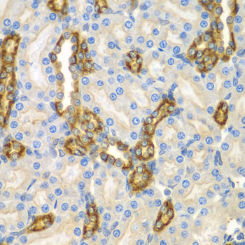
A10971 is listed for IHC and human/mouse reactivity (catalog: A10971 applications/reactivity). Its IHC figure shows paraffin-embedded mouse kidney (A10971 image caption).

Real IHC data Immunohistochemistry of paraffin-embedded mouse kidney using CNPY3 antibody at dilution of 1:100 .Perform microwave antigen retrieval with 10 mM PBS buffer pH 7.2 before commencing with IHC staining protocol.
Anti-Protein canopy homolog 3 CNPY3 Antibody
Cat # A10971

A10971 is the sole rendered SKU, listed for IHC and human/mouse reactivity (catalog: A10971 applications/reactivity). Its own IHC image shows paraffin-embedded mouse kidney stained at 1:100 after microwave retrieval in 10 mM PBS at pH 7.2 (A10971 image caption).

Which to pick: For paraffin-section tissue IHC, choose A10971: this rabbit antibody is listed for IHC at 1:50–1:200, and its own image documents paraffin-embedded mouse kidney (catalog: A10971 host/applications/IHC dilution; A10971 image caption). No SKU here has an IF/ICC application listing, dilution, or image (catalog: A10971 applications/IF fields). For human/mouse work, A10971 lists both species, but its IHC image documents mouse kidney only; the fixative is unreported (catalog: A10971 reactivity; A10971 image caption).

Each figure is that product's own IHC / IF validation image from its datasheet.

References

  1. UniProt Consortium. UniProt entry Q9BT09 (CNPY3_HUMAN, Protein canopy homolog 3).
  2. Human Protein Atlas. CNPY3 tissue IHC expression (reliability: Uncertain).
  3. Human Protein Atlas. CNPY3 subcellular location (ICC-IF): Mainly localized to the plasma membrane and endoplasmic reticulum..
  4. Human Protein Atlas. CNPY3 antibody validation summary (1 antibodies).
  5. CNPY3's regulation of tumor microenvironment and its impact on colon cancer aggressiveness. Molecular medicine (Cambridge, Mass.) 2025 — PMC11887163.
  6. Canopy FGF signaling regulator 3 affects prognosis, immune infiltration, and PI3K/AKT pathway in colon adenocarcinoma. World journal of gastrointestinal oncology 2024 — PMC11271795.
  7. CNPY3 Promotes Human Breast Cancer Progression and Metastasis via Modulation of the Tumor Microenvironment. Current issues in molecular biology 2025 — PMC12651230.
  8. RNA-Binding Protein HuR Regulates Paneth Cell Function by Altering Membrane Localization of TLR2 via Post-transcriptional Control of CNPY3. Gastroenterology 2019 — PMC6707881.
  9. PubMed PMID:12975309 — UniProt-cited evidence.
  10. PubMed PMID:14574404 — UniProt-cited evidence.
  11. PubMed PMID:15489334 — UniProt-cited evidence.