COL17A1 / Collagen alpha-1(XVII) chain · IHC design guide

Design Immunohistochemistry for COL17A1

Plan COL17A1 chromogenic IHC in paraffin sections around cytoplasmic and membranous staining in basal squamous epithelial cells (HPA tissue IHC). Use this guide to choose controls and interpret staining in light of COL17A1 topology and processing (UniProt).

Evidence assembled Oct 2026 · For research use; verify linked source records and product datasheet before use
Immunohistochemistry protocol sheet for COL17A1 (IHC for COL17A1): expected localisation Basal squamous cells: cytoplasmic and membranous (HPA tissue IHC), antibody M03031, validated IHC image, and IHC protocol steps
Printable COL17A1 IHC protocol sheet — expected localisation Basal squamous cells: cytoplasmic and membranous (HPA tissue IHC), antibody M03031, controls and protocol steps. Open the full COL17A1 IHC guide →

COL17A1 Immunohistochemistry Experimental Design Guide

Expected localisation, validated protocols, controls and antibodies — the at-a-glance facts below, then the full design guide.

Must know before staining
Expected localisation Basal squamous cells: cytoplasmic and membranous (HPA tissue IHC)
Staining pattern Basal squamous epithelial cells show cytoplasmic and membrane staining (HPA tissue IHC)
Antigen retrieval Citrate pH 6.0 HIER, 95–98 °C, 20 min (rule: cytoplasmic / membrane antigen)
Positive control ⓘ Testis+1 more · see all
Negative control ⓘ Adipose tissue+4 more · see all
Important caveats
Reasons your staining may differ from the expected pattern.
Fixation Keep fixation consistent across paraffin sections. (standard IHC practice; not target-specific)
Caveat Processed extracellular fragments may alter the pattern (UniProt)
Regulation No expression regulator specified (UniProt)
Isoform / epitope 2 isoforms; epitope side matters after processing (UniProt)
Section 1

Recommended COL17A1 IHC & IF Protocols

The catalog antibody’s IHC-P protocol is accompanied by three published COL17A1 paraffin-section protocols (PMC7758145; PMC13511157; PMC12367868).

Recommended immunohistochemistry (IHC-P) protocol parameters
SampleParaffin-embedded mouse skin tissue; fixative not specified (datasheet M03031)
FixationImage fixative and duration unreported (datasheet M03031); verify before use.
Sectioning4–5 µm sections on charged slides (standard)
DeparaffinisationXylene, graded ethanol series to water (standard)
Antigen retrievalHeat-induced epitope retrieval in citrate buffer, pH 6.0, 20 min at 95–98 °C (standard rule: cytoplasmic / membrane antigen)
Peroxidase block3% H2O2, 10 min, room temperature (standard)
Blocking10% normal serum of the secondary host, 30 min, room temperature (standard)
Primary antibodyRabbit monoclonal (clone AA-3) anti-COL17A1, 1:50 (datasheet M03031)
Primary incubationOvernight at 4 °C (standard)
DetectionHRP-polymer secondary, DAB chromogen 5–10 min (standard)
CounterstainHematoxylin, blue, dehydrate and mount (standard)
Expected resultCOL17A1-positive staining in elongated or late spermatids of testis (HPA tissue IHC: High). HPA tissue profile: Cytoplasmic and membranous expression in basal layer of squamous epithelia. No signal in the no-primary control.
💡Decision noteStart with citrate HIER at pH 6.0, 95–98 °C for 20 min (page retrieval rule); published protocols also use high-pH retrieval (PMC13511157; PMC12367868).
Section 2

What Is the Expected COL17A1 Staining Pattern?

COL17A1 is a transmembrane hemidesmosomal protein with a cytoplasmic region and a large extracellular region (UniProt Q9UMD9 topology). In paraffin-section IHC, expect basal squamous epithelial cells to stain at membranes, with some cytoplasmic staining (HPA tissue IHC: Enhanced; basal-layer profile). Interpret extracellular staining cautiously because COL17A1 also has a secreted variant (UniProt Q9UMD9 processing; HPA reliability description).

What am I looking at on my slide?
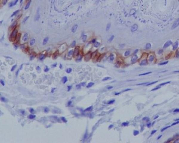
Basal epidermal cells show membranous staining with some cytoplasmic signal; skin basal cells have medium staining (HPA tissue IHC).This matches the reported basal-layer profile (HPA tissue IHC). Signal along the basal cell surface is consistent with hemidesmosomal localization, though chromogenic IHC alone cannot resolve individual hemidesmosomes (UniProt Q9UMD9 subcellular location; standard IHC practice).
Predominantly nuclear staining, or equally strong staining throughout all epidermal layers.Neither is the reported basal-cell membranous and cytoplasmic profile (HPA tissue IHC). Check the positive control, staining distribution and detection controls before calling it COL17A1; an unexpected compartment can reflect nonspecific staining (standard IHC practice).
Strong staining appears in cells expected to be negative, such as adipocytes or bronchial respiratory epithelial cells (HPA tissue IHC: not detected).Investigate antibody cross-reactivity or endogenous detection activity with appropriate controls (standard IHC practice). Do not treat every non-squamous signal as false: elongated or late spermatids show high staining (HPA tissue IHC).
A broad, diffuse haze covers epithelium, stroma and blank areas of the section.Diffuse background obscures the basal-cell pattern reported for COL17A1 (HPA tissue IHC). Review antibody concentration, blocking, washes and chromogen development with a negative control; these are general IHC checks, not reported COL17A1-specific effects (standard IHC practice).
No basal-cell staining is visible in an otherwise intact skin positive control.Skin basal cells normally show medium staining (HPA tissue IHC). Check that the section contains basal epithelium and that retrieval, primary-antibody incubation and detection worked; the supplied sources give no COL17A1-specific retrieval or fixation sensitivity (standard IHC practice).
💡Expected COL17A1 appearanceCall positive a focused basal squamous-cell pattern with membrane emphasis and some cytoplasmic staining, approximately medium in skin (HPA tissue IHC); diffuse field-wide or predominantly nuclear color is a suspect result requiring controls (standard IHC practice).
How each factor affects the staining
Tissue and cell selectionSkin basal cells are a documented medium IHC positive (HPA tissue IHC). Esophageal, vaginal and cervical squamous epithelial cells are reported at low levels, so a faint result there has a different expectation (HPA tissue IHC).
Antibody validationHPA lists Enhanced IHC validation for HPA043673 and HPA052963, and reports tissue-level consistency with RNA expression (HPA antibodies; HPA tissue IHC). HPA also notes presumed off-target binding was disregarded; assess the actual slide pattern with controls.
Membrane topology and unknown epitopeResidues 1–467 are cytoplasmic, 468–488 span the membrane, and 489–1497 are extracellular (UniProt Q9UMD9 topology). The supplied record does not locate the IHC antibody epitope, so do not infer which domain its stain detects.
Processing and extracellular materialUniProt lists the full chain and extracellular processed chains beginning at residues 524 and 531 (UniProt Q9UMD9 processing). Extracellular signal can therefore merit review, but this record cannot assign a particular IHC deposit to a processed form.
Isoforms and localizationUniProt lists 2 isoforms and localization at hemidesmosomes and basement membrane (UniProt Q9UMD9 isoforms; subcellular location). Neither fact establishes an isoform-specific chromogenic pattern without epitope and antibody data.
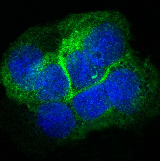
IF/ICC Q: What localization is reported?A: Mainly plasma membrane, with additional Golgi localization (HPA subcellular ICC-IF). This is an ICC-IF observation, so use the tissue IHC basal-layer profile to judge paraffin-section chromogenic staining (HPA tissue IHC).
Why is my staining missing, weak or wrong?
SituationLikely causeNext action
Skin positive control lacks basal staining.The expected medium basal-cell signal is absent (HPA tissue IHC); the failed step is unknown.Confirm basal epithelium is present, then review retrieval, primary incubation and detection against run controls (standard IHC practice). No COL17A1-specific retrieval sensitivity is supplied.
All tissue compartments are brown.The broad pattern differs from basal epithelial enrichment (HPA tissue IHC); excess background or endogenous detection activity is possible (standard IHC practice).Compare a no-primary control, check endogenous-activity blocking where relevant, and review washes and chromogen development (standard IHC practice).
Signal is mostly nuclear.Nuclear localization is outside the reported hemidesmosomal and membrane profile (UniProt Q9UMD9 subcellular location; HPA tissue IHC).Check nuclear counterstain versus chromogen, examine negative controls, and repeat with an IHC-validated antibody if the unexpected signal persists (standard IHC practice; HPA antibodies: IHC Enhanced).
A low-level squamous tissue looks weak beside skin.Esophageal, vaginal and cervical squamous epithelial staining is reported as low, while skin basal-cell staining is medium (HPA tissue IHC).Judge each tissue against its reported level and the same-run skin control; avoid increasing development solely to make a low-level tissue resemble skin (HPA tissue IHC; standard IHC practice).
Extracellular staining accompanies a plausible basal-cell pattern.Basement-membrane localization and extracellular processed forms are documented (UniProt Q9UMD9 subcellular location; processing), but their contribution to this slide is unproven.Record extracellular and cellular staining separately; compare distribution with controls before assigning the signal to COL17A1 or a specific processed form (standard IHC practice).
A presumed negative tissue shows distinct cells staining.Some sampled cell types are reported as not detected, but elongated or late spermatids are high (HPA tissue IHC); tissue-wide labels can mislead.Identify the stained cell type first, compare it with HPA's cell-specific profile, then assess cross-reactivity and detection controls if the pattern remains unexpected (HPA tissue IHC; standard IHC practice).

Sample controls for COL17A1 IHC & IF

🧪Run skin first: basal-layer cells should show COL17A1 staining at the epidermal basement membrane (HPA: Medium in skin basal-layer cells; UniProt Q9UMD9: hemidesmosome localization). Use adrenal gland as the negative tissue, assessing glandular cells (HPA: Not detected in adrenal gland glandular cells); on the skin slide, compare non-basal epidermal cells for absence of the basal-junctional staining pattern (HPA: staining reported in basal-layer cells).
Positive control tissue: Testis (Elongated or late spermatids, HPA High)
Negative control tissue: Adipose tissue (HPA Not detected)
ICC-IF cell lines (HPA subcellular resource): HPA ICC-IF images show COL17A1 in A-431, U2OS, hTCEpi, with annotated localisation: Plasma membrane (supported) (HPA subcellular).
Technical controls: Include a no-primary, secondary-only control; an isotype control matched to the primary antibody’s host species, immunoglobulin class, and concentration; and COL17A1 knockout tissue as a biological negative if available (standard IHC practice). For chromogenic skin IHC, quench endogenous peroxidase and inspect pigment or other background on the control slide; for skin IF, check tissue autofluorescence with an unstained control (standard IHC/IF practice).
⚠️Feasibility: A target-specific fixation window and antigen-retrieval dependency are unreported in the supplied evidence; optimize retrieval on matched paraffin sections (standard IHC practice). The selected M03031 caption shows paraffin-embedded mouse skin but does not report a fixative, so fixation cannot be assumed (selected M03031 tissue-IHC caption). The evidence does not establish whether frozen sections or IF are easier; skin IF requires attention to autofluorescence, while the expected basal hemidesmosomal pattern provides a localization check (standard IF practice; UniProt Q9UMD9: hemidesmosome localization). The selected M03031 tissue-IHC caption documents paraffin sections, but does not specify the fixative (selected-SKU IHC image M03031).

HPA tissue IHC evidence for COL17A1

Comprehensive Human Protein Atlas IHC scoring per tissue (reliability: Enhanced — High consistency between antibody staining and RNA expression data. At least one protein variant secreted, tissue location of RNA and protein might differ and correlation is complex. Presumed off target binding observed and disregarded.). Rows are taken directly from the HPA tissue chart — click any row's HPA link to view the source.

Positive expression · recommended positive controls

TissueCell typeLevelEvidenceSource
Testis Elongated or late spermatids High Protein (IHC) HPA →
Skin Cells in basal layer Medium Protein (IHC) HPA →

Undetected expression · recommended negative controls

TissueCell typeLevelEvidenceSource
Adipose tissue Adipocytes Not detected Protein (IHC) HPA →
Adrenal gland Glandular cells Not detected Protein (IHC) HPA →
Appendix Glandular cells Not detected Protein (IHC) HPA →
Bone marrow Hematopoietic cells Not detected Protein (IHC) HPA →
Breast Adipocytes Not detected Protein (IHC) HPA →
Section 3

Advanced COL17A1 IHC Tips

Troubleshoot COL17A1 chromogenic IHC by checking retrieval, basal epithelial localisation, antibody epitope, and section-level controls.

How should I troubleshoot weak COL17A1 staining after antigen retrieval?
Begin with citrate pH 6.0 heat-induced retrieval at 95–98 °C for 20 min (page retrieval setting). The catalog antibody has a paraffin-embedded mouse skin image, but its caption gives neither a fixative nor retrieval conditions (catalog antibody M03031 caption). If basal staining is weak, check deparaffinisation, temperature at the slide, and controlled cooling before changing retrieval chemistry (standard IHC practice). Compare adjacent sections while holding primary antibody concentration and chromogen development constant (standard IHC practice). Assess improvement at the basal epithelial interface, where COL17A1 is associated with hemidesmosomes (UniProt Q9UMD9 localisation).
Could fixation explain variable COL17A1 staining between paraffin sections?
Target-specific fixation sensitivity is unknown from the supplied evidence; the selected mouse skin caption identifies paraffin embedding but does not state a fixative (catalog antibody M03031 caption). Record each specimen’s fixative and fixation duration, then compare sections processed together under the same retrieval and staining conditions (standard IHC practice). If staining differs, examine section integrity and preservation of the epithelial basement membrane before attributing the difference to antigen loss (standard IHC practice). A matched positive-control section helps distinguish a run-wide failure from specimen variation (standard IHC practice). Do not infer fixation tolerance from the reported tissue staining pattern (HPA: basal squamous epithelial staining).
Where should convincing COL17A1 chromogenic staining appear in skin?
Look for staining associated with basal epithelial cells and their interface with the underlying basement membrane, consistent with hemidesmosomal localisation (UniProt Q9UMD9 localisation). The tissue survey describes cytoplasmic and membranous staining in the basal layer of squamous epithelia and medium staining in skin basal cells (HPA: tissue IHC profile). Evaluate the pattern across intact epithelium rather than relying on one strongly stained field (standard IHC practice). Some intracellular signal can be considered alongside the basal pattern, since an additional Golgi location is reported in cells (HPA: subcellular localisation). Predominantly diffuse staining in unrelated compartments warrants control review before scoring (standard IHC practice).
How does an unknown antibody epitope affect interpretation of COL17A1 staining?
First obtain the catalog antibody’s epitope information, because the supplied caption does not identify the region recognised (catalog antibody M03031 caption). COL17A1 has a cytoplasmic segment at residues 1–467, a transmembrane segment at 468–488, and an extracellular segment at 489–1497 (UniProt Q9UMD9 topology). An extracellular epitope could recognise the annotated 524–1497 processed chain, whereas a cytoplasmic epitope would not be present in that chain (UniProt Q9UMD9 processing and topology). Two isoforms are recorded, but their epitope coverage cannot be assigned from this payload (UniProt Q9UMD9 isoforms). Compare staining patterns cautiously until epitope coverage is established (standard IHC practice).
How should I adapt COL17A1 localisation checks for multiplex immunofluorescence?
For IF, pair COL17A1 with a marker of basal epithelial cells and inspect their spatial relationship at the epithelial basement membrane (UniProt Q9UMD9 localisation; standard IF practice). Choose spectrally separated fluorophores after checking tissue autofluorescence, and include single-stain controls for bleed-through (standard IF practice). If the antibody recognises the cytoplasmic 1–467 region, test permeabilisation; an extracellular epitope may be accessible without it, depending on preparation (UniProt Q9UMD9 topology; standard IF practice). Because the catalog epitope is unspecified, compare these conditions on matched samples and keep acquisition settings fixed (catalog antibody M03031 caption; standard IF practice). Interpret puncta alongside the expected basal membrane pattern (UniProt Q9UMD9 localisation).
What should I check when COL17A1 DAB staining looks widespread?
Start with a section lacking primary antibody to assess signal from detection reagents, endogenous enzyme activity, and chromogen deposition (standard IHC practice). Include a peroxidase block before DAB development and check whether excessive development or concentrated primary antibody increases diffuse colour (standard IHC practice). Examine tissue folds, cut edges, and damaged regions separately from intact basal epithelium, where the expected COL17A1 pattern is concentrated (HPA: tissue IHC profile; standard IHC practice). Use the same counterstain and development settings across comparisons so changes in contrast do not masquerade as improved specificity (standard IHC practice). Review background before adjusting retrieval (standard IHC practice).
How can I quantify COL17A1 IHC without overstating weak staining? ⚠ ANSWER MARKED FOR VERIFICATION
Define an intact basal epithelial region before scoring and exclude folds, necrosis, and section edges using consistent criteria (standard IHC practice). Report the percentage of positive basal cells and an H-score from 0–300 using intensity categories 0–3, or measure positive interface length per millimetre of evaluable basement membrane (standard IHC practice). Normalise cell counts to the number of evaluable basal cells, or interface measurements to intact basement membrane length (standard IHC practice). Keep threshold, counterstain, illumination, and DAB development consistent across sections (standard IHC practice). Document whether membranous and cytoplasmic signals were scored separately (HPA: tissue IHC profile; standard IHC practice).
How do I distinguish true COL17A1 positivity from staining artefact?
A plausible skin result follows basal epithelial cells and the epithelial basement membrane, matching the reported tissue pattern and hemidesmosomal location (HPA: tissue IHC profile; UniProt Q9UMD9 localisation). Verify that the pattern persists through intact areas rather than appearing only at cut edges, folds, or necrotic regions (standard IHC practice). Diffuse colour in unrelated cells, or signal in a no-primary control, calls for review of antibody background and endogenous peroxidase activity (standard IHC practice). Compare a concurrently stained positive-control section before interpreting an apparently negative specimen (standard IHC practice). Record localisation as well as intensity when reporting the result (standard IHC practice).
Boster reagents

Best COL17A1 / Collagen alpha-1(XVII) chain IHC Antibodies

M03031 has IHC data from paraffin-embedded mouse skin and IF data from A431 cells (catalog image captions), with listed Human, Mouse and Rat reactivity (catalog reactivity).

Real IHC data Immunohistochemical analysis of paraffin-embedded mouse skin, using Collagen XVII Antibody.
Anti-Collagen XVII Rabbit Monoclonal Antibody
Cat # M03031

M03031 shows IHC staining of paraffin-embedded mouse skin (M03031 IHC image caption). It also shows IF staining of A431 cells and lists Human, Mouse and Rat reactivity (M03031 IF image caption; catalog reactivity).

Which to pick: For tissue IHC, choose M03031 based on its paraffin-embedded mouse skin image; the fixative is unreported (M03031 IHC image caption). For IF/ICC, M03031 lists both applications and shows IF staining of A431 cells (M03031 applications; IF image caption). For cross-species work, M03031 is a rabbit monoclonal, clone AA-3, with listed Human, Mouse and Rat reactivity (M03031 host, clone and reactivity).

Each figure is that product's own IHC / IF validation image from its datasheet.

References

  1. UniProt Consortium. UniProt entry Q9UMD9 (COHA1_HUMAN, Collagen alpha-1(XVII) chain).
  2. Human Protein Atlas. COL17A1 tissue IHC expression (reliability: Enhanced).
  3. Human Protein Atlas. COL17A1 subcellular location (ICC-IF): Mainly localized to the plasma membrane. In addition localized to the Golgi apparatus..
  4. Human Protein Atlas. COL17A1 antibody validation summary (2 antibodies).
  5. High Expression of COL17A1 Predicts Poor Prognosis and Promotes the Tumor Progression via NF-κB Pathway in Pancreatic Adenocarcinoma. Journal of oncology 2020 — PMC7758145.
  6. Transcriptomic Profiling Identifies a Subset of Renal Tumors with Overlapping Features of Clear Cell Papillary Renal Cell Tumor and Renal Cell Carcinoma with Fibromyomatous Stroma. Cancers 2026 — PMC13511157.
  7. Whole exome sequencing study of adamantinomatous craniopharyngioma reveals the mutational characteristics of recurrent cases. Journal of neuro-oncology 2025 — PMC12367868.
  8. Extracellular vesicles from human urine-derived stem cells delay aging through the transfer of PLAU and TIMP1. Acta pharmaceutica Sinica. B 2024 — PMC10935484.
  9. PubMed PMID:1324962 — UniProt-cited evidence.
  10. PubMed PMID:9012408 — UniProt-cited evidence.
  11. PubMed PMID:15164054 — UniProt-cited evidence.