This website uses cookies to ensure you get the best experience on our website.


- Table of Contents
Plan paraffin section IHC for COL4A3 using its extracellular basement membrane location (UniProt) and observed cytoplasmic and extracellular tissue staining (HPA tissue IHC). Compare tissue controls and interpret staining with the protein’s secreted location in mind (UniProt).

Expected localisation, validated protocols, controls and antibodies — the at-a-glance facts below, then the full design guide.
| Expected localisation | Extracellular basement membrane (UniProt); cytoplasmic tissue staining (HPA tissue IHC) | |
| Staining pattern | Cytoplasmic and extracellular positivity in several tissues (HPA tissue IHC) | |
| Antigen retrieval | EDTA pH 8.0 HIER, heat-mediated (datasheet A01755-3) | |
| Positive control | Appendix+4 more · see all | |
| Negative control | Adipose tissue+4 more · see all |
| Fixation | Keep fixation consistent across sections (standard IHC practice; not target-specific). Selected-image fixative and duration unreported (datasheet A01755-3); verify before use. | |
| Caveat | Secreted COL4A3 may stain beyond its producing cells (UniProt) | |
| Regulation | Staining intensity varies by tissue (HPA tissue IHC) | |
| Isoform / epitope | 5 isoforms; no cytoplasmic segment; check chain/tumstatin epitope (UniProt) |
The catalog antibody has a datasheet IHC-P protocol (datasheet: A01755-3). The published options below cover human lung multiplex IHC and mouse inner ear chromogenic IHC (PMC13452252; PMC7619435).
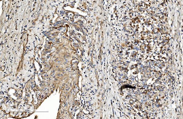
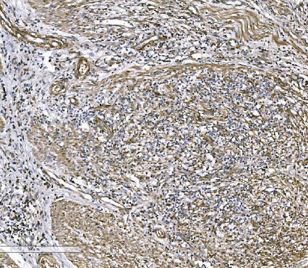
| Sample | Paraffin-embedded human gastric cancer tissue; fixative not specified (datasheet A01755-3) |
| Fixation | Image fixative and duration unreported (datasheet A01755-3); verify before use. |
| Sectioning | 4–5 µm sections on charged slides (standard) |
| Deparaffinisation | Xylene, graded ethanol series to water (standard) |
| Antigen retrieval | Heat retrieval: EDTA pH 8.0 (datasheet A01755-3); 20 min, 95–100 °C (standard) |
| Peroxidase block | 3% H2O2, 10 min, room temperature (standard) |
| Blocking | 10% goat serum (datasheet A01755-3) |
| Primary antibody | Rabbit anti-COL4A3, 2-5 μg/ml (datasheet A01755-3) |
| Primary incubation | Overnight at 4 °C (datasheet A01755-3) |
| Detection | Streptavidin-biotin complex (SABC), DAB chromogen (datasheet A01755-3) |
| Counterstain | Hematoxylin, blue, dehydrate and mount (standard) |
| Expected result | COL4A3-positive staining in glandular cells of appendix (HPA tissue IHC: Medium). HPA tissue profile: Cytoplasmic expression and extracellular positivity in several tissues. No signal in the no-primary control. |
COL4A3 is a secreted protein found in extracellular matrix and basement membranes, with no transmembrane segment (UniProt Q01955 localization/topology). On paraffin sections, expect staining associated with extracellular structures as well as cytoplasmic staining in some cells: HPA describes both patterns and rates its tissue IHC reliability Approved (HPA tissue IHC). Appendix glandular cells are one documented medium-positive cell population (HPA tissue IHC).
| Extracellular staining follows a basement-membrane outline, with staining in the relevant cells. | This fits COL4A3 matrix localization (UniProt Q01955 localization). HPA also reports cytoplasmic and extracellular positivity across several tissues (HPA tissue IHC). Judge the pattern against the particular tissue and cell population; extracellular staining need not appear equally strong beside every cell. |
| Nuclei are the dominant stained compartment, without a convincing extracellular pattern. | A predominantly nuclear result conflicts with the reported extracellular and cytoplasmic locations (UniProt Q01955 localization; HPA tissue IHC). Treat it as a possible staining artifact and review control sections before assigning it to COL4A3. |
| Strong staining appears in a cell population documented as undetected. | For example, HPA reports no staining in bone-marrow hematopoietic cells (HPA tissue IHC). Staining there warrants checks for cross-reactivity or endogenous chromogen-generating activity; a negative cell-level report does not establish that every extracellular structure in that tissue must be negative. |
| Brown signal spreads across tissue and background with little anatomical definition. | The pattern cannot be confidently assigned to the reported cytoplasmic or extracellular distribution (HPA tissue IHC). Compare a section processed without primary antibody and review blocking and washing as general chromogenic IHC practice. |
| A documented medium-positive cell population has no visible signal. | Absent appendix glandular-cell staining, for example, disagrees with HPA's medium IHC observation (HPA tissue IHC). First verify the positive control and tissue preservation; one failed section does not establish that COL4A3 is absent from the specimen. |
| Extracellular deposition and cellular signal | COL4A3 is secreted and belongs to basement-membrane matrix (UniProt Q01955 localization). HPA nevertheless describes cytoplasmic expression alongside extracellular positivity (HPA tissue IHC); cytoplasmic staining alone should be interpreted in its tissue context, not automatically rejected. |
| Anatomical context | UniProt places COL4A3 with COL4A4 and COL4A5 in glomerular and tubular basement membranes (UniProt Q01955 localization). HPA's supplied IHC summary does not score kidney, so that anatomical expectation is not an HPA-confirmed positive-control result. |
| Tissue-source disagreement | HPA reports medium staining in placenta trophoblastic cells and skin keratinocytes (HPA tissue IHC), while UniProt cites reports of absent expression in placenta and skin (UniProt Q01955 tissue specificity). Record the sampled cells and pattern instead of treating either source as a universal tissue-wide rule. |
| Antibody validation | HPA042064 is rated Approved for IHC and Supported for ICC (HPA antibodies). Approved is the supplied IHC status; the record does not provide an Enhanced IHC rating or establish that every staining feature was reproduced by independent antibodies. |
| Processing and isoforms | UniProt lists 5 isoforms, a signal peptide at residues 1–28, and a tumstatin region at 1426–1670 (UniProt Q01955 processing/isoforms). The supplied record gives no epitope for the IHC-validated antibody, so it cannot establish which forms or processed regions this stain detects. |
| ICC/IF Q&A: should intracellular fluorescence match tissue IHC? | HPA reports mainly vesicular ICC/IF localization, with additional endoplasmic-reticulum signal, and lists HeLa and U2OS images (HPA subcellular). That cell-based observation provides context for intracellular signal; tissue IHC also has an extracellular pattern (HPA tissue IHC). |
| Situation | Likely cause | Next action |
|---|---|---|
| No stain in appendix glandular cells | This conflicts with their documented medium IHC level (HPA tissue IHC); the section or detection run may have failed. | Check a documented positive-control section, tissue integrity, primary-antibody step, and detection reagents (general IHC-P practice). |
| No glomerular basement-membrane outline | UniProt places COL4A3 in the GBM (UniProt Q01955 localization), but the supplied HPA IHC summary gives no kidney score (HPA tissue IHC). | Check whether basement membranes are intact and compare the run with a documented HPA-positive tissue; avoid calling the kidney result definitive from this summary alone. |
| Predominantly nuclear staining | The compartment conflicts with UniProt extracellular localization and HPA's cytoplasmic/extracellular profile (UniProt Q01955 localization; HPA tissue IHC). | Compare a section processed without primary antibody, then review blocking, detection, and antibody concentration (general IHC-P practice). |
| Strong staining in lung macrophages | HPA lists lung macrophages as Low, so strong signal needs confirmation; chromogenic background or cross-reactivity is possible (HPA tissue IHC). | Compare the no-primary control and review endogenous enzyme blocking if enzyme-based detection is used (general chromogenic IHC practice). |
| Diffuse brown background obscures structure | The result cannot be matched confidently to HPA's cytoplasmic and extracellular profile (HPA tissue IHC). | Inspect control sections, washing, blocking, and detection exposure before scoring the specimen (general IHC-P practice). |
| Placenta or skin staining seems to contradict a tissue-specificity note | HPA reports medium staining in selected cells there (HPA tissue IHC); UniProt also records reports of absent expression in those tissues (UniProt Q01955 tissue specificity). | Document the stained cell type and compartment, compare controls, and report the source disagreement rather than reclassifying the result solely by tissue name. |
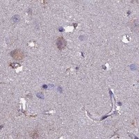


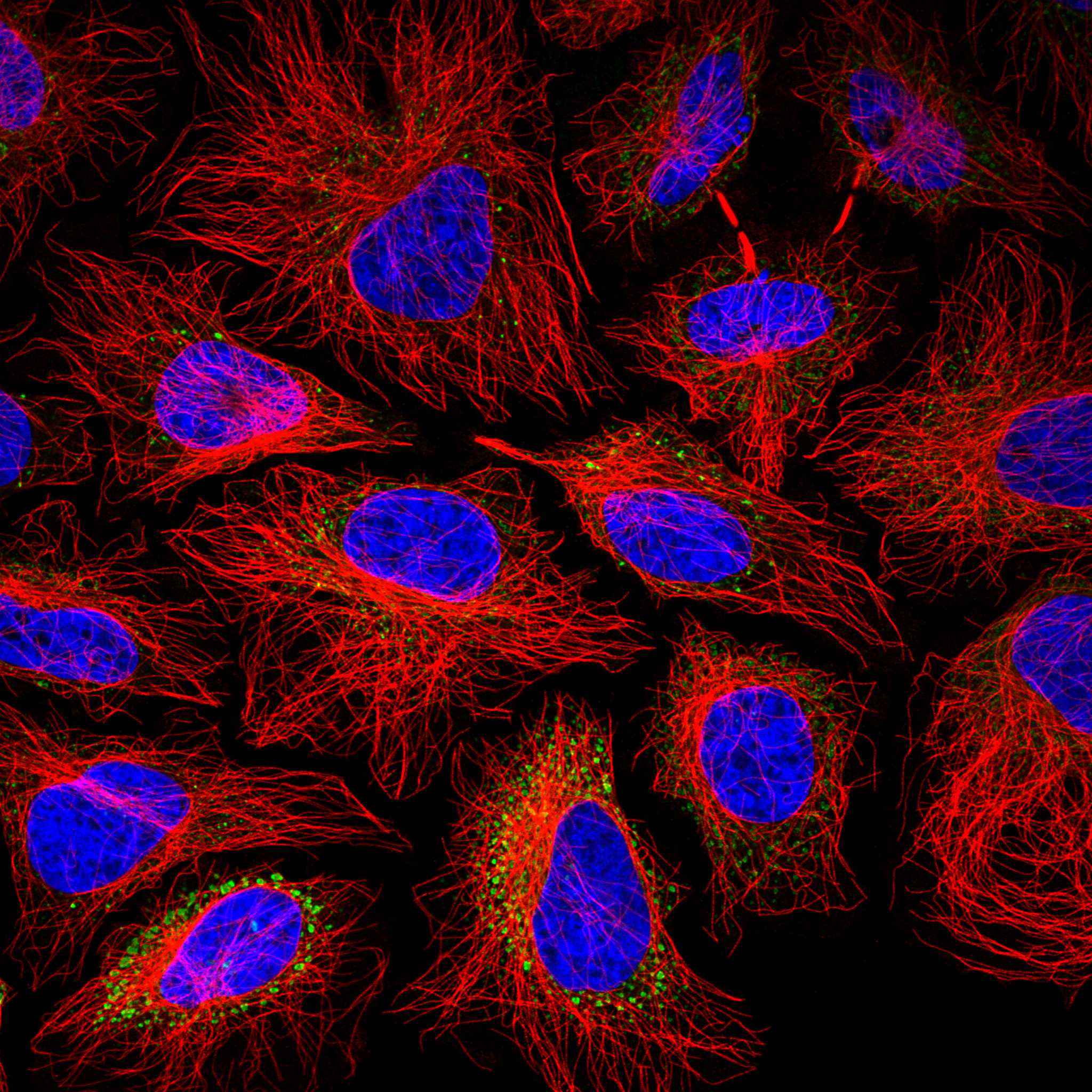
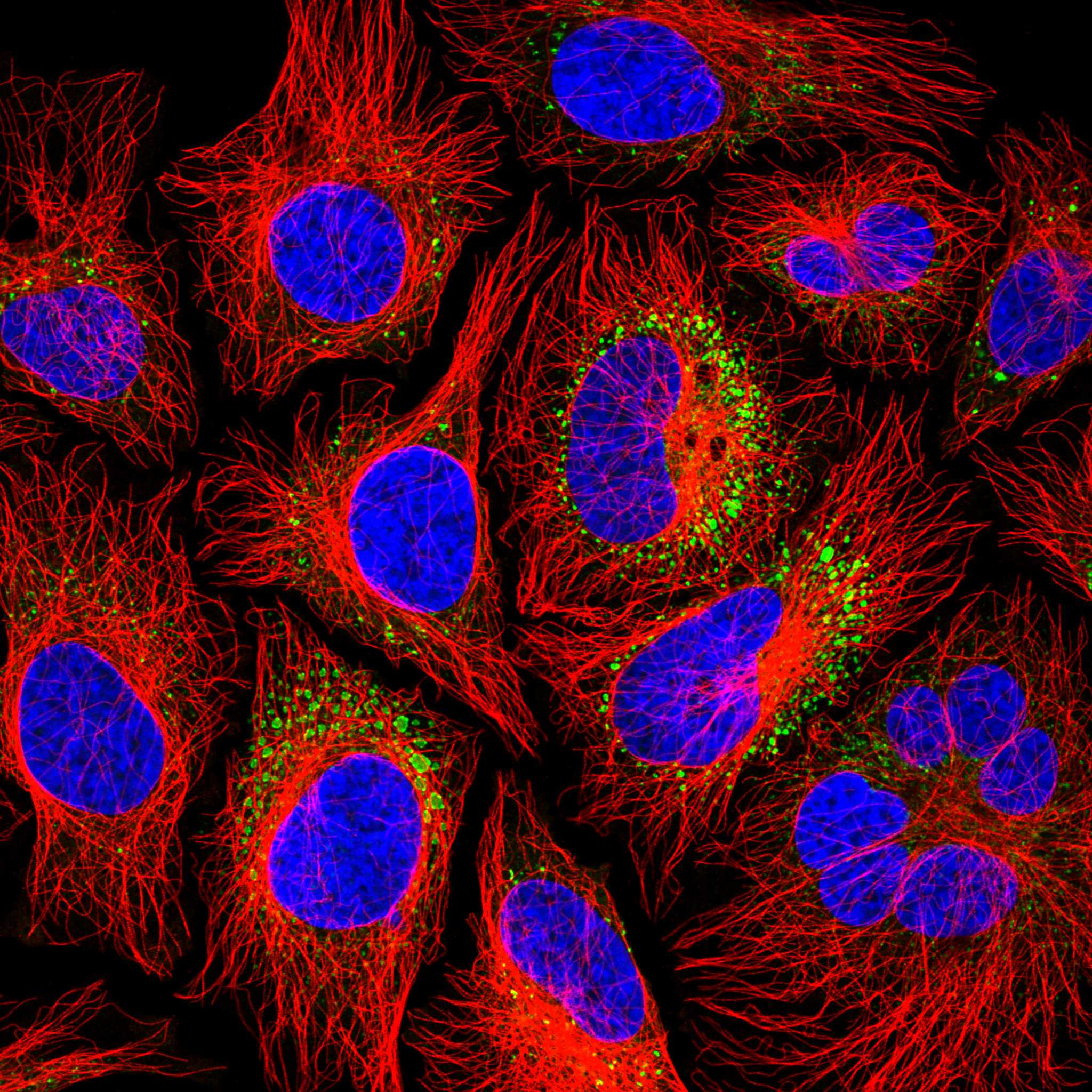
Comprehensive Human Protein Atlas IHC scoring per tissue (reliability: Approved — At least one protein variant secreted, tissue location of RNA and protein might differ and correlation is complex.). Rows are taken directly from the HPA tissue chart — click any row's HPA link to view the source.
| Tissue | Cell type | Level | Evidence | Source |
|---|---|---|---|---|
| Appendix | Glandular cells | Medium | Protein (IHC) | HPA → |
| Cervix | Glandular cells | Medium | Protein (IHC) | HPA → |
| Gallbladder | Glandular cells | Medium | Protein (IHC) | HPA → |
| Placenta | Trophoblastic cells | Medium | Protein (IHC) | HPA → |
| Skin | Keratinocytes | Medium | Protein (IHC) | HPA → |
| Tissue | Cell type | Level | Evidence | Source |
|---|---|---|---|---|
| Adipose tissue | Adipocytes | Not detected | Protein (IHC) | HPA → |
| Adrenal gland | Glandular cells | Not detected | Protein (IHC) | HPA → |
| Bone marrow | Hematopoietic cells | Not detected | Protein (IHC) | HPA → |
| Breast | Adipocytes | Not detected | Protein (IHC) | HPA → |
| Caudate | Glial cells | Not detected | Protein (IHC) | HPA → |
Troubleshoot COL4A3 chromogenic IHC by assessing basement-membrane staining, secretory compartments and the processing details documented for the catalog antibody.
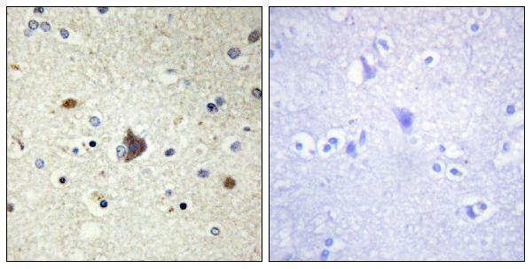
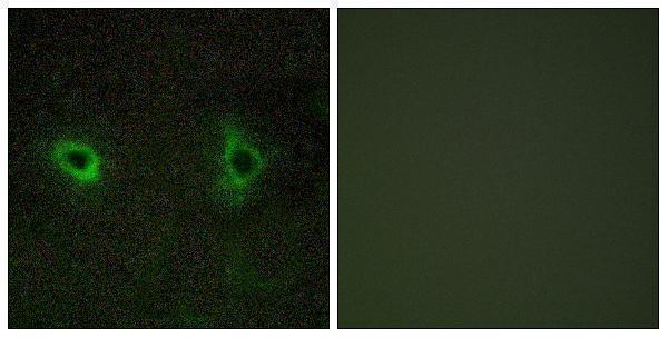
Two anti-COL4A3 antibodies have paraffin-section IHC images from human tissue (catalog image captions); one also has an IF image from COS7 cells (A01755-1 image caption).
A01755-3 will render with IHC data from paraffin-embedded human gastric cancer tissue (A01755-3 image caption). A01755-1 will render with IHC data from paraffin-embedded human brain tissue; its separate IF image shows COS7 cells (A01755-1 image captions).
Which to pick: For human tissue IHC, choose A01755-3 when its documented paraffin-section procedure is useful: 2 μg/ml primary antibody after EDTA retrieval at pH 8.0 (A01755-3 image caption). For IF or mouse samples, choose A01755-1 because IF and IHC are listed applications and Human and Mouse are listed reactivities; its IHC image shows paraffin-embedded human brain tissue (A01755-1 catalog and image caption). The fixative is unreported in both IHC captions (A01755-1 and A01755-3 image captions).