COL7A1 / Collagen alpha-1(VII) chain · IHC design guide

Design Immunohistochemistry for COL7A1

This paraffin IHC guide focuses on COL7A1 staining at squamous epithelial basal membranes (HPA tissue IHC). Start with the catalog antibody at 2–5 μg/mL (datasheet A01170-1) and assess DAB staining against a no-primary control (standard IHC practice).

Evidence assembled Oct 2026 · For research use; verify linked source records and product datasheet before use
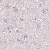
Immunohistochemistry protocol sheet for COL7A1 (IHC for COL7A1): expected localisation Basal membranes of squamous epithelia (HPA tissue IHC), antibody A01170-1, validated IHC image, and IHC protocol steps
Printable COL7A1 IHC protocol sheet — expected localisation Basal membranes of squamous epithelia (HPA tissue IHC), antibody A01170-1, controls and protocol steps. Open the full COL7A1 IHC guide →

COL7A1 Immunohistochemistry Experimental Design Guide

Expected localisation, validated protocols, controls and antibodies — the at-a-glance facts below, then the full design guide.

Must know before staining
Expected localisation Basal membranes of squamous epithelia (HPA tissue IHC)
Staining pattern Basal-membrane staining beneath squamous epithelia (HPA tissue IHC)
Antigen retrieval EDTA pH 8.0 HIER, heat-mediated (datasheet A01170-1)
Positive control ⓘ Placenta+1 more · see all
Negative control ⓘ Adipose tissue+4 more · see all
Important caveats
Reasons your staining may differ from the expected pattern.
Fixation Keep fixation consistent across sections (standard IHC practice; not target-specific)
Caveat Secreted protein may stain away from source-cell RNA (HPA tissue IHC)
Regulation No expression regulation annotated (UniProt)
Isoform / epitope 2 isoforms; epitope differences unreported (UniProt)
Section 1

Recommended COL7A1 IHC & IF Protocols

The catalog antibody protocol uses heat-mediated EDTA pH 8.0 retrieval (datasheet A01170-1). Two published COL7A1 IHC protocols provide tissue-specific alternatives (PMC8815231; PMC11415666).

Recommended immunohistochemistry (IHC-P) protocol parameters
SampleParaffin-embedded human melanoma tissue; fixative not specified (datasheet A01170-1)
FixationImage fixative and duration unreported (datasheet A01170-1); verify before use.
Sectioning4–5 µm sections on charged slides (standard)
DeparaffinisationXylene, graded ethanol series to water (standard)
Antigen retrievalHeat retrieval: EDTA pH 8.0 (datasheet A01170-1); 20 min, 95–100 °C (standard)
Peroxidase block3% H2O2, 10 min, room temperature (standard)
Blocking10% goat serum (datasheet A01170-1)
Primary antibodyRabbit anti-COL7A1, 2-5μg/ml (datasheet A01170-1)
Primary incubationOvernight at 4 °C (datasheet A01170-1)
DetectionStreptavidin-biotin complex (SABC), DAB chromogen (datasheet A01170-1)
CounterstainHematoxylin, blue, dehydrate and mount (standard)
Expected resultCOL7A1-positive staining in endothelial cells of placenta (HPA tissue IHC: Medium). HPA tissue profile: Expression in basal membranes of squamous epithelia. No signal in the no-primary control.
💡Decision noteStart with EDTA pH 8.0 retrieval for the catalog antibody (datasheet A01170-1); consider proteinase K for stomach tumor sections (PMC8815231).
Section 2

What Is the Expected COL7A1 Staining Pattern?

COL7A1 is a secreted extracellular matrix protein of stratified squamous epithelial basement membranes, with no transmembrane segment (UniProt Q02388). In paraffin-section IHC, expect staining along the basal membrane beneath squamous epithelial cells, rather than a uniform signal inside those cells (HPA tissue IHC: basal membranes of squamous epithelia). HPA rates its tissue IHC evidence Enhanced, while noting that secretion can complicate comparisons between the cells expressing RNA and the location of protein (HPA tissue IHC).

What am I looking at on my slide?
A distinct line follows the basal membrane beneath squamous epithelium.This fits the expected extracellular basement-membrane pattern (HPA tissue IHC; UniProt Q02388 localization). Judge the stained structure in its tissue context: a secreted protein can appear outside the cells that produced it (HPA tissue IHC reliability note).
Strong staining fills epithelial cytoplasm or nuclei, with little basal-membrane signal.That is the wrong dominant compartment for tissue IHC and should be treated as possible artefact until checked (HPA tissue IHC; UniProt Q02388 localization). Intracellular production does not, by itself, establish that diffuse cellular staining is a valid tissue result.
Adipocytes, respiratory epithelial cells or hematopoietic cells stain prominently.HPA reports these cell populations as Not detected in adipose tissue, bronchus and bone marrow, respectively (HPA tissue IHC). Investigate cross-reactivity or endogenous detection activity before assigning the signal to COL7A1 (general IHC practice).
Color spreads broadly across tissue or appears in the negative detection control.Diffuse color without a basal-membrane pattern is background rather than a convincing COL7A1 result (HPA tissue IHC pattern; general IHC practice). A matching control signal points toward detection-system background; it cannot establish target localization (general IHC practice).
No basal-membrane signal is visible in a squamous epithelial section used as a positive control.The expected structure has failed to stain (HPA tissue IHC). First assess section integrity and whether the detection run worked (general IHC practice). A negative run alone cannot establish biological absence, and HPA supplies no target-specific fixation-sensitivity finding.
💡Expected COL7A1 appearanceCall IHC positive when a clear basal-membrane line follows squamous epithelium (HPA tissue IHC; UniProt Q02388 localization); no numeric intensity cutoff is supplied, and broad nuclear or diffuse cellular color is suspicious (general IHC practice).
How each factor affects the staining
Extracellular location and secretionCOL7A1 forms anchoring fibrils at stratified squamous epithelial basement membranes (UniProt Q02388 function). Interpret the stained membrane as the primary tissue landmark; do not require uniform staining of the overlying cell bodies (HPA tissue IHC).
Antibody evidenceHPA lists IHC as Enhanced for HPA042420 and CAB016357, while HPA018877 has no listed IHC validation (HPA antibodies). The tissue profile is also rated Enhanced (HPA tissue IHC); those ratings support pattern interpretation, not every signal in every tissue.
Additional reported tissue stainingHPA reports Medium staining in placental endothelial cells and testicular Sertoli cells (HPA tissue IHC). These observations can aid comparison, but they do not redefine the reported basal-membrane profile of squamous epithelia (HPA tissue IHC).
Isoforms and epitope limitsUniProt records two isoforms and a secreted processed chain spanning residues 17–2944 (UniProt Q02388). The supplied evidence gives no antibody epitope or isoform-specific IHC result, so do not infer which form a stain detects.
IF/ICC Q: should cultured cells show the same pattern as tissue IHC?A: HPA places the main ICC-IF signal in endoplasmic reticulum, with additional vesicles, in images from HeLa, SiHa and U2OS cells (HPA subcellular). That intracellular observation should be interpreted separately from the extracellular tissue IHC pattern (HPA tissue IHC; UniProt Q02388).
Why is my staining missing, weak or wrong?
SituationLikely causeNext action
The squamous epithelial positive-control section has no basal-membrane signal.The stain may have failed, or the expected structure may be missing from the examined section (HPA tissue IHC pattern; general IHC practice). The supplied sources do not identify a COL7A1-specific fixation effect.Confirm the basal membrane is present on the section, then review antibody, detection and run controls (general IHC practice). Treat the result as unresolved until a positive control stains in the expected location.
Color covers much of the section without following tissue structure.Nonspecific staining or detection background may obscure the reported basal-membrane pattern (HPA tissue IHC; general IHC practice). The appearance alone does not identify which step caused it.Compare a negative detection control; review blocking, washes and chromogen development using standard IHC controls (general IHC practice). Reassess whether a distinct basal-membrane line remains.
Epithelial nuclei or most cell bodies stain more strongly than the basal membrane.The dominant location conflicts with the reported tissue pattern and extracellular localization (HPA tissue IHC; UniProt Q02388). A cellular signal alone is insufficient to assign COL7A1 specificity.Check the tissue landmarks and control staining, then compare the distribution with HPA's basal-membrane profile (HPA tissue IHC; general IHC practice). Report the compartment discrepancy if it persists.
Adipocytes or bronchial respiratory epithelial cells are strongly positive.HPA reports those cell types as Not detected (HPA tissue IHC). Cross-reactivity or endogenous detection activity is possible, but the stain alone cannot distinguish them (general IHC practice).Compare matched negative controls and an expected positive squamous epithelial section; verify that color remains tied to the basal membrane (HPA tissue IHC; general IHC practice). Avoid scoring unrelated cellular color as COL7A1.
Placental endothelial or testicular Sertoli cell staining differs from a squamous epithelial control.HPA records Medium staining in those cell populations, while its overall tissue profile describes basal membranes of squamous epithelia (HPA tissue IHC). Their cellular observations need not have the same visual pattern.Score each tissue against its reported HPA observation and the structure actually present; retain the squamous epithelial basal membrane as the main localization check (HPA tissue IHC). Do not infer an intensity cutoff across tissues.

Sample controls for COL7A1 IHC & IF

🧪Run placenta first and assess its endothelial cells for staining (HPA: placenta endothelial cells, Medium). Use adipose tissue adipocytes as the negative comparator (HPA: adipocytes, Not detected); on the placenta slide, compare non-endothelial cells for background while scoring basement-membrane staining separately (UniProt Q02388 localization).
Positive control tissue: Placenta (Endothelial cells, HPA Medium)
Negative control tissue: Adipose tissue (HPA Not detected)
ICC-IF cell lines (HPA subcellular resource): HPA ICC-IF images show COL7A1 in HeLa, SiHa, U2OS, with annotated localisation: Endoplasmic reticulum (approved) (HPA subcellular).
Technical controls: Include a no-primary, secondary-only control and a concentration-matched rabbit isotype control; match clonality if known (caption: rabbit primary antibody; clonality unreported). A COL7A1 knockout specimen can serve as a biological negative (standard IHC practice). Quench endogenous peroxidase and check endogenous biotin when using the caption’s biotin–SABC/DAB detection on placenta (caption: biotinylated secondary, SABC and DAB; standard IHC practice).
⚠️Feasibility: A target-specific fixation window or fixation effect is unreported, and the selected A01170-1 paraffin-section caption does not state the fixative (caption: fixative not stated). Heat retrieval in EDTA at pH 8.0 was used for that IHC example, but whether retrieval is required is unreported (caption: EDTA retrieval, pH 8.0). The supplied evidence does not establish that frozen sections or IF are easier; for placental IHC, check blood-associated peroxidase background and endogenous biotin with the reported SABC detection (caption: SABC/DAB; standard IHC practice).

HPA tissue IHC evidence for COL7A1

Comprehensive Human Protein Atlas IHC scoring per tissue (reliability: Enhanced — High consistency between antibody staining and RNA expression data. At least one protein variant secreted, tissue location of RNA and protein might differ and correlation is complex. Antibody staining in cells/structures not annotated, view images.). Rows are taken directly from the HPA tissue chart — click any row's HPA link to view the source.

Positive expression · recommended positive controls

TissueCell typeLevelEvidenceSource
Placenta Endothelial cells Medium Protein (IHC) HPA →
Testis Sertoli cells Medium Protein (IHC) HPA →

Undetected expression · recommended negative controls

TissueCell typeLevelEvidenceSource
Adipose tissue Adipocytes Not detected Protein (IHC) HPA →
Adrenal gland Glandular cells Not detected Protein (IHC) HPA →
Appendix Glandular cells Not detected Protein (IHC) HPA →
Bone marrow Hematopoietic cells Not detected Protein (IHC) HPA →
Breast Adipocytes Not detected Protein (IHC) HPA →
Section 3

Advanced COL7A1 IHC Tips

Troubleshoot COL7A1 staining by evaluating basement membrane localisation, section processing, and controls alongside the catalog antibody’s IHC evidence.

Which retrieval conditions should I start with for paraffin-section COL7A1 IHC?
Start with heat-mediated antigen retrieval in EDTA at pH 8.0 (datasheet A01170-1). The catalog antibody’s paraffin-section image used that retrieval, followed by 10% goat-serum blocking and 2 μg/mL primary antibody overnight at 4°C (datasheet A01170-1). If staining is weak, first check that the section stayed attached, the retrieval buffer retained its intended pH, and a known positive section developed as expected (standard IHC practice). Compare any adjusted heating conditions on adjacent sections, because a weak signal alone cannot distinguish inadequate epitope exposure from low COL7A1 abundance (standard IHC practice; HPA: tissue-enhanced RNA expression in skin).
Could fixation explain weak or uneven COL7A1 staining?
The selected paraffin-section caption does not state a fixative, so target-specific fixation sensitivity is unknown (datasheet A01170-1). Record the actual fixative and fixation duration for each specimen, then compare similarly processed sections before attributing differences to COL7A1 expression (standard IHC practice). Include a positive reference section in the same staining run and inspect tissue preservation, section adhesion, and retrieval consistency when signal is patchy (standard IHC practice). The catalog image documents EDTA retrieval at pH 8.0 and overnight primary incubation at 4°C, but those conditions do not establish which fixation duration preserves its epitope (datasheet A01170-1).
Where should convincing COL7A1 chromogenic staining appear?
Look for a defined basement membrane pattern beneath squamous epithelium, consistent with COL7A1’s role in anchoring fibrils (HPA: basal membranes of squamous epithelia; UniProt Q02388: function). COL7A1 is secreted into extracellular matrix and basement membrane and has no transmembrane segment, so a sharp extracellular interface is more persuasive than diffuse staining throughout epithelial cells (UniProt Q02388: subcellular location and topology). Assess the interface in intact tissue away from folds and torn edges, using the same counterstain and microscope settings across comparisons (standard IHC practice). Intracellular staining may reflect protein handling, but it should be reported separately from basement membrane deposition (HPA: endoplasmic reticulum and vesicles; UniProt Q02388: secretion).
How can isoforms and collagen modifications affect epitope interpretation?
COL7A1 has 2 listed isoforms, while the supplied catalog caption does not identify the antibody’s epitope or establish isoform selectivity (UniProt Q02388: isoforms; datasheet A01170-1). Its sequence includes fibronectin type-III and VWFA domains, annotated glycosylation sites, and hydroxyproline residues; these annotations alone do not show which feature this antibody recognises (UniProt Q02388: domains, glycosylation and modified residues). Obtain an epitope map or independent validation before calling a staining difference isoform-specific (standard IHC practice). For tissue comparisons, keep retrieval at EDTA pH 8.0 and primary incubation conditions matched, so processing differences do not masquerade as epitope-dependent biology (datasheet A01170-1; standard IHC practice).
How should I assess COL7A1 in a complementary IF experiment?
Treat IF as a separate validation experiment: the selected antibody evidence documents paraffin-section chromogenic IHC, not an IF protocol (datasheet A01170-1). Multiplex COL7A1 with a marker that identifies the expected epithelial compartment, then judge whether extracellular signal follows its basal boundary (HPA: basal membranes of squamous epithelia; UniProt Q02388: basement membrane location). Choose fluorophores after checking the specimen’s autofluorescence and include single-label controls to assess channel bleed-through (standard IF practice). Because COL7A1 has no transmembrane segment, prioritise accessible extracellular deposits; consider permeabilisation only when testing an intracellular epitope or the reported endoplasmic reticulum and vesicle signal (UniProt Q02388: topology and secretion; HPA: subcellular location; standard IF practice).
What should I check when DAB obscures the basement membrane?
Compare a no-primary control with the stained section to identify detection-system or tissue background before changing the COL7A1 antibody concentration (standard IHC practice). The catalog image used 2 μg/mL primary antibody, a biotinylated goat anti-rabbit secondary, an avidin–biotin detection complex, and DAB; each detection step should be checked against its control (datasheet A01170-1; standard IHC practice). Apply a peroxidase block for chromogenic detection and inspect whether signal persists in the no-primary control (standard IHC practice). If broad brown staining remains, adjust blocking, washing, or detection exposure systematically while preserving an intact basement membrane reference for comparison (standard IHC practice; HPA: basal membrane pattern).
How should I quantify COL7A1 when staining follows a basement membrane? ⚠ ANSWER MARKED FOR VERIFICATION
Measure staining along an annotated epithelial basement membrane, using positive membrane length or DAB-positive area per unit basement membrane length as the primary readout (HPA: basal membranes of squamous epithelia; standard image-analysis practice). Record the fraction of evaluable interface that stains and its intensity using the same threshold and counterstain handling across sections (standard IHC practice). Normalise to intact, assessable basement membrane length, rather than total tissue area or all nucleated cells, because COL7A1 is an extracellular matrix protein (UniProt Q02388: subcellular location; standard image-analysis practice). An H-score or percent-positive-cell score may describe cellular staining separately, but it should not replace the interface measurement (standard IHC practice).
When does COL7A1 staining indicate biology rather than artefact?
Give greatest weight to reproducible staining at intact squamous epithelial basement membranes, the reported tissue pattern for COL7A1 (HPA: basal membranes of squamous epithelia). Evaluate intracellular signal separately: endoplasmic reticulum and vesicle localisation is reported in cell imaging, whereas secreted COL7A1 functions in extracellular anchoring fibrils (HPA: subcellular location; UniProt Q02388: location and function). Treat edge-only deposits, necrotic areas, and brown signal in a no-primary control as reasons to investigate section or detection artefact before assigning biological meaning (standard IHC practice). Compare matched positive and negative tissue regions and review morphology, since chromogenic signal alone cannot establish the producing cell or a specific isoform (standard IHC practice; UniProt Q02388: isoforms and secretion).
Boster reagents

Best COL7A1 / Collagen alpha-1(VII) chain IHC Antibodies

The catalog lists one human-reactive anti-COL7A1 antibody with real IHC data from paraffin sections of human melanoma and tonsil (catalog: A01170-1; IHC image captions).

Real IHC data IHC analysis of Collagen VII/COL7A1 using anti-Collagen VII/COL7A1 antibody (A01170-1). Collagen VII/COL7A1 was detected in paraffin-embedded section of human melanoma tissue. Heat mediated antigen retrieval was performed in EDTA buffer (pH8.0, epitope retrieval solution). The tissue section was blocked with 10% goat serum. The tissue section was then incubated with 2μg/ml rabbit anti-Collagen VII/COL7A1 Antibody (A01170-1) overnight at 4°C. Biotinylated goat anti-rabbit IgG was used as secondary antibody and incubated for 30 minutes at 37°C. The tissue section was developed using Strepavidin-Biotin-Complex (SABC) (Catalog # SA1022) with DAB as the chromogen.
Anti-Collagen VII/COL7A1 Antibody ®
Cat # A01170-1

A01170-1 will render with an IHC figure showing detection in a paraffin section of human melanoma (A01170-1 IHC image caption). Its second IHC image documents detection in a paraffin section of human tonsil (A01170-1 IHC image caption).

Which to pick: Choose A01170-1 for human paraffin-section IHC; it is rabbit-derived and listed for human IHC at 2–5 μg/ml (catalog: A01170-1 applications, host, reactivity and dilution). No listed SKU supports an IF/ICC or cross-species recommendation (catalog: A01170-1 applications and reactivity). The IHC captions report paraffin sections and EDTA retrieval at pH 8.0, but do not report the fixative (A01170-1 IHC image captions).

Each figure is that product's own IHC / IF validation image from its datasheet.

References

  1. UniProt Consortium. UniProt entry Q02388 (CO7A1_HUMAN, Collagen alpha-1(VII) chain).
  2. Human Protein Atlas. COL7A1 tissue IHC expression (reliability: Enhanced).
  3. Human Protein Atlas. COL7A1 subcellular location (ICC-IF): Mainly localized to the endoplasmic reticulum. In addition localized to vesicles..
  4. Human Protein Atlas. COL7A1 antibody validation summary (3 antibodies).
  5. Therapeutic potential of recombinant human collagen XVII in blue light-induced skin photoaging: preserving epidermal-dermal structural integrity and functional homeostasis. Frontiers in bioengineering and biotechnology 2026 — PMC13161066.
  6. Prognostic Value of Highly Expressed Type VII Collagen (COL7A1) in Patients With Gastric Cancer. Pathology oncology research : POR 2021 — PMC8426344.
  7. Somatic mutations in collagens are associated with a distinct tumor environment and overall survival in gastric cancer. BMC cancer 2022 — PMC8815231.
  8. The COL7A1/PI3K/AKT axis regulates the progression of cholangiocarcinoma. Heliyon 2024 — PMC11415666.
  9. PubMed PMID:8088784 — UniProt-cited evidence.
  10. PubMed PMID:8051117 — UniProt-cited evidence.
  11. PubMed PMID:1307247 — UniProt-cited evidence.