COL9A3 / Collagen alpha-3(IX) chain · IHC design guide

Design Immunohistochemistry for COL9A3

Plan chromogenic COL9A3 IHC in paraffin sections using cartilage chondrocytes as a positive tissue reference (HPA tissue IHC). Interpret staining with its expected extracellular matrix localization in mind (UniProt), and start antibody optimization within the 1:100–1:300 IHC range (datasheet).

Evidence assembled Oct 2026 · For research use; verify linked source records and product datasheet before use
Immunohistochemistry protocol sheet for COL9A3 (IHC for COL9A3): expected localisation Extracellular matrix expected; tissue compartment unreported (UniProt; HPA tissue IHC), antibody A05963, validated IHC image, and IHC protocol steps
Printable COL9A3 IHC protocol sheet — expected localisation Extracellular matrix expected; tissue compartment unreported (UniProt; HPA tissue IHC), antibody A05963, controls and protocol steps. Open the full COL9A3 IHC guide →

COL9A3 Immunohistochemistry Experimental Design Guide

Expected localisation, validated protocols, controls and antibodies — the at-a-glance facts below, then the full design guide.

Must know before staining
Expected localisation Extracellular matrix expected; tissue compartment unreported (UniProt; HPA tissue IHC)
Staining pattern High in cartilage chondrocytes; hyaloid membrane expression (HPA tissue IHC)
Antigen retrieval Citrate pH 6.0 HIER, 95–98 °C, 20 min (rule: cytoplasmic / membrane antigen)
Positive control ⓘ Cartilage
Negative control ⓘ Adipose tissue+4 more · see all
Important caveats
Reasons your staining may differ from the expected pattern.
Fixation Keep fixation consistent across sections (standard IHC practice; not target-specific)
Caveat Secreted protein may stain away from its producing cells (UniProt)
Regulation Regulation not annotated (UniProt)
Isoform / epitope No isoforms annotated; mature chain spans aa 26–684 (UniProt)
Section 1

Recommended COL9A3 IHC & IF Protocols

The catalog antibody’s IHC-P protocol is accompanied by published COL9A3 protocols for tissue samples and gastric cancer tissue microarrays (PMC13576891; PMC8688524).

Recommended immunohistochemistry (IHC-P) protocol parameters
SampleParaffin-embedded human tonsil tissue; fixative not specified (datasheet A05963)
FixationImage fixative and duration unreported (datasheet A05963); verify before use.
Sectioning4–5 µm sections on charged slides (standard)
DeparaffinisationXylene, graded ethanol series to water (standard)
Antigen retrievalHeat-induced epitope retrieval in citrate buffer, pH 6.0, 20 min at 95–98 °C (standard rule: cytoplasmic / membrane antigen)
Peroxidase block3% H2O2, 10 min, room temperature (standard)
Blocking10% normal serum of the secondary host, 30 min, room temperature (standard)
Primary antibodyRabbit anti-COL9A3, 1:100 - 1:300 (datasheet A05963)
Primary incubationOvernight at 4 °C (standard)
DetectionHRP-polymer secondary, DAB chromogen 5–10 min (standard)
CounterstainHematoxylin, blue, dehydrate and mount (standard)
Expected resultCOL9A3-positive staining in chondrocytes of cartilage (HPA tissue IHC: High). HPA tissue profile: Expression in hyaline cartilage and hyaloid membrane in eye. No signal in the no-primary control.
💡Decision noteStart with citrate pH 6.0 HIER at 95–98 °C for 20 min (page retrieval). CC1 for 32 min is a published alternative (PMC13576891).
Section 2

What Is the Expected COL9A3 Staining Pattern?

COL9A3 is a secreted collagen chain with no transmembrane segment and is assigned to the extracellular matrix (UniProt Q14050). In paraffin sections, expect staining associated with cartilage and its chondrocytes; HPA reports High staining in chondrocytes and expression in hyaline cartilage and the eye’s hyaloid membrane (HPA tissue IHC). HPA rates its tissue IHC evidence Supported and cautions that protein and RNA locations may differ for a secreted protein (HPA tissue IHC).

What am I looking at on my slide?
Cartilage stains around chondrocytes, with signal in adjacent matrix.This fits the High chondrocyte result and extracellular assignment (HPA tissue IHC; UniProt Q14050). Score the cell-associated and surrounding signal together, since a secreted chain need not remain within the cell that produced it (UniProt Q14050).
Strong, sharply defined nuclear staining dominates the cartilage section.Reassess this as an IHC-P result: UniProt places COL9A3 outside cells (UniProt Q14050). HPA reports nucleoplasmic staining in a separate ICC-IF dataset, so that observation alone does not establish nuclear localisation in paraffin cartilage sections (HPA subcellular ICC-IF).
A cell type reported as unstained shows strong signal, while cartilage also stains.For example, HPA reports adipocytes in adipose tissue as Not detected (HPA tissue IHC). Check for cross-reactivity or endogenous chromogen activity before calling the unexpected staining COL9A3 (general IHC practice); a positive cartilage control does not resolve that discrepancy.
Brown colour spreads across tissue and blank areas without anatomical definition.Treat this as background until it can be separated from cartilage-associated staining (HPA tissue IHC). In chromogenic IHC, excess antibody, incomplete blocking, or residual detection reagent can produce diffuse signal (general IHC practice); review a no-primary control.
Cartilage chondrocytes and their surrounding tissue show no detectable signal.This conflicts with HPA’s High chondrocyte staining (HPA tissue IHC), but one negative section does not establish absence of COL9A3. Check that cartilage is present and review antibody and detection performance using a known-positive section (general IHC practice).
💡Expected COL9A3 appearanceCall a result positive when cartilage shows clear chondrocyte-associated and extracellular staining consistent with HPA’s High chondrocyte signal and UniProt’s matrix localisation; isolated strong nuclear staining or equally strong staining in an HPA-negative cell type warrants review (HPA tissue IHC; UniProt Q14050).
How each factor affects the staining
Secreted location and cell attributionThe chain is extracellular and lacks a transmembrane segment (UniProt Q14050). Interpret matrix signal near chondrocytes alongside the cells; do not require a membrane outline to count a cartilage-associated result.
Tissue choice and validationCartilage chondrocytes are High, while adipose adipocytes are Not detected (HPA tissue IHC). HPA calls tissue IHC Supported and lists HPA040125 as IHC Supported; these labels do not certify every compartment-level feature (HPA antibodies).
Processing and epitope limitsUniProt annotates a signal peptide at 1–25 and a chain at 26–684 (UniProt Q14050). The supplied record gives no antibody epitope, so it cannot predict whether the IHC-validated antibody recognises the signal peptide or mature chain.
IF/ICC Q&A: should nuclear signal guide IHC scoring?No direct transfer is justified: HPA’s ICC-IF record lists nucleoplasm as the main approved location and intermediate filaments as additional, whereas its tissue IHC record identifies cartilage chondrocytes (HPA subcellular ICC-IF; HPA tissue IHC).
Fixation and retrieval evidenceTarget-specific fixation sensitivity and antigen retrieval conditions are unreported in the supplied UniProt and HPA records. Retrieval optimisation can be assessed with matched positive and negative controls as general IHC practice; no COL9A3-specific effect is established.
Why is my staining missing, weak or wrong?
SituationLikely causeNext action
Cartilage gives no signal across the section.The result disagrees with High chondrocyte staining (HPA tissue IHC); tissue sampling or an IHC workflow failure remains possible (general IHC practice).Confirm cartilage morphology, then check the IHC-validated antibody, detection reagents, and a known-positive cartilage section under the same conditions (general IHC practice).
Only nuclei stain strongly in paraffin cartilage.Nucleoplasm is an approved HPA ICC-IF location, while UniProt assigns the chain to extracellular space (HPA subcellular ICC-IF; UniProt Q14050).Interpret the nuclear IHC signal cautiously; compare its distribution with cartilage-associated signal and review a no-primary control (general IHC practice).
Adipocytes in adipose tissue stain strongly.That conflicts with HPA’s Not detected adipocyte result; cross-reactivity or endogenous detection activity may explain it (HPA tissue IHC; general IHC practice).Review tissue identity and no-primary staining, then assess antibody dilution and endogenous enzyme blocking as applicable to the chromogen (general IHC practice).
Colour is diffuse, including tissue edges or empty spaces.A pattern lacking cell or matrix definition may reflect nonspecific antibody binding or excess detection reagent (general IHC practice).Inspect the no-primary control; optimise blocking, washes, and antibody dilution while retaining a positive cartilage control (general IHC practice).
Signal appears confined to chondrocyte interiors, with no surrounding pattern.HPA identifies stained chondrocytes, but the protein is secreted and extracellular (HPA tissue IHC; UniProt Q14050); the records do not require one exact microscopic distribution.Review morphology and the negative control, then report the observed compartment precisely rather than treating intracellular staining alone as proof of matrix deposition (general IHC practice).
A negative comparison section stains as strongly as cartilage.This weakens tissue discrimination when the comparison contains a cell type HPA reports as Not detected (HPA tissue IHC).Verify the compared cell type and examine no-primary staining; reassess antibody concentration and detection background before scoring either section (general IHC practice).

Sample controls for COL9A3 IHC & IF

🧪Run cartilage first and assess staining in chondrocytes (HPA: High in cartilage chondrocytes); use adipose tissue as a negative tissue (HPA: Not detected in adipocytes). On the cartilage slide, morphologically distinct non-chondrocytes that lack specific staining can serve as internal negatives if present; HPA does not identify a specific internal-negative cell type there (HPA: cartilage row).
Positive control tissue: Cartilage (Chondrocytes, HPA High)
Negative control tissue: Adipose tissue (HPA Not detected)
ICC-IF cell lines (HPA subcellular resource): HPA ICC-IF images show COL9A3 in HeLa, Hep-G2, U2OS, with annotated localisation: Nucleoplasm (approved) (HPA subcellular).
Technical controls: Include a no-primary, secondary-only control and an isotype control matched to the primary antibody’s host species and clonality (standard IHC practice). Use knockout tissue, if available, or a peptide competition control as a biological specificity check (catalog antibody A05963 caption: peptide-blocked tonsil image), and quench endogenous peroxidase for chromogenic detection (standard IHC practice).
⚠️Feasibility: No target-specific fixation window or fixation effect is reported; the A05963 paraffin-section tonsil caption does not state a fixative (catalog antibody A05963 caption). Antigen-retrieval dependence is unreported, so compare retrieval conditions on cartilage sections (standard IHC practice; HPA: High in cartilage chondrocytes). The supplied evidence does not establish that frozen sections or IF are easier; extracellular matrix localization can complicate cell-level scoring (UniProt Q14050: secreted, extracellular matrix), and cartilage autofluorescence should be checked with an unstained section if IF is attempted (standard IF practice).

HPA tissue IHC evidence for COL9A3

Comprehensive Human Protein Atlas IHC scoring per tissue (reliability: Supported — At least one protein variant secreted, tissue location of RNA and protein might differ and correlation is complex. External characterization data supports antibody staining but no internal RNA data available for correlation.). Rows are taken directly from the HPA tissue chart — click any row's HPA link to view the source.

Positive expression · recommended positive controls

TissueCell typeLevelEvidenceSource
Cartilage Chondrocytes High Protein (IHC) HPA →

Undetected expression · recommended negative controls

TissueCell typeLevelEvidenceSource
Adipose tissue Adipocytes Not detected Protein (IHC) HPA →
Adrenal gland Glandular cells Not detected Protein (IHC) HPA →
Appendix Glandular cells Not detected Protein (IHC) HPA →
Bone marrow Hematopoietic cells Not detected Protein (IHC) HPA →
Breast Adipocytes Not detected Protein (IHC) HPA →
Section 3

Advanced COL9A3 IHC Tips

Troubleshoot COL9A3 chromogenic IHC by checking retrieval, matrix localisation and controls before scoring; IF/ICC requires separate validation.

What retrieval should I try first if COL9A3 staining is weak in paraffin sections?
Start with citrate buffer at pH 6.0 for heat-induced retrieval at 95–98 °C for 20 min (page retrieval setting). Cool the slides in retrieval buffer, then compare a dilution series of the catalog antibody on adjacent sections while keeping detection conditions constant (standard IHC practice). If staining remains weak, test an alternative retrieval buffer or shorter heating on matched sections, because retrieval can expose an epitope or damage tissue (standard IHC practice). Judge the result against chondrocyte-associated cartilage staining and matrix distribution, rather than intensity alone (HPA tissue IHC: High in cartilage chondrocytes; UniProt Q14050: extracellular matrix).
Could fixation explain weak or uneven COL9A3 staining?
Target-specific fixation sensitivity is unknown from the supplied evidence, so record the fixative and fixation duration for every specimen before comparing staining (selected A05963 caption: fixative not stated; standard IHC practice). The A05963 image shows paraffin-embedded human tonsil, but its caption does not establish how that tissue was fixed (selected A05963 caption). Compare sections processed with documented, consistent fixation and the same pH 6.0 retrieval conditions to assess whether handling tracks with signal loss (page retrieval setting; standard IHC practice). Check morphology and section adhesion after retrieval, since damaged or detached tissue makes staining comparisons unreliable (standard IHC practice).
Where should convincing COL9A3 chromogenic staining appear?
Prioritise extracellular, chondrocyte-associated cartilage staining when assessing tissue sections (UniProt Q14050: secreted extracellular matrix protein; HPA tissue IHC: High in cartilage chondrocytes). COL9A3 is a component of hyaline cartilage and vitreous, and its annotated chain has no transmembrane segment, so a crisp cell-surface rim is not an expected requirement (UniProt Q14050: function and topology). HPA reports nucleoplasmic and intermediate-filament localisation in ICC/IF images, which differs from the secreted-protein annotation and should be checked independently before applying it to paraffin IHC (HPA subcellular; UniProt Q14050: localisation). Compare tissue context and staining pattern across sections, including a negative control, before assigning an unexpected intracellular signal to COL9A3 (standard IHC practice).
How can epitope processing complicate interpretation of the catalog antibody?
COL9A3 has a 1–25 signal peptide and a 26–684 mature chain, with no annotated isoforms (UniProt Q14050: processing and isoforms). The record also lists one glycosylation site at residue 483, but it does not identify the catalog antibody’s epitope (UniProt Q14050: glycosylation; supplied A05963 caption). Therefore, do not attribute a weak stain to signal-peptide removal or glycosylation without antibody-specific epitope evidence (UniProt Q14050: processing; standard IHC interpretation). The caption’s synthesized-peptide blocking image supports competition in that paraffin tonsil image, while a cartilage control and matched negative control help assess the pattern in your assay (selected A05963 caption; HPA tissue IHC; standard IHC practice).
How should I cross-check COL9A3 staining by multiplex IF?
Use an independently validated chondrocyte marker in a separate channel to locate the expected cartilage cell population, while assessing COL9A3 signal in the surrounding matrix (HPA tissue IHC: High in cartilage chondrocytes; UniProt Q14050: extracellular matrix; standard IF practice). Choose a COL9A3 fluorophore away from the specimen’s strongest autofluorescence and include an unstained section to identify that background (standard IF practice). Because the annotated protein is secreted and has no transmembrane segment, begin without detergent permeabilisation for accessible extracellular epitopes; test permeabilisation only if epitope evidence or assay controls warrant it (UniProt Q14050: localisation and topology; standard IF practice). The paraffin tonsil IHC caption provides no IF/ICC validation or fixation conditions (selected A05963 caption).
What should I check when cartilage sections stain diffusely brown?
Inspect a no-primary control and an isotype control, where appropriate, to separate detection-system background from antibody-dependent signal (standard IHC practice). Block endogenous peroxidase before chromogenic development and keep the DAB development time consistent across matched sections (standard chromogenic IHC practice). Matrix staining can be biologically plausible for secreted COL9A3, but diffuse brown colour alone does not establish specificity (UniProt Q14050: extracellular matrix; standard IHC interpretation). Compare the pattern with the documented peptide-blocked A05963 tonsil image and with cartilage and negative-control sections, while recognising that the caption reports paraffin embedding without a fixative (selected A05963 caption; HPA tissue IHC; standard IHC practice).
How should I score COL9A3 when much of the signal lies outside cells? ⚠ ANSWER MARKED FOR VERIFICATION
Define cartilage regions and exclude folds, detached tissue and section edges before measuring stain (HPA tissue IHC: cartilage expression; standard image-analysis practice). For chromogenic matrix staining, measure DAB-positive area or optical density per mm² of evaluable cartilage, using the same colour-deconvolution and threshold settings for every slide (UniProt Q14050: extracellular matrix; standard IHC quantification practice). If the question concerns chondrocyte-associated signal, also report the percentage of positive chondrocytes or an H-score, with the cell-counting rule stated in advance (HPA tissue IHC: High in cartilage chondrocytes; standard IHC scoring practice). Normalise to cartilage area or total scored chondrocytes, and compare matched negative controls to account for nonspecific colour (standard IHC quantification practice).
How can I distinguish genuine COL9A3 staining from artefact?
A persuasive result follows cartilage and its chondrocyte-associated matrix, supported by appropriate controls and section morphology (HPA tissue IHC: High in cartilage chondrocytes; UniProt Q14050: extracellular matrix; standard IHC practice). Treat isolated nuclear or intermediate-filament-like DAB signal cautiously: HPA lists those locations from subcellular imaging, whereas UniProt annotates COL9A3 as secreted (HPA subcellular; UniProt Q14050: localisation). Recheck intense section-edge deposits, necrotic areas and residual endogenous-enzyme colour against a no-primary control before calling them positive (standard chromogenic IHC practice). The peptide-blocked A05963 tonsil image can inform antibody competition, but it does not by itself establish the expected pattern or fixation conditions in another specimen (selected A05963 caption; standard IHC interpretation).
Boster reagents

Best COL9A3 / Collagen alpha-3(IX) chain IHC Antibodies

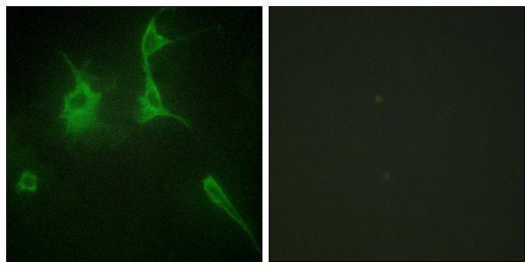
The catalog antibody has IHC data from paraffin-embedded human tonsil and IF data from NIH/3T3 cells (image captions); its listed reactivity is human, mouse, and rat (catalog).

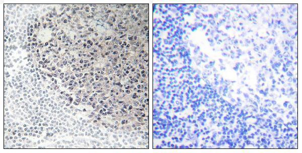
Real IHC data Immunohistochemistry analysis of paraffin-embedded human tonsil tissue, using Collagen IX alpha3 Antibody. The picture on the right is blocked with the synthesized peptide.
Anti-Collagen IX alpha3 COL9A3 Antibody
Cat # A05963

A05963 will render with IHC data from paraffin-embedded human tonsil, including a peptide-blocked comparison (IHC image caption). Its IF image shows NIH/3T3 cells with a peptide-blocked comparison (IF image caption); the catalog lists human, mouse, and rat reactivity and IHC, IF, and ICC applications (catalog).

Which to pick: Choose A05963 for tissue IHC when paraffin sections fit the experiment: its IHC image shows paraffin-embedded human tonsil (IHC image caption), but the fixative is unreported (IHC image caption). Choose A05963 for IF/ICC because both applications are listed and its IF image shows NIH/3T3 cells (catalog; IF image caption). It is also the listed cross-species choice for human, mouse, and rat samples and is polyclonal; the supplied images document human tonsil IHC and NIH/3T3 IF, respectively (catalog; image captions).

Each figure is that product's own IHC / IF validation image from its datasheet.

References

  1. UniProt Consortium. UniProt entry Q14050 (CO9A3_HUMAN, Collagen alpha-3(IX) chain).
  2. Human Protein Atlas. COL9A3 tissue IHC expression (reliability: Supported).
  3. Human Protein Atlas. COL9A3 subcellular location (ICC-IF): Mainly localized to the nucleoplasm. In addition localized to the intermediate filaments..
  4. Human Protein Atlas. COL9A3 antibody validation summary (2 antibodies).
  5. Functional validation of driver mutation-specific uveal melanoma biomarkers: role of COL9A3 in cancer cell plasticity. The Journal of pathology 2026 — PMC13576891.
  6. Transcriptomic alterations underlying metaplasia into specific metaplastic components in metaplastic breast carcinoma. Breast cancer research : BCR 2023 — PMC9883935.
  7. USP3 promotes gastric cancer progression and metastasis by deubiquitination-dependent COL9A3/COL6A5 stabilisation. Cell death & disease 2021 — PMC8688524.
  8. PubMed PMID:8586434 — UniProt-cited evidence.
  9. PubMed PMID:10428822 — UniProt-cited evidence.
  10. PubMed PMID:11780052 — UniProt-cited evidence.