This website uses cookies to ensure you get the best experience on our website.


- Table of Contents
Plan chromogenic COPB2 IHC in paraffin sections using the catalog antibody at 1:50–1:200 (datasheet A07493). Colon glandular cells show high staining, while adipocytes have no detected staining (HPA tissue IHC).

Expected localisation, validated protocols, controls and antibodies — the at-a-glance facts below, then the full design guide.
| Expected localisation | General cytoplasmic staining (HPA tissue IHC) | |
| Staining pattern | Cytoplasmic staining in many tissue cell types (HPA tissue IHC) | |
| Antigen retrieval | Citrate pH 6.0 HIER, heat-mediated (datasheet A07493) | |
| Positive control | Bronchus+4 more · see all | |
| Negative control | Adipose tissue+3 more · see all |
| Fixation | Keep paraffin-section fixation consistent across samples. (standard IHC practice; not target-specific) | |
| Caveat | Staining and RNA show medium consistency (HPA tissue IHC) | |
| Regulation | Low tissue specificity (HPA tissue RNA) | |
| Isoform / epitope | 2 isoforms; epitope coverage is unspecified (UniProt) |
The catalog antibody protocol (datasheet: A07493) is accompanied by published COPB2 IHC protocols for gastric cancer, hepatocellular carcinoma, and gastric cancer xenograft sections (PMC12867441; PMC11964868; PMC6411345).
| Sample | Paraffin-embedded human breast cancer tissue; fixative not specified (datasheet A07493) |
| Fixation | Image fixative and duration unreported (datasheet A07493); verify before use. |
| Sectioning | 4–5 µm sections on charged slides (standard) |
| Deparaffinisation | Xylene, graded ethanol series to water (standard) |
| Antigen retrieval | Heat retrieval: Citrate pH 6.0 (datasheet A07493); 20 min, 95–100 °C (standard) |
| Peroxidase block | 3% H2O2, 10 min, room temperature (standard) |
| Blocking | 10% normal serum of the secondary host, 30 min, room temperature (standard) |
| Primary antibody | Rabbit anti-COPB2, 1:50-1:200 (datasheet A07493) |
| Primary incubation | Overnight at 4 °C (standard) |
| Detection | HRP-polymer secondary, DAB chromogen 5–10 min (standard) |
| Counterstain | Hematoxylin, blue, dehydrate and mount (standard) |
| Expected result | COPB2-positive staining in respiratory epithelial cells of bronchus (HPA tissue IHC: High). HPA tissue profile: General cytoplasmic expression. No signal in the no-primary control. |
COPB2 should produce cytoplasmic staining in many cell types, with high staining documented in bronchial respiratory epithelium and colon, duodenal, and pancreatic glandular cells (HPA tissue IHC: general cytoplasmic expression; High in these cells). It is cytosolic and associates with the cytoplasmic face of Golgi and COPI vesicle membranes; it has no transmembrane segment (UniProt P35606 topology). The HPA tissue IHC assessment is Approved, with medium consistency between staining and RNA expression (HPA tissue IHC: reliability).
| Cytoplasmic chromogen in bronchial respiratory epithelial cells or colon glandular cells. | This matches the reported high staining in those cells (HPA tissue IHC: High). Judge the signal within the identified cell population: COPB2 is associated with cytosol, Golgi membranes, and COPI vesicles (UniProt P35606 subcellular location). A visibly concentrated Golgi region can be compatible, but tissue IHC does not establish a required Golgi pattern (HPA tissue IHC: general cytoplasmic expression). |
| Signal appears confined to nuclei or cell surfaces, without convincing cytoplasmic staining. | Treat that distribution as discordant with the reported general cytoplasmic tissue pattern (HPA tissue IHC: profile) and cytosolic or cytoplasmic membrane association (UniProt P35606 subcellular location; topology). Review morphology and control staining before assigning it to COPB2; localization alone cannot identify the source of an unexpected signal. |
| Strong staining in adipocytes or ovarian stromal cells while expected epithelial cells remain weak. | These cell populations are reported as not detected, whereas several epithelial and glandular populations are High (HPA tissue IHC: adipose tissue, ovary, bronchus, colon). Check cell identification and detection controls; cross-reactivity or endogenous detection activity are possible explanations under standard IHC practice. An unexpected signal does not by itself prove either cause. |
| Haze or chromogen covers stroma and empty spaces, obscuring cell boundaries. | A field-wide deposit is difficult to reconcile with a cell-resolved cytoplasmic pattern (HPA tissue IHC: general cytoplasmic expression). Under standard IHC practice, assess blocking, antibody concentration, washing, and chromogen development with an appropriate negative detection control. Do not score obscured cells as positive. |
| No detectable signal in bronchial respiratory epithelium or pancreatic exocrine glandular cells. | Both are reported High, so a blank result warrants a technical check (HPA tissue IHC: bronchus, pancreas). Confirm that the selected antibody is validated for IHC and that the detection controls worked (HPA antibodies: HPA036867 IHC Approved). The HPA reliability rating is Approved with medium RNA–staining consistency, so a single negative section cannot establish true absence (HPA tissue IHC: reliability). |
| Compartment assignment | COPB2 is cytosolic or bound on the cytoplasmic side of Golgi and COPI vesicle membranes, with no transmembrane segment (UniProt P35606 topology; subcellular location). Interpret a cytoplasmic signal in that context. The supplied evidence does not define an antibody epitope, so it cannot support a claim that a particular retrieval condition exposes it. |
| Choice of reference tissue and cell population | Bronchial respiratory epithelial, colon and duodenal glandular, and pancreatic exocrine glandular cells are High; adipocytes, ovarian stromal cells, smooth muscle cells, and splenic red-pulp cells are Not detected (HPA tissue IHC). Compare the same cell populations when interpreting a section; a tissue name alone can conceal different cell-level results. |
| Evidence strength for tissue IHC | The tissue profile is Approved but has medium consistency with RNA expression data, and RNA specificity is low across tissues (HPA tissue IHC: reliability; RNA specificity). Use the reported cell-level observations as comparators, while allowing for uncertainty in an individual specimen. This evidence supplies no target-specific fixation or antigen-retrieval effect. |
| Assay-specific antibody validation | HPA036867 is listed as IHC Approved, while HPA058180 is listed as ICC Approved without an IHC status in the supplied record (HPA antibodies). An ICC result does not establish IHC performance. The supplied antibody record gives no dilution, retrieval setting, or epitope for this section. |
| Situation | Likely cause | Next action |
|---|---|---|
| Known High epithelial cells are blank. | The result conflicts with the HPA High observations; the supplied data do not identify a COPB2-specific technical cause (HPA tissue IHC: bronchus, colon, pancreas). | Check the IHC antibody's validation status, section integrity, and positive detection control; then review the established staining workflow (HPA antibodies: HPA036867 IHC Approved; standard IHC practice). |
| Nearly every cell and tissue space stains. | Diffuse deposit prevents assessment of the reported cell-resolved cytoplasmic pattern (HPA tissue IHC: general cytoplasmic expression). Background from detection or chromogen handling is possible under standard IHC practice. | Run a negative detection control and review blocking, washes, antibody concentration, and development time (standard IHC practice). Re-score only cells with interpretable morphology. |
| Staining is mainly nuclear or at the cell surface. | That distribution disagrees with COPB2's cytosolic and cytoplasmic-face membrane locations (UniProt P35606 subcellular location; topology). Its origin cannot be determined from appearance alone. | Check cell boundaries and counterstain, compare a documented High tissue, and inspect negative controls before assigning specificity (HPA tissue IHC: High examples; standard IHC practice). |
| Adipocytes, smooth muscle, or ovarian stromal cells appear strongly positive. | HPA reports these cell populations as Not detected (HPA tissue IHC: adipose tissue, smooth muscle, ovary). Misidentified cells, cross-reactivity, or endogenous detection activity are possible under standard IHC practice. | Verify cell identity and compare negative detection controls with nearby expected positive cells; report discordant staining without calling its cause proven (standard IHC practice; HPA tissue IHC: High epithelial and glandular cells). |
| The same tissue contains both strong and blank areas. | HPA results are cell-specific, and its overall tissue IHC assessment has medium RNA–staining consistency (HPA tissue IHC: cell levels; reliability). Tissue-wide scoring can therefore obscure which cells carry the signal. | Score identifiable cell populations separately, using morphology and a counterstain; compare each population with its HPA entry (standard IHC practice; HPA tissue IHC: cell-level observations). |
| Why does ICC-IF show an ER-associated signal alongside Golgi staining? | HPA ICC-IF lists the ER as the approved main location and Golgi as an approved additional location; UniProt describes cytosol and the cytoplasmic side of Golgi and COPI vesicles (HPA subcellular ICC-IF; UniProt P35606 subcellular location). | Interpret the ICC-IF result within its own assay and antibody validation: HPA058180 is ICC Approved, while HPA036867 is IHC Approved (HPA antibodies). Use the tissue IHC profile for chromogenic-section expectations (HPA tissue IHC: general cytoplasmic expression). |


Comprehensive Human Protein Atlas IHC scoring per tissue (reliability: Approved — Medium consistency between antibody staining and RNA expression data.). Rows are taken directly from the HPA tissue chart — click any row's HPA link to view the source.
| Tissue | Cell type | Level | Evidence | Source |
|---|---|---|---|---|
| Bronchus | Respiratory epithelial cells | High | Protein (IHC) | HPA → |
| Colon | Glandular cells | High | Protein (IHC) | HPA → |
| Duodenum | Glandular cells | High | Protein (IHC) | HPA → |
| Epididymis | Glandular cells | High | Protein (IHC) | HPA → |
| Fallopian tube | Glandular cells | High | Protein (IHC) | HPA → |
| Tissue | Cell type | Level | Evidence | Source |
|---|---|---|---|---|
| Adipose tissue | Adipocytes | Not detected | Protein (IHC) | HPA → |
| Ovary | Ovarian stroma cells | Not detected | Protein (IHC) | HPA → |
| Smooth muscle | Smooth muscle cells | Not detected | Protein (IHC) | HPA → |
| Spleen | Cells in red pulp | Not detected | Protein (IHC) | HPA → |
Use compartment, cell type, and matched controls to evaluate COPB2 staining in paraffin sections; treat fixation effects and epitope specificity as unresolved.
A07493 has real COPB2 IHC data from paraffin-embedded human breast cancer (IHC image caption). Its catalog lists IF/ICC and human, mouse, and rat reactivity (catalog).
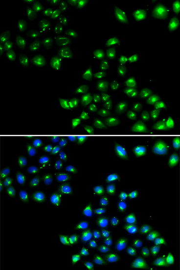
A07493 will render with an IHC image of paraffin-embedded human breast cancer stained at 1:50 after citrate retrieval (IHC image caption). IF/ICC and human, mouse, and rat reactivity are catalog-listed; no IF image is supplied (catalog payload).
Which to pick: For tissue IHC, choose A07493 for paraffin sections: its image shows human breast cancer after high-pressure retrieval in 10 mM citrate buffer at pH 6.0; the fixative is unreported (IHC image caption). For IF/ICC, A07493 is listed at 1:50–1:100, but has no supplied IF image (catalog payload). A07493 is a rabbit polyclonal listed as reactive with human, mouse, and rat; the supplied IHC image demonstrates staining only in human breast cancer (catalog; IHC image caption).