CORO1A / Coronin-1A · IHC design guide

Design Immunohistochemistry for CORO1A

Plan CORO1A staining in paraffin sections around cytoplasmic lymphocytes in lymphoid tissue (HPA tissue IHC). Use the catalog antibody’s IHC conditions and score staining by cell population (datasheet A04245-2; standard IHC practice).

Evidence assembled Oct 2026 · For research use; verify linked source records and product datasheet before use
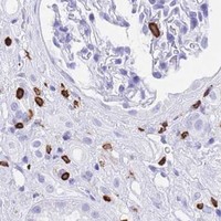
Immunohistochemistry protocol sheet for CORO1A (IHC for CORO1A): expected localisation Cytoplasmic staining in lymphocytes (HPA tissue IHC), antibody A04245-2, validated IHC image, and IHC protocol steps
Printable CORO1A IHC protocol sheet — expected localisation Cytoplasmic staining in lymphocytes (HPA tissue IHC), antibody A04245-2, controls and protocol steps. Open the full CORO1A IHC guide →

CORO1A Immunohistochemistry Experimental Design Guide

Expected localisation, validated protocols, controls and antibodies — the at-a-glance facts below, then the full design guide.

Must know before staining
Expected localisation Cytoplasmic staining in lymphocytes (HPA tissue IHC)
Staining pattern Cytoplasmic lymphocytes, most abundant in lymphoid tissue (HPA tissue IHC)
Antigen retrieval EDTA pH 8.0 HIER, heat-mediated (datasheet A04245-2)
Positive control ⓘ Appendix+4 more · see all
Negative control ⓘ Adipose tissue+4 more · see all
Important caveats
Reasons your staining may differ from the expected pattern.
Fixation Matched tissue-IHC evidence does not establish the fixation claim. Validate the specimen-specific method before use. (selected-SKU IHC image A04245-2)
Caveat Lymphocyte-rich areas can dominate tissue staining (HPA tissue IHC)
Regulation Marrow and lymphoid tissue enriched (HPA tissue IHC)
Isoform / epitope No annotated isoforms; one 2–461 chain (UniProt)
Section 1

Recommended CORO1A IHC & IF Protocols

The catalog antibody’s IHC-P protocol (datasheet: A04245-2) is accompanied by three published CORO1A IHC protocols (PMC10418749; PMC13293819; PMC12535887).

Recommended immunohistochemistry (IHC-P) protocol parameters
SampleParaffin-embedded human colonic adenocarcinoma tissue; fixative not specified (datasheet A04245-2)
FixationImage fixative and duration unreported (datasheet A04245-2); verify before use.
Sectioning4–5 µm sections on charged slides (standard)
DeparaffinisationXylene, graded ethanol series to water (standard)
Antigen retrievalHeat retrieval: EDTA pH 8.0 (datasheet A04245-2); 20 min, 95–100 °C (standard)
Peroxidase block3% H2O2, 10 min, room temperature (standard)
Blocking10% goat serum (datasheet A04245-2)
Primary antibodyRabbit anti-CORO1A, 1-2μg/ml (datasheet A04245-2)
Primary incubationOvernight at 4 °C (datasheet A04245-2)
DetectionStreptavidin-biotin complex (SABC), DAB chromogen (datasheet A04245-2)
CounterstainHematoxylin, blue, dehydrate and mount (standard)
Expected resultCORO1A-positive staining in non-germinal center cells of appendix (HPA tissue IHC: High). HPA tissue profile: Cytoplasmic expression of lymphocytes, most abundant in lymphoid tissue, and of subsets of cells in brain. No signal in the no-primary control.
💡Decision noteStart with heat-mediated EDTA retrieval at pH 8.0 (datasheet: A04245-2). Citrate retrieval is reported for mouse knee sections (PMC13293819).
Section 2

What Is the Expected CORO1A Staining Pattern?

CORO1A is a cytoplasmic protein associated with the cytoskeleton and cell cortex; it has no transmembrane segment (UniProt P31146 topology and subcellular location). In paraffin sections, expect staining chiefly in lymphocytes and other cells of lymphoid tissue, with staining also reported in subsets of brain cells (HPA: tissue IHC profile). HPA rates its tissue IHC pattern Enhanced, citing consistency between antibody staining and RNA expression (HPA: IHC reliability).

What am I looking at on my slide?
Strong cytoplasmic staining in lymph node germinal center cells, tonsil germinal center cells, or spleen red pulp cells.This fits the reported high staining in those cell populations (HPA: tissue IHC). Judge the signal within the named cells; a positive tissue does not imply that every cell in its section should stain (HPA: tissue IHC profile).
Cytoplasmic or peripheral staining is visible, with nuclei comparatively unstained.This agrees with CORO1A's cytoskeletal and cell cortex locations (UniProt P31146: subcellular location). Predominantly nuclear staining would conflict with the reported location and warrants checking the assay before calling it specific (UniProt P31146: subcellular location).
Strong staining dominates a cell population reported as undetected, such as adipocytes or bronchial respiratory epithelium.HPA reports no staining in those named populations (HPA: tissue IHC). Consider antibody cross-reactivity or endogenous chromogen-generating activity as possible explanations; inspect a matched negative control and the location of positive cells (general IHC practice).
Brown signal is spread across cells and extracellular areas without a discernible cytoplasmic pattern.This is difficult to reconcile with HPA's cell-specific cytoplasmic profile (HPA: tissue IHC profile). Uneven washing, detection background, or endogenous activity can produce diffuse staining; compare the no-primary control (general IHC practice).
A lymph node germinal center or bone marrow hematopoietic population shows no detectable signal.Both are reported as high-staining populations (HPA: tissue IHC). A blank result in either is a reason to check assay performance and tissue preservation before concluding that CORO1A is absent from the specimen (general IHC practice).
💡Expected CORO1A appearanceCall a result consistent with CORO1A when distinct cytoplasmic staining is high in reported lymphoid or hematopoietic populations (HPA: tissue IHC); predominantly nuclear signal or strong staining of HPA-undetected adipocytes is a warning to investigate specificity (UniProt P31146: subcellular location; HPA: tissue IHC).
How each factor affects the staining
Choice of tissue and cellsHPA reports high staining in appendix non-germinal center cells, bone marrow hematopoietic cells, lymph node and tonsil germinal center cells, and spleen red pulp cells (HPA: tissue IHC). Specify the cell population when scoring; tissue-wide scores can obscure that distribution (general IHC practice).
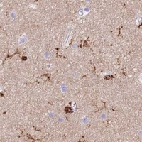
Brain and skin interpretationCerebellar granular cells are reported high; cerebral cortex glial cells and skin basal-layer cells are reported medium, while hippocampal and caudate glial cells are low (HPA: tissue IHC). Interpret these compartments against their own reported levels (HPA: tissue IHC).
Subcellular contextCORO1A has no transmembrane segment and is associated with cytoplasm, cytoskeleton, and cell cortex (UniProt P31146: topology and subcellular location). UniProt's phagosome-retention note concerns viable mycobacterial infection and is marked By Similarity; it is not a routine paraffin-section expectation (UniProt P31146: subcellular note).
Antibody evidence and scopeHPA lists 2 antibodies with Enhanced IHC status (HPA: antibody validation). That supports the reported tissue pattern but does not establish how fixation or antigen retrieval affects this target; target-specific fixation sensitivity is unreported in the supplied sources (HPA: tissue IHC and antibody validation).
IF/ICC Q: What location should a separate IF/ICC assessment check?A: HPA approves cytosol as the main ICC-IF location (HPA: subcellular). UniProt also reports cell cortex and cytoskeletal association (UniProt P31146: subcellular location). Interpret IF/ICC with its own controls and guide; the IHC tissue pattern alone does not validate an IF/ICC result (general assay practice).
Why is my staining missing, weak or wrong?
SituationLikely causeNext action
The known-positive lymph node or bone marrow control is blank.Those cell populations are reported high (HPA: tissue IHC); a blank control suggests an assay or specimen problem, although this record does not identify a CORO1A-specific fixation effect.Check the section and control integrity, antibody application, detection reagents, and counterstain; review the validated IHC-P procedure before changing retrieval conditions (general IHC practice).
Signal is concentrated in nuclei rather than cytoplasm.A nuclear-dominant pattern conflicts with reported cytoplasmic, cytoskeletal, and cortical locations (UniProt P31146: subcellular location).Compare a known-positive tissue and no-primary control, then reassess staining specificity and imaging or scoring boundaries (general IHC practice).
Adipocytes or bronchial respiratory epithelial cells stain strongly.Those specific populations are reported undetected (HPA: tissue IHC); cross-reactivity or endogenous detection activity is possible (general IHC practice).Check a no-primary control and examine whether the signal tracks cell boundaries and expected positive populations; investigate endogenous activity in the detection system (general IHC practice).
Background obscures the lymphoid-cell pattern.Diffuse signal prevents assessment of the reported cytoplasmic lymphocyte staining (HPA: tissue IHC profile); nonspecific binding or detection background may contribute (general IHC practice).Review blocking, washing, antibody concentration, and the no-primary control using the IHC-P procedure as the starting point (general IHC practice).
Brain staining is weaker or more limited than lymphoid-tissue staining.HPA reports cell-dependent brain levels, including medium cerebral cortex glial staining and low hippocampal or caudate glial staining (HPA: tissue IHC).Score the identified brain cell population against its reported level and include a reported high-staining lymphoid control in the run (HPA: tissue IHC; general IHC practice).
A cortical or phagosome-like rim is treated as the sole required positive pattern.UniProt's phagosomal retention description is specific to viable mycobacterial infection and marked By Similarity (UniProt P31146: subcellular note).For routine IHC, assess the reported cytoplasmic staining in the appropriate cells; reserve an infection-dependent interpretation for a separately documented infection experiment (HPA: tissue IHC profile; UniProt P31146: subcellular note).

Sample controls for CORO1A IHC & IF

🧪Run bone marrow first: hematopoietic cells should stain (HPA: High in hematopoietic cells). Use adipose tissue adipocytes as the negative comparison (HPA: Not detected in adipocytes); on the positive slide, cells without specific signal should show counterstain without cytoplasmic DAB staining, but no internal negative cell type is established by the supplied HPA row.
Positive control tissue: Appendix (Non-germinal center cells, HPA High)
Negative control tissue: Adipose tissue (HPA Not detected)
ICC-IF cell lines (HPA subcellular resource): HPA ICC-IF images show CORO1A in Hep-G2, MCF-7, U2OS, KOLF2.1J, NIH 3T3, with annotated localisation: Cytosol (approved) (HPA subcellular).
Technical controls: Include a no-primary, secondary-only control; a rabbit IgG isotype control matched to the primary antibody’s clonality; and CORO1A knockout tissue or cells as a biological specificity control (caption: rabbit primary antibody; standard IHC practice). Quench endogenous peroxidase in bone marrow before DAB detection, particularly in hematopoietic cells (standard IHC practice).
⚠️Feasibility: A target-specific fixation window or fixation effect is unreported, and the selected A04245-2 tissue-IHC caption does not state a fixative. The caption uses heat-mediated EDTA retrieval at pH 8.0, but does not establish that retrieval is required; the supplied evidence also does not establish whether frozen sections or IF are easier (A04245-2 caption; HPA: ICC-IF images available). Endogenous peroxidase in bone marrow can create chromogenic background, so interpret staining against the no-primary control (standard IHC practice).

HPA tissue IHC evidence for CORO1A

Comprehensive Human Protein Atlas IHC scoring per tissue (reliability: Enhanced — High consistency between antibody staining and RNA expression data.). Rows are taken directly from the HPA tissue chart — click any row's HPA link to view the source.

Positive expression · recommended positive controls

TissueCell typeLevelEvidenceSource
Appendix Non-germinal center cells High Protein (IHC) HPA →
Bone marrow Hematopoietic cells High Protein (IHC) HPA →
Cerebellum Granular cells - cytoplasm/membrane High Protein (IHC) HPA →
Lymph node Germinal center cells High Protein (IHC) HPA →
Spleen Cells in red pulp High Protein (IHC) HPA →

Undetected expression · recommended negative controls

TissueCell typeLevelEvidenceSource
Adipose tissue Adipocytes Not detected Protein (IHC) HPA →
Adrenal gland Glandular cells Not detected Protein (IHC) HPA →
Breast Adipocytes Not detected Protein (IHC) HPA →
Bronchus Respiratory epithelial cells Not detected Protein (IHC) HPA →
Cervix Glandular cells Not detected Protein (IHC) HPA →
Section 3

Advanced CORO1A IHC Tips

Troubleshoot CORO1A staining in paraffin sections by checking retrieval, cell identity, cytoplasmic localisation, and detection controls.

What retrieval should I try when CORO1A staining is weak in paraffin sections?
Start with heat-mediated retrieval in EDTA at pH 8.0 for paraffin sections (datasheet A04245-2). The selected tissue image used this retrieval before overnight incubation at 4°C with 2 µg/mL primary antibody; its caption does not report the fixative (datasheet A04245-2). If staining remains weak, compare retrieval times on adjacent sections while holding antibody concentration and detection constant, and watch for tissue damage (standard IHC practice). Include a lymph node or tonsil control, where germinal center cells stain strongly, so a failed run can be distinguished from low expression in the test area (HPA: High in germinal center cells).
How should I troubleshoot variable staining when the specimen fixative is uncertain?
The selected paraffin-section caption does not state a fixative, so target-specific CORO1A sensitivity to fixation is unknown (datasheet A04245-2). Record the fixative and processing history for each specimen, then compare sections with matched processing, retrieval, antibody incubation, and detection conditions (standard IHC practice). Use the documented EDTA retrieval at pH 8.0 as the starting condition, and change only one processing variable at a time when troubleshooting weak staining (datasheet A04245-2; standard IHC practice). Check a concurrently processed positive control before attributing a negative specimen to biology; lymph node germinal center cells provide a reported high-staining reference (HPA: High in germinal center cells).
Where should CORO1A staining appear, and how should membrane-adjacent signal be judged?
Expect predominantly cytoplasmic staining in lymphocytes in tissue sections, with abundant signal in lymphoid tissue (HPA: tissue IHC profile). CORO1A is associated with the cytoskeleton and cell cortex and binds actin, so peripheral cytoplasmic accentuation can be biologically plausible (UniProt P31146: subcellular location and subunit). It has no annotated transmembrane segment; a sharp outline alone should therefore be checked against cell morphology and staining controls before being called a membrane pattern (UniProt P31146: topology; standard IHC practice). Compare adjacent cells and a high-staining lymphoid control, and assess whether the chromogen remains within identifiable cytoplasm rather than collecting along section edges (HPA: tissue IHC profile; standard IHC practice).
Could epitope accessibility or modification explain discrepant CORO1A staining?
The supplied record lists 0 isoforms, no annotated domains, and no transmembrane segment, so these annotations do not identify an alternative isoform or membrane-facing epitope to explain the discrepancy (UniProt P31146: isoforms, domains, topology). It records modifications near residues 2, 412, 422, and 449, but the selected antibody's epitope is not specified in the payload (UniProt P31146: modified residues; datasheet A04245-2). Do not assign a staining change to a modification without epitope information or direct validation (standard IHC interpretation). Compare matched sections using the documented EDTA pH 8.0 retrieval and fixed detection settings, then evaluate morphology and the positive control together (datasheet A04245-2; standard IHC practice).
How can I assess CORO1A by multiplex IF alongside the chromogenic IHC result?
Treat IF as a separate assay requiring its own validation; the selected product evidence describes paraffin-section chromogenic IHC with EDTA retrieval at pH 8.0, not an IF protocol (datasheet A04245-2). Multiplex CORO1A with a validated lymphocyte marker when examining lymphoid tissue, and assess signal within marker-positive cells against appropriate single-stain controls (HPA: cytoplasmic lymphocyte expression; standard IF practice). Choose fluorophores after checking tissue autofluorescence in unstained sections, favoring channels with better separation from that background (standard IF practice). For intracellular access, optimise permeabilisation for the cytosolic or cortical epitope being tested; CORO1A has no transmembrane segment, and the antibody epitope is unspecified here (UniProt P31146: subcellular location and topology; standard IF practice).
What should I check when CORO1A DAB staining is diffuse or widespread?
First compare the suspect section with a no-primary control and a high-staining lymphoid control to separate detection background from expected cellular signal (HPA: high staining in lymphoid tissue; standard IHC practice). The selected image used 10% goat serum blocking, 2 µg/mL primary antibody overnight at 4°C, and a biotin-based DAB detection system (datasheet A04245-2). Check peroxidase blocking and the biotin-based detection controls when DAB appears outside identifiable cells, then adjust blocking, antibody concentration, or development one variable at a time (standard IHC practice). Judge signal against cytoplasmic lymphocyte staining rather than treating uniform colour across unrelated cell types as CORO1A expression (HPA: tissue IHC profile; standard IHC interpretation).
How should I quantify CORO1A staining across sections with different cell compositions? ⚠ ANSWER MARKED FOR VERIFICATION
Define the tissue region and cell population before scoring, because reported high staining occurs in specific populations such as lymph node germinal center cells and bone marrow hematopoietic cells (HPA: tissue IHC). For those defined cells, report the percentage positive and an H-score from intensity categories 0–3; alternatively report positive-cell density per mm² of viable tissue (standard IHC scoring practice). Normalise cell-based results to the number of eligible cells and area-based results to the analysed viable area, while excluding folds and necrosis by a prespecified rule (standard IHC scoring practice). Keep retrieval, DAB development, imaging, and positivity thresholds consistent across sections, and include the same positive control in each run (standard IHC practice).
How can I distinguish genuine CORO1A staining from section or detection artefacts?
A credible result follows the expected cytoplasmic pattern in identifiable lymphocytes, with strong staining in suitable lymphoid controls (HPA: tissue IHC profile and High in germinal center cells). Inspect unexpected nuclear-only or uniform epithelial staining against morphology and the no-primary control before assigning it to CORO1A (HPA: cytoplasmic lymphocyte expression; standard IHC interpretation). Signal confined to section edges, folds, or necrotic areas warrants repeat assessment on intact tissue, while colour in the no-primary control calls for a detection and endogenous-peroxidase check (standard IHC practice). CORO1A can associate with the cell cortex and, in mycobacteria-infected macrophages, phagosome membranes; that specialised pattern requires matching biological context rather than interpretation from DAB localisation alone (UniProt P31146: subcellular location).
Boster reagents

Best CORO1A / Coronin-1A IHC Antibodies

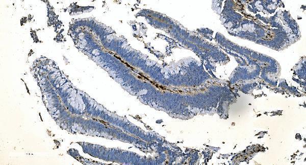
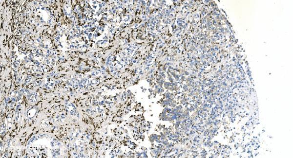
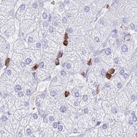
A04245-2 has real IHC images from paraffin-embedded human colonic adenocarcinoma, gastric, liver, and lung cancer sections (catalog IHC captions). No IF/ICC data are supplied (catalog applications/images).

Real IHC data IHC analysis of Coronin 1a/TACO/CORO1A using anti-Coronin 1a/TACO/CORO1A antibody (A04245-2). Coronin 1a/TACO/CORO1A was detected in paraffin-embedded section of human colonic adenocarcinoma tissue. Heat mediated antigen retrieval was performed in EDTA buffer (pH8.0, epitope retrieval solution). The tissue section was blocked with 10% goat serum. The tissue section was then incubated with 2μg/ml rabbit anti-Coronin 1a/TACO/CORO1A Antibody (A04245-2) overnight at 4°C. Biotinylated goat anti-rabbit IgG was used as secondary antibody and incubated for 30 minutes at 37°C. The tissue section was developed using Strepavidin-Biotin-Complex (SABC) (Catalog # SA1022) with DAB as the chromogen.
Anti-Coronin 1a/TACO/CORO1A Antibody ®
Cat # A04245-2

A04245-2 is listed for IHC and human, mouse, and rat reactivity (catalog applications/reactivity). Its IHC captions show staining of paraffin-embedded human colonic adenocarcinoma, gastric cancer, liver cancer, and lung cancer sections at 2 μg/ml (catalog IHC captions).

Which to pick: Choose A04245-2 for human paraffin-section IHC based on its own image captions; the fixative is unreported (catalog IHC captions). No listed SKU has IF/ICC validation or an IF image (catalog applications/images). A04245-2 is the only cross-species candidate because human, mouse, and rat reactivity are listed, although its IHC images show human tissue only; its host is rabbit and clonality is unreported (catalog reactivity/IHC captions/host/clone).

Each figure is that product's own IHC / IF validation image from its datasheet.

References

  1. UniProt Consortium. UniProt entry P31146 (COR1A_HUMAN, Coronin-1A).
  2. Human Protein Atlas. CORO1A tissue IHC expression (reliability: Enhanced).
  3. Human Protein Atlas. CORO1A subcellular location (ICC-IF): Localized to the cytosol..
  4. Human Protein Atlas. CORO1A antibody validation summary (2 antibodies).
  5. Proteomic Analysis of Dysfunctional Liver Sinusoidal Endothelial Cells Reveals Substantial Differences in Most Common Experimental Models of Chronic Liver Diseases. International journal of molecular sciences 2023 — PMC10418749.
  6. CORO1A links inflammatory chondrocyte subpopulations to immune microenvironment alterations in osteoarthritis: an integrative multi-omics and single-cell study. Frontiers in immunology 2026 — PMC13293819.
  7. Identification of key biomarkers and immune infiltration in renal interstitial fibrosis. Annals of translational medicine 2022 — PMC8908133.
  8. CORO1A: a pan-cancer prognosis, diagnostic and immune biomarker based on breast cancer validation. Frontiers in oncology 2025 — PMC12535887.
  9. PubMed PMID:7758584 — UniProt-cited evidence.
  10. PubMed PMID:14702039 — UniProt-cited evidence.
  11. PubMed PMID:15489334 — UniProt-cited evidence.