This website uses cookies to ensure you get the best experience on our website.


- Table of Contents
Plan chromogenic COX5B IHC in paraffin sections using its inner mitochondrial membrane location (UniProt) and widespread tissue staining (HPA tissue IHC). Use the catalog antibody’s paraffin-section protocol (datasheet A06090-2), and account for low staining in chondrocytes when comparing cell types (HPA tissue IHC).

Expected localisation, validated protocols, controls and antibodies — the at-a-glance facts below, then the full design guide.
| Expected localisation | Inner mitochondrial membrane; tissue signal is widespread (UniProt; HPA tissue IHC) | |
| Staining pattern | Mitochondrial staining across tissue cell types (HPA tissue IHC) | |
| Antigen retrieval | EDTA pH 8.0 HIER, heat-mediated (datasheet A06090-2) | |
| Positive control | Adrenal gland+4 more · see all | |
| Negative control | None in HPA (detected in all 45 tissues); use no-primary + isotype controls |
| Fixation | Keep fixation consistent across paraffin sections. (standard IHC practice; not target-specific) | |
| Caveat | Chondrocytes show low staining despite broad expression (HPA tissue IHC) | |
| Regulation | Heart and skeletal muscle have enhanced RNA (HPA tissue RNA) | |
| Isoform / epitope | No isoforms annotated; mature chain spans residues 32–129 (UniProt) |
Compare the catalog antibody’s IHC-P protocol (datasheet: A06090-2) with two published COX5B chromogenic IHC protocols (PMC12774556; PMC11588429).
| Sample | Paraffin-embedded human breast cancer tissue; fixative not specified (datasheet A06090-2) |
| Fixation | Image fixative and duration unreported (datasheet A06090-2); verify before use. |
| Sectioning | 4–5 µm sections on charged slides (standard) |
| Deparaffinisation | Xylene, graded ethanol series to water (standard) |
| Antigen retrieval | Heat retrieval: EDTA pH 8.0 (datasheet A06090-2); 20 min, 95–100 °C (standard) |
| Peroxidase block | 3% H2O2, 10 min, room temperature (standard) |
| Blocking | 10% goat serum (datasheet A06090-2) |
| Primary antibody | Rabbit anti-COX5B, 2-5 μg/ml (datasheet A06090-2) |
| Primary incubation | Overnight at 4 °C (datasheet A06090-2) |
| Detection | HRP-conjugated secondary, DAB chromogen (datasheet A06090-2) |
| Counterstain | Hematoxylin, blue, dehydrate and mount (standard) |
| Expected result | COX5B-positive staining in glandular cells of adrenal gland (HPA tissue IHC: High). HPA tissue profile: Ubiquitous mitochondrial expression. No signal in the no-primary control. |
COX5B is a component of mitochondrial complex IV in the inner membrane, with no annotated transmembrane segment (UniProt P10606 topology and subunit). Expect predominantly cytoplasmic mitochondrial staining across many cell types; HPA describes ubiquitous mitochondrial expression and high IHC staining in several glandular, epithelial, hematopoietic and neural cell populations (HPA: tissue IHC). Treat intensity comparisons cautiously: HPA rates tissue IHC reliability as Supported, with medium consistency between staining and RNA data (HPA: reliability).
| Granular or punctate cytoplasmic staining in expected cells. | This fits a mitochondrial pattern (UniProt P10606: inner membrane; HPA: ubiquitous mitochondrial expression). Judge the pattern within cells as well as its overall intensity; chromogenic IHC cannot resolve the inner membrane itself (standard IHC interpretation). |
| Dominant nuclear staining or a sharply defined cell-surface rim. | These compartments do not fit the annotated mitochondrial location (UniProt P10606: inner membrane; HPA: mitochondria enhanced in ICC-IF). Review controls and the staining workflow before calling such a pattern COX5B specific (standard IHC practice). |
| Strong staining only in cells outside the expected reference population. | Compare adjacent morphology and a matched reference section before suspecting cross-reactivity or endogenous detection activity (standard IHC practice). HPA reports broad expression, so an unlisted cell type alone is not evidence of a false positive (HPA: ubiquitous mitochondrial expression). |
| Uniform haze across cells and surrounding section. | Diffuse staining obscures the granular mitochondrial pattern expected for COX5B (HPA: ubiquitous mitochondrial expression; HPA: mitochondria enhanced in ICC-IF). It can reflect background from the antibody or detection system; assess a matched negative control (standard IHC practice). |
| Little or no signal in a reference population reported as High. | Check tissue preservation, antibody and detection controls before interpreting absence as biology (standard IHC practice). HPA lists High staining in adrenal gland glandular cells and bronchus respiratory epithelial cells, but its IHC reliability is Supported with medium RNA agreement (HPA: tissue IHC; HPA: reliability). |
| Cell and tissue context | HPA reports High IHC staining in breast glandular cells, bone marrow hematopoietic cells, caudate neuronal cells and other listed populations, but Low staining in soft tissue chondrocytes (HPA: tissue IHC). Use those observations as reference patterns, not absolute thresholds. |
| Strength of tissue evidence | The tissue profile is Supported, with medium consistency between antibody staining and RNA expression (HPA: reliability). HPA supplies no negative tissue example, so absence in an unlisted tissue has no validated negative-reference status (HPA: tissue IHC). |
| Antibody validation | HPA034517 has Supported IHC status and Enhanced ICC status (HPA: antibody validation). These are application-specific assessments; the ICC rating does not upgrade the IHC evidence or establish a staining threshold for every tissue (HPA: antibody validation). |
| Protein location and processing | UniProt places COX5B in the inner membrane, annotates no transmembrane segment and identifies a mature chain spanning residues 32–129 (UniProt P10606: topology and processing). Epitope location is unspecified, so these annotations cannot predict retrieval requirements (UniProt P10606: processing). |
| Antigen retrieval in paraffin IHC | Use a retrieval condition appropriate to the IHC-validated antibody and compare it with the supplied positive reference and controls (standard IHC-P practice). No COX5B-specific retrieval condition or fixation-sensitivity result is supplied (UniProt P10606; HPA: tissue IHC). |
| IF/ICC Q&A: what localisation should I expect? | Predominantly mitochondrial signal; HPA also reports acrosome localisation in ICC-IF (HPA: subcellular ICC-IF). That additional finding is an ICC-IF observation, not an expected feature of every paraffin tissue section or an IHC protocol instruction (HPA: subcellular ICC-IF). |
| Situation | Likely cause | Next action |
|---|---|---|
| A High-reference section is blank. | An ineffective antibody, retrieval or chromogenic detection step is possible; blank staining alone does not establish absent COX5B (standard IHC practice; HPA: High in adrenal gland glandular cells). | Verify the IHC-validated antibody and detection controls, then assess retrieval using its IHC-P instructions (standard IHC-P practice). Compare with a matched HPA High reference population (HPA: tissue IHC). |
| Signal is mostly nuclear. | Nuclear dominance conflicts with mitochondrial localisation (UniProt P10606: inner membrane; HPA: mitochondria enhanced in ICC-IF). | Check the negative control and whether counterstain or chromogen has been mistaken for specific signal; reassess the cytoplasmic pattern at higher magnification (standard IHC practice). |
| Color appears throughout the section. | Diffuse background can arise from nonspecific binding or the detection chemistry (standard IHC practice). It cannot be assigned to COX5B from intensity alone (HPA: ubiquitous mitochondrial expression). | Compare a no-primary control, review blocking and washes, and adjust antibody concentration according to the IHC-P instructions (standard IHC-P practice). Retain the condition that best preserves cellular detail. |
| Strong signal appears in unexpected cells. | Cross-reactivity or endogenous detection activity is possible (standard IHC practice). HPA's ubiquitous profile means an unlisted cell population is not automatically negative (HPA: tissue IHC). | Compare morphology with an HPA High reference and a no-primary control; if using enzyme detection, check the corresponding endogenous-activity block (standard IHC practice; HPA: tissue IHC). |
| Chondrocytes stain weakly. | Low staining in soft tissue chondrocytes is reported by HPA and need not indicate a failed run (HPA: Low in chondrocytes). | Assess the same run in a High-reference population before changing conditions; use controls to distinguish weak specific staining from background (HPA: tissue IHC; standard IHC practice). |
| IF/ICC and tissue IHC patterns differ. | The applications have separate evidence ratings, and ICC-IF includes an additional acrosome observation (HPA: antibody validation; HPA: subcellular ICC-IF). | Judge paraffin IHC against tissue IHC references and its own controls; use the separate IF/ICC guide for fluorescence-specific setup (HPA: tissue IHC; standard IHC practice). |
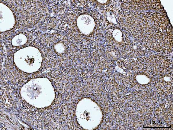
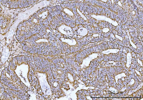
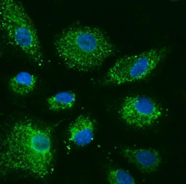


Comprehensive Human Protein Atlas IHC scoring per tissue (reliability: Supported — Medium consistency between antibody staining and RNA expression data.). Rows are taken directly from the HPA tissue chart — click any row's HPA link to view the source.
| Tissue | Cell type | Level | Evidence | Source |
|---|---|---|---|---|
| Adrenal gland | Glandular cells | High | Protein (IHC) | HPA → |
| Appendix | Endocrine cells | High | Protein (IHC) | HPA → |
| Bone marrow | Hematopoietic cells | High | Protein (IHC) | HPA → |
| Breast | Glandular cells | High | Protein (IHC) | HPA → |
| Bronchus | Respiratory epithelial cells | High | Protein (IHC) | HPA → |
| Tissue | Cell type | Level | Evidence | Source |
|---|---|---|---|---|
| None in HPA: COX5B is detected in all 45 scored tissues. Use a no-primary (secondary-only) and an isotype control instead. | ||||
Troubleshoot COX5B chromogenic IHC in paraffin sections using the catalog antibody’s tissue staining conditions and mitochondrial localisation as reference points.
A06090-2 has IHC and IF images of human paraffin sections and an IF/ICC image of A549 cells (image captions). Human, mouse, and rat reactivity is listed (catalog).
A06090-2 has IHC images of paraffin sections from human breast, endometrial, colorectal, and lung cancers (IHC image captions). Its IF images show A549 cells and paraffin sections from human ovary and breast cancers (IF image captions).
Which to pick: Choose A06090-2 for chromogenic IHC of paraffin sections: its breast cancer caption reports EDTA pH 8 retrieval, 2 μg/ml primary antibody, and HRP/DAB detection (IHC image caption). The same SKU has IF/ICC examples and a listed IF dilution of 5 μg/ml (IF image captions; catalog). A06090-2 lists mouse and rat reactivity, although its shown IHC sections are human; the fixative used for those paraffin sections is unreported (catalog reactivity; IHC image captions).