This website uses cookies to ensure you get the best experience on our website.


- Table of Contents
Plan chromogenic paraffin-section IHC for COX6A1 using the granular cytoplasmic tissue pattern as a readout (HPA tissue IHC). This guide covers the catalog antibody’s IHC dilution (datasheet A09572-1), fixation consistency and staining controls (standard IHC practice), with a caution about possible detection of protein from more than one gene (HPA tissue IHC).

Expected localisation, validated protocols, controls and antibodies — the at-a-glance facts below, then the full design guide.
| Expected localisation | Granular cytoplasm in tissue (HPA tissue IHC) | |
| Staining pattern | Granular cytoplasmic staining in most tissues (HPA tissue IHC) | |
| Antigen retrieval | EDTA pH 8.0 HIER, heat-mediated (datasheet A09572-1) | |
| Positive control | Adrenal gland+4 more · see all | |
| Negative control | Adipose tissue+4 more · see all |
| Fixation | Keep fixation consistent across sections (standard IHC practice; not target-specific) | |
| Caveat | Antibody may detect protein from more than one gene (HPA tissue IHC) | |
| Regulation | Expression regulation not annotated (UniProt) | |
| Isoform / epitope | No isoforms annotated; mature chain spans aa 25–109 (UniProt) |
The catalog antibody’s IHC-P protocol (datasheet: A09572-1) is paired with one published mouse liver IHC protocol (PMC12581582).
| Sample | Paraffin-embedded human liver cancer tissue; fixative not specified (datasheet A09572-1) |
| Fixation | Image fixative and duration unreported (datasheet A09572-1); verify before use. |
| Sectioning | 4–5 µm sections on charged slides (standard) |
| Deparaffinisation | Xylene, graded ethanol series to water (standard) |
| Antigen retrieval | Heat retrieval: EDTA pH 8.0 (datasheet A09572-1); 20 min, 95–100 °C (standard) |
| Peroxidase block | 3% H2O2, 10 min, room temperature (standard) |
| Blocking | 10% goat serum (datasheet A09572-1) |
| Primary antibody | Rabbit anti-COX6A1, 1:50 recommended; image 2 μg/ml (datasheet A09572-1) |
| Primary incubation | Overnight at 4 °C (datasheet A09572-1) |
| Detection | HRP-conjugated secondary, DAB chromogen (datasheet A09572-1) |
| Counterstain | Hematoxylin, blue, dehydrate and mount (standard) |
| Expected result | COX6A1-positive staining in glandular cells of adrenal gland (HPA tissue IHC: High). HPA tissue profile: Cytoplasmic expression with a granular pattern in most tissues. No signal in the no-primary control. |
COX6A1 is a cytochrome c oxidase subunit in the mitochondrial inner membrane, with one transmembrane segment at residues 35–59 (UniProt P12074 topology). In paraffin section IHC, expect granular cytoplasmic staining in many tissues, including kidney tubular and colon glandular cells, where HPA reports high staining (HPA tissue IHC). HPA rates its tissue IHC antibody Approved, pending external verification, and cautions that it targets proteins from more than one gene (HPA tissue IHC).
| Granular cytoplasmic stain in kidney tubules or colon glands, with little nuclear stain. | This matches HPA's high staining in those cell types and its granular cytoplasmic tissue profile (HPA tissue IHC). The appearance is consistent with COX6A1's inner mitochondrial membrane location (UniProt P12074 topology), but morphology alone cannot establish target specificity because the HPA antibody may recognize proteins from more than one gene (HPA tissue IHC). |
| Predominantly nuclear or sharply cell-surface stain in an otherwise interpretable section. | That compartment differs from the granular cytoplasmic IHC profile (HPA tissue IHC) and the mitochondrial inner membrane assignment (UniProt P12074 topology). Treat it as a localization mismatch; review the detection controls, morphology, and antibody specificity before scoring it as COX6A1. |
| Strong stain in adipocytes or skeletal myocytes while known-positive cells also stain. | HPA reports COX6A1 staining as not detected in those sampled cell types (HPA tissue IHC). Investigate nonspecific binding or detection background before interpreting the signal. An HPA 'not detected' result is an assay observation; it does not prove biological absence in every specimen. |
| Diffuse color across several compartments, including areas without a discernible granular cellular pattern. | The distribution does not match HPA's granular cytoplasmic profile (HPA tissue IHC). Background from the staining workflow is a possible explanation under general IHC practice; use reagent controls and inspect tissue morphology before assigning cell-specific positivity. |
| No stain in kidney tubules or colon glandular cells on the same run. | These are reported high-staining cell types (HPA tissue IHC), so a negative result calls for a run-level check of tissue preservation, retrieval, antibody application, and chromogen detection under general IHC practice. It does not, by itself, establish that the specimen lacks COX6A1. |
| Target location and membrane topology | COX6A1 occupies the inner mitochondrial membrane; residues 25–34 face the matrix, 35–59 span the membrane, and 60–109 face the intermembrane space (UniProt P12074 topology). These facts support a mitochondrial interpretation but do not identify the antibody's epitope or establish which antigen retrieval condition works best. |
| Protein processing | UniProt annotates the mature chain as residues 25–109 and lists no glycosylation sites or modified residues (UniProt P12074 processing/PTMs). Without an antibody epitope map, these annotations cannot predict whether processing changes staining in a section. |
| Tissue and cell selection | HPA reports high staining in kidney tubules, colon glands, caudate neurons, and cerebral cortex neuropil, among other sites; it reports no detectable staining in adipocytes and skeletal myocytes (HPA tissue IHC). Compare the named cells, not whole-tissue averages, when assessing a control section. |
| Strength of the IHC evidence | The HPA antibody is rated Approved, with external verification pending and a warning that it targets proteins from more than one gene (HPA tissue IHC). Its reported pattern is useful for comparison, but staining with that antibody alone cannot uniquely identify COX6A1; no IHC Enhanced validation is supplied (HPA antibodies; HPA tissue IHC). |
| IF/ICC Q&A: what localization should be expected? | HPA reports supported mitochondrial localization and an additional approved principal-piece localization in ICC-IF (HPA subcellular). The principal-piece finding belongs to that imaging context; it should not be generalized to ordinary tissue IHC. HPA also cautions that the IF antibodies target proteins from multiple genes (HPA subcellular). |
| Situation | Likely cause | Next action |
|---|---|---|
| Kidney tubules and colon glands are blank in a completed IHC run. | These cells are high-staining references (HPA tissue IHC); the blank result may reflect a staining-run problem or specimen quality under general IHC practice. | Check section integrity, the run's positive control, retrieval record, primary-antibody application, and chromogen development. Resolve any run failure before interpreting the specimen as negative. |
| Stain is mainly nuclear despite acceptable morphology. | The localization conflicts with HPA's granular cytoplasmic pattern (HPA tissue IHC) and the UniProt inner mitochondrial membrane assignment (UniProt P12074 topology). | Review reagent controls and antibody specificity, then score only a reproducible cellular pattern consistent with the expected compartment. |
| Adipocytes or skeletal myocytes stain prominently. | HPA reports staining as not detected in those cell types (HPA tissue IHC); nonspecific binding or detection background is possible under general IHC practice. | Compare the same run with a high-staining cell type and appropriate reagent controls. Record the discordance instead of assigning the unexpected signal to COX6A1. |
| Color spreads through the section without cell boundaries or granules. | The appearance differs from the HPA granular cytoplasmic profile (HPA tissue IHC) and may reflect nonspecific workflow background under general IHC practice. | Examine a no-primary control and review blocking, washes, detection reagent, and chromogen development; reassess the stain only after the background is controlled. |
| A granular cytoplasmic pattern appears, but target identity is uncertain. | The HPA antibody can target proteins from more than one gene, and its IHC status remains pending external verification (HPA tissue IHC). | Describe the pattern as consistent with the reported COX6A1 distribution, and seek independent specificity evidence before claiming that the stain uniquely identifies COX6A1. |
| IF/ICC images show a principal-piece signal that is absent from the IHC section. | HPA lists the principal piece as an additional approved ICC-IF location alongside supported mitochondrial localization (HPA subcellular); the two observations have different specimen contexts. | Interpret each result within its application and sampled cell type. Use the HPA tissue IHC profile for paraffin section scoring (HPA tissue IHC). |
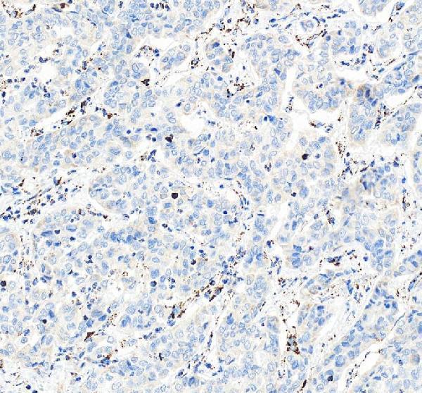
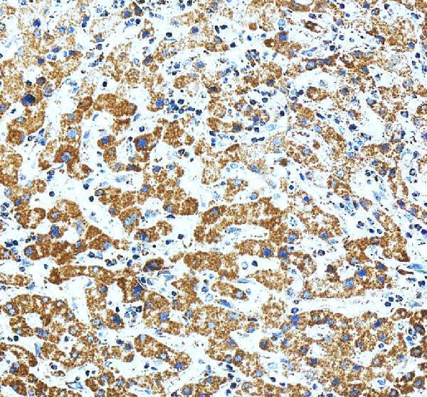
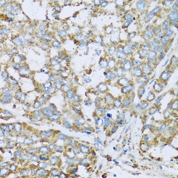
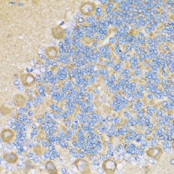


Comprehensive Human Protein Atlas IHC scoring per tissue (reliability: Approved — Pending external verification. Caution, targets protein from more than one gene. Pending external verification.). Rows are taken directly from the HPA tissue chart — click any row's HPA link to view the source.
| Tissue | Cell type | Level | Evidence | Source |
|---|---|---|---|---|
| Adrenal gland | Glandular cells | High | Protein (IHC) | HPA → |
| Appendix | Glandular cells | High | Protein (IHC) | HPA → |
| Caudate | Neuronal cells | High | Protein (IHC) | HPA → |
| Cerebral cortex | Neuropil | High | Protein (IHC) | HPA → |
| Colon | Glandular cells | High | Protein (IHC) | HPA → |
| Tissue | Cell type | Level | Evidence | Source |
|---|---|---|---|---|
| Adipose tissue | Adipocytes | Not detected | Protein (IHC) | HPA → |
| Bone marrow | Hematopoietic cells | Not detected | Protein (IHC) | HPA → |
| Ovary | Follicle cells | Not detected | Protein (IHC) | HPA → |
| Placenta | Decidual cells | Not detected | Protein (IHC) | HPA → |
| Skeletal muscle | Myocytes | Not detected | Protein (IHC) | HPA → |
Troubleshoot COX6A1 staining in paraffin sections by checking retrieval, compartmental pattern, controls and scoring before interpreting chromogenic signal.
Both anti-COX6A1 antibodies have paraffin-section IHC images (catalog IHC captions), list IF/ICC applications (catalog applications), and list human, mouse and rat reactivity (catalog reactivity).
A09572-1 will render with an IHC image from paraffin-embedded human liver cancer tissue; another caption documents human liver tissue (A09572-1 IHC captions). A09572 will render with an IHC image from paraffin-embedded rat cerebellum; its other captions document human liver cancer and mouse liver (A09572 IHC captions).
Which to pick: For tissue IHC using an EDTA pH 8.0 retrieval and DAB example, choose A09572-1 (A09572-1 IHC caption); the fixative is unreported (A09572-1 IHC caption). For IF/ICC, either SKU is listed for both applications, and both are rabbit polyclonals with human, mouse and rat reactivity (catalog applications, dilution_raw and reactivity); no IF image is supplied for either (catalog IF image alts). For cross-species tissue IHC, choose A09572 because its paraffin-section captions cover rat, human and mouse samples (A09572 IHC captions); the fixative is unreported (A09572 IHC captions).