CPNE8 / Copine-8 · IHC design guide

Design Immunohistochemistry for CPNE8

Plan CPNE8 paraffin IHC around cytoplasmic and membranous tissue staining (HPA tissue IHC). Kidney tubules provide a high-staining reference (HPA tissue IHC); titrate the catalog antibody at 1:100–1:300 (datasheet).

Evidence assembled Oct 2026 · For research use; verify linked source records and product datasheet before use
Immunohistochemistry protocol sheet for CPNE8 (IHC for CPNE8): expected localisation Cytoplasmic and membranous tissue staining (HPA tissue IHC), antibody A15136-2, validated IHC image, and IHC protocol steps
Printable CPNE8 IHC protocol sheet — expected localisation Cytoplasmic and membranous tissue staining (HPA tissue IHC), antibody A15136-2, controls and protocol steps. Open the full CPNE8 IHC guide →

CPNE8 Immunohistochemistry Experimental Design Guide

Expected localisation, validated protocols, controls and antibodies — the at-a-glance facts below, then the full design guide.

Must know before staining
Expected localisation Cytoplasmic and membranous tissue staining (HPA tissue IHC)
Staining pattern High in kidney tubules; cytoplasmic/membranous tissue pattern (HPA tissue IHC)
Antigen retrieval Citrate pH 6.0 HIER, 95–98 °C, 20 min (rule: cytoplasmic / membrane antigen)
Positive control ⓘ Adipose tissue+4 more · see all
Negative control ⓘ Caudate+2 more · see all
Important caveats
Reasons your staining may differ from the expected pattern.
Fixation Keep formalin fixation consistent across sections (standard IHC practice; not target-specific)
Caveat Staining shows medium consistency with RNA data (HPA tissue IHC)
Regulation Expression regulation unreported (UniProt)
Isoform / epitope 2 isoforms; epitope coverage unreported (UniProt)
Section 1

Recommended CPNE8 IHC & IF Protocols

Compare the catalog antibody’s IHC-P protocol with the published CPNE8 xenograft tumor IHC protocol (PMC9379401: IHC methods).

Recommended immunohistochemistry (IHC-P) protocol parameters
SampleParaffin-embedded human lung carcinoma tissue; fixative not specified (datasheet A15136-2)
FixationImage fixative and duration unreported (datasheet A15136-2); verify before use.
Sectioning4–5 µm sections on charged slides (standard)
DeparaffinisationXylene, graded ethanol series to water (standard)
Antigen retrievalHeat-induced epitope retrieval in citrate buffer, pH 6.0, 20 min at 95–98 °C (standard rule: cytoplasmic / membrane antigen)
Peroxidase block3% H2O2, 10 min, room temperature (standard)
Blocking10% normal serum of the secondary host, 30 min, room temperature (standard)
Primary antibodyRabbit anti-CPNE8, 1:100-1:300 (datasheet A15136-2)
Primary incubationOvernight at 4 °C (standard)
DetectionHRP-polymer secondary, DAB chromogen 5–10 min (standard)
CounterstainHematoxylin, blue, dehydrate and mount (standard)
Expected resultCPNE8-positive staining in adipocytes of adipose tissue (HPA tissue IHC: High). HPA tissue profile: Cytoplasmic and membranous expression in several tissues. No signal in the no-primary control.
💡Decision noteStart with citrate pH 6.0 HIER at 95–98 °C for 20 min (page antigen-retrieval rule); the article reports antigen repair without conditions (PMC9379401: IHC methods).
Section 2

What Is the Expected CPNE8 Staining Pattern?

In paraffin-section IHC, expect CPNE8 staining in the cytoplasm and along cell membranes, particularly in kidney tubule cells, hepatocytes, adipocytes and cardiomyocytes (HPA: cytoplasmic and membranous expression; High in these cells). UniProt reports no signal peptide or transmembrane segment (UniProt Q86YQ8 topology). HPA rates its tissue staining Approved, with medium consistency between antibody staining and RNA expression; interpret unexpected patterns cautiously (HPA: reliability).

What am I looking at on my slide?
Distinct cytoplasmic and membranous staining in kidney tubule cells or hepatocytes.This matches the reported compartment and High staining in those cells (HPA: tissue IHC). Compare intensity within the same run; HPA levels describe observed tissue patterns, not a required numerical staining score.
Predominantly nuclear staining, with little cytoplasmic or membranous signal.A nuclear-only pattern differs from the reported tissue pattern (HPA: cytoplasmic and membranous expression). Check staining controls and image interpretation before assigning it to CPNE8; the supplied sources do not establish a nuclear location.
Strong signal in tonsil germinal center cells, while expected positive cells are unstained.Those cells are listed as Not detected (HPA: tonsil germinal center cells). Cross-reactivity or endogenous detection activity is possible; use a no-primary control and review where staining sits within the tissue (general IHC practice).
Diffuse color covers cells and surrounding tissue without clear compartment boundaries.That pattern cannot reliably establish the reported cytoplasmic and membranous localization (HPA: tissue IHC). Assess the no-primary control, blocking and wash conditions to distinguish background from cell-associated signal (general IHC practice).
No staining in kidney tubule cells on a section expected to be positive.This conflicts with their High staining designation (HPA: kidney cells in tubules). First check section quality and detection controls, then review retrieval and antibody conditions (general IHC practice); absence alone does not establish biological loss.
💡Expected CPNE8 appearanceCall a section positive when identifiable cells show cytoplasmic and membranous signal, with conspicuous staining in a reported High population such as kidney tubule cells (HPA: tissue IHC); nuclear-only or tissue-wide haze is an unexpected pattern (HPA: cytoplasmic and membranous expression).
How each factor affects the staining
Tissue and cell selectionHigh staining is reported in adipocytes, adrenal and breast glandular cells, cardiomyocytes, kidney tubule cells, hepatocytes, respiratory epithelial cells and pancreatic exocrine glandular cells (HPA: tissue IHC). Select a named cell population when comparing slides.
Reference for low or absent stainingHPA lists low staining in cerebral cortex endothelial cells, splenic red-pulp cells and lymph-node non-germinal-center cells; caudate glia, ovarian stroma and tonsil germinal centers are Not detected (HPA: tissue IHC). These are cell-specific comparisons, not whole-organ negatives.
Location and protein topologyThe observed cytoplasmic and membranous pattern is a tissue-IHC finding (HPA: tissue IHC). CPNE8 has no annotated transmembrane segment or signal peptide (UniProt Q86YQ8 topology); membrane-adjacent staining does not establish membrane insertion.
Antibody evidence and isoformsThe listed antibody has IHC status Approved, and the tissue profile has medium antibody-to-RNA consistency (HPA: antibody and tissue IHC). UniProt lists two isoforms (UniProt Q86YQ8 isoforms); epitope location and isoform recognition are not supplied.
IF/ICC: is the IHC location confirmed in cells?HPA supplies no ICC-IF image set or main subcellular location for CPNE8 (HPA: subcellular record). Treat the tissue-IHC pattern as a starting expectation, not an independently confirmed IF/ICC localization.
Why is my staining missing, weak or wrong?
SituationLikely causeNext action
Known positive tissue gives no visible signal.Possible staining-run failure; kidney tubule cells are reported High (HPA: tissue IHC).Confirm the detection control and tissue integrity, then review the antibody conditions and antigen retrieval used in the run (general IHC practice). No CPNE8-specific retrieval setting is supplied.
No-primary control develops color.Signal may arise from endogenous detection activity or the detection reagents (general IHC practice).Review the applicable endogenous-activity block and detection controls before interpreting CPNE8 staining (general IHC practice).
The whole section has diffuse color.Diffuse background can obscure the expected cell-associated pattern (HPA: cytoplasmic and membranous expression).Check blocking, washes and detection exposure against the no-primary control (general IHC practice). Score only signal with clear cellular boundaries.
Strong signal is confined to nuclei.This differs from the reported cytoplasmic and membranous pattern (HPA: tissue IHC).Verify the counterstain and detection controls, then compare a named High cell population (HPA: tissue IHC; general IHC practice). Do not assign nuclear localization from this result alone.
A presumed negative organ contains stained cells.HPA's Not detected entries refer to specified cells, such as tonsil germinal center cells, not every cell in that organ (HPA: tissue IHC).Identify the stained cell type before judging specificity; compare like cell populations on positive and negative sections (general IHC practice).
Staining intensity varies across tissues.HPA reports High, low and Not detected staining in different named cell populations, while rating overall tissue-IHC consistency as medium (HPA: tissue IHC).Record cell type, compartment and intensity separately. Compare sections processed in the same run and avoid treating intensity differences alone as evidence of altered CPNE8 expression (general IHC practice).

Sample controls for CPNE8 IHC & IF

🧪Start with kidney: cells in tubules should stain (HPA: High in kidney cells in tubules). Run ovarian stroma as a negative tissue (HPA: Not detected in ovarian stroma cells); on the kidney slide, use non-tubular cells to assess background without assuming they are CPNE8-negative (HPA: kidney result specifies cells in tubules).
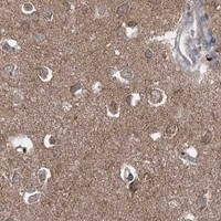
Positive control tissue: Adipose tissue (Adipocytes, HPA High)
Negative control tissue: Caudate (HPA Not detected)
ICC-IF cell lines (HPA subcellular resource): HPA carries no ICC-IF cell line for CPNE8; derive a cell-line control from the positive tissue's cell type (Adipocytes) and confirm it by RNA or western blot first.
Technical controls: Include a no-primary, secondary-only control and an isotype control matched to the primary antibody’s host species and, for a monoclonal antibody, its subclass; use a CPNE8 knockout or peptide-block control for biological specificity (selected A15136-2 tissue-IHC caption: peptide block). For kidney chromogenic IHC, block endogenous peroxidase and assess endogenous biotin if using avidin-biotin detection (standard IHC practice).
⚠️Feasibility: A CPNE8-specific fixation window and antigen-retrieval dependency are unreported; the A15136-2 paraffin-section caption does not state the fixative (selected SKU tissue-IHC caption). The supplied evidence does not establish whether frozen sections or IF would be easier, and HPA supplies no ICC-IF images or assigned subcellular location (HPA subcellular). In kidney, endogenous biotin can cause background with avidin-biotin detection (standard IHC practice).

HPA tissue IHC evidence for CPNE8

Comprehensive Human Protein Atlas IHC scoring per tissue (reliability: Approved — Medium consistency between antibody staining and RNA expression data.). Rows are taken directly from the HPA tissue chart — click any row's HPA link to view the source.

Positive expression · recommended positive controls

TissueCell typeLevelEvidenceSource
Adipose tissue Adipocytes High Protein (IHC) HPA →
Adrenal gland Glandular cells High Protein (IHC) HPA →
Breast Glandular cells High Protein (IHC) HPA →
Heart muscle Cardiomyocytes High Protein (IHC) HPA →
Kidney Cells in tubules High Protein (IHC) HPA →

Undetected expression · recommended negative controls

TissueCell typeLevelEvidenceSource
Caudate Glial cells Not detected Protein (IHC) HPA →
Ovary Ovarian stroma cells Not detected Protein (IHC) HPA →
Tonsil Germinal center cells Not detected Protein (IHC) HPA →
Section 3

Advanced CPNE8 IHC Tips

Troubleshoot CPNE8 staining in paraffin sections by checking retrieval, controls, cell identity and compartment before comparing staining intensity.

How should I adjust retrieval when CPNE8 staining is weak or uneven?
Start with citrate buffer at pH 6.0 for heat-induced antigen retrieval at 95–98 °C for 20 min (page retrieval rule). Keep section thickness, cooling time and antibody incubation consistent while comparing a positive-control section with the test section (standard IHC practice; HPA: high in kidney tubule cells). If signal remains weak, test a more alkaline retrieval buffer on adjacent sections as a fallback, watching for tissue damage or increased background (standard IHC practice). Score cytoplasmic and membranous staining separately so a retrieval change does not appear successful merely because nonspecific surface staining increased (HPA: cytoplasmic and membranous expression; standard IHC practice).
Could fixation explain weak CPNE8 staining in paraffin sections?
CPNE8-specific sensitivity to fixation is unknown because no target-specific fixation evidence is supplied, and the selected product caption states only that the human lung carcinoma section was paraffin embedded (caption: A15136-2). Record the actual fixative and fixation duration for each specimen, then compare sections processed under matched conditions before changing retrieval or antibody concentration (standard IHC practice). Include a consistently processed positive-control tissue, such as kidney with high staining in tubule cells, to distinguish a run-wide failure from a specimen issue (HPA: high in kidney tubule cells; standard IHC practice). Treat poor morphology and diffuse staining as reasons to inspect processing and detection controls, without assigning them a CPNE8-specific fixation mechanism (standard IHC practice).
Which compartments should show credible CPNE8 staining?
Assess cytoplasmic and membranous staining in the relevant cell population, since that is the reported tissue-IHC pattern (HPA: cytoplasmic and membranous expression in several tissues). CPNE8 has no annotated transmembrane segment, so membranous staining should be interpreted as a staining pattern rather than proof that it spans a membrane (UniProt Q86YQ8 topology; HPA: tissue-IHC profile). Its probable calcium-dependent phospholipid binding offers a biological context for that pattern but does not establish a fixed subcellular location in every specimen (UniProt Q86YQ8 function; UniProt Q86YQ8: subcellular location not annotated). Compare adjacent sections and cell morphology before accepting isolated nuclear or extracellular signal as specific (standard IHC practice; HPA: tissue-IHC profile).
Can isoforms or epitope accessibility change the CPNE8 staining pattern?
CPNE8 has 2 listed isoforms, but the supplied product caption does not identify the antibody epitope or establish isoform recognition (UniProt Q86YQ8: isoforms 1 and 2; caption: A15136-2). Map any documented immunogen against both isoform sequences before using staining differences to infer isoform expression (standard antibody-validation practice; UniProt Q86YQ8: alternative splicing). The protein contains C2 domains at residues 1–133 and 142–265, plus a VWFA domain at 309–510; phosphoserine 260 is listed (UniProt Q86YQ8 domains and modified residues). If epitope information remains unavailable, compare retrieval conditions and use independent validation instead of assigning a negative section to a particular isoform or modification (standard IHC practice).
How can I check CPNE8 localisation by multiplex IF?
Pair CPNE8 with a validated marker for the expected cell type, such as a tubule-cell marker when examining kidney, and confirm overlap at the single-cell level (HPA: high in kidney tubule cells; standard IF practice). Choose fluorophores after inspecting unstained tissue for autofluorescence, reserving a channel with low tissue background for the weaker signal (standard IF practice). Because tissue IHC reports cytoplasmic and membranous CPNE8 while UniProt lists no transmembrane segment, test mild permeabilisation for access to intracellular epitopes and compare it with an unpermeabilised control (HPA: tissue-IHC profile; UniProt Q86YQ8 topology; standard IF practice). Use single-label and secondary-only controls to check bleed-through and nonspecific fluorescence before interpreting colocalisation (standard IF practice).
What should I change when CPNE8 IHC has diffuse background?
Inspect a no-primary control and the section margins to distinguish detection-system background from a broadly distributed target signal (standard IHC practice). Check endogenous peroxidase blocking before chromogenic development, and shorten development or titrate the primary antibody if background rises across both expected and unexpected cell populations (standard chromogenic IHC practice). The selected paraffin-section image includes a synthesized-peptide blocking comparison, which supports an antibody-specific check for that image but does not establish specificity in every tissue (caption: A15136-2). Compare the test section with an HPA-reported low or undetected cell population, such as ovarian stroma cells, while preserving the same processing and exposure conditions (HPA: not detected in ovarian stroma cells; standard IHC practice).
How should I quantify CPNE8 across differently composed tissue sections? ⚠ ANSWER MARKED FOR VERIFICATION
Define the cell population and compartment before scoring; HPA reports cytoplasmic and membranous staining in several tissues and high staining in kidney tubule cells (HPA: tissue-IHC profile; HPA: kidney tubule cells high). For a cell-based comparison, report the percentage of positive cells and an H-score using intensity categories 0–3, with the scoring rules fixed before reviewing groups (standard IHC scoring practice). When architecture makes cell counts unreliable, measure positive area or cell density per mm² within annotated viable regions (standard image-analysis practice). Normalise to the area or number of eligible cells in that same compartment, and compare batches using a consistently processed reference section (standard IHC practice).
How do I distinguish true CPNE8 staining from artefact?
A credible result combines appropriate cell identity with cytoplasmic or membranous staining, rather than relying on staining intensity alone (HPA: tissue-IHC profile; standard IHC interpretation). High staining in hepatocytes or kidney tubule cells is consistent with reported tissue patterns, whereas staining confined to cells reported as undetected, such as tonsil germinal center cells, warrants additional checks (HPA: high in hepatocytes and kidney tubule cells; HPA: not detected in tonsil germinal center cells). Examine whether signal follows cut edges, damaged or necrotic regions, or areas with strong no-primary staining (standard IHC practice). Review the peroxidase-block control and peptide-blocking comparison where available, while treating the latter as evidence limited to the selected paraffin-section image (standard chromogenic IHC practice; caption: A15136-2).
Boster reagents

Best CPNE8 / Copine-8 IHC Antibodies

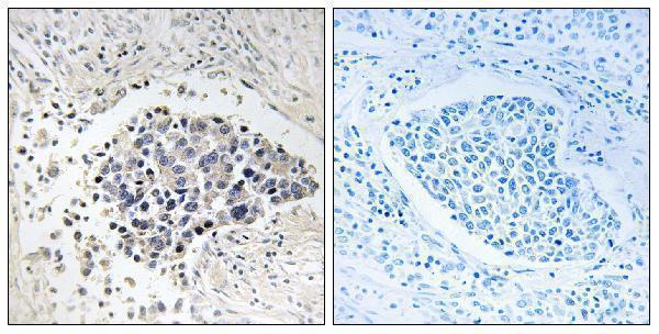
A15136-2 has a human lung carcinoma paraffin-section IHC image; IF is listed without an image (catalog: IHC image caption; applications). Catalog reactivity includes human, mouse, and rat (catalog: reactivity).

Real IHC data Immunohistochemistry analysis of paraffin-embedded human lung carcinoma tissue, using CPNE8 Antibody. The picture on the right is blocked with the synthesized peptide.
Anti-Copine 8 CPNE8 Antibody
Cat # A15136-2

A15136-2 will render with an IHC image of paraffin-embedded human lung carcinoma tissue, including a peptide-blocked comparison (catalog: IHC image caption). The catalog lists IHC and IF applications and human, mouse, and rat reactivity, but provides no IF image (catalog: applications/reactivity; IF image alts).

Which to pick: Choose A15136-2 for paraffin-section tissue IHC because its own image shows staining in paraffin-embedded human lung carcinoma tissue; the fixative is unreported (catalog: IHC image caption). For IF, A15136-2 is listed at 1:50, but ICC is not separately listed and no IF image is supplied (catalog: applications/IF dilution; IF image alts). A15136-2 is also the listed cross-species option for human, mouse, and rat; its image documents human tissue only (catalog: reactivity; IHC image caption).

Each figure is that product's own IHC / IF validation image from its datasheet.