CPS1 / Carbamoyl-phosphate synthase [ammonia], mitochondrial · IHC design guide

Design Immunohistochemistry for CPS1

Plan CPS1 chromogenic IHC in paraffin sections using liver hepatocytes as a positive tissue reference (HPA tissue IHC). This guide covers staining pattern, fixation consistency, controls, and interpretation.

Evidence assembled Oct 2026 · For research use; verify linked source records and product datasheet before use
Immunohistochemistry protocol sheet for CPS1 (IHC for CPS1): expected localisation Granular cytoplasm in hepatocytes (HPA tissue IHC), antibody A01320-2, validated IHC image, and IHC protocol steps
Printable CPS1 IHC protocol sheet — expected localisation Granular cytoplasm in hepatocytes (HPA tissue IHC), antibody A01320-2, controls and protocol steps. Open the full CPS1 IHC guide →

CPS1 Immunohistochemistry Experimental Design Guide

Expected localisation, validated protocols, controls and antibodies — the at-a-glance facts below, then the full design guide.

Must know before staining
Expected localisation Granular cytoplasm in hepatocytes (HPA tissue IHC)
Staining pattern Granular cytoplasm in hepatocytes and intestinal endocrine cells (HPA tissue IHC)
Antigen retrieval EDTA pH 8.0 HIER, heat-mediated (datasheet A01320-2)
Positive control ⓘ Duodenum+2 more · see all
Negative control ⓘ Adipose tissue+4 more · see all
Important caveats
Reasons your staining may differ from the expected pattern.
Fixation Matched tissue-IHC evidence does not establish the fixation claim. Validate the specimen-specific method before use. (selected-SKU IHC image A01320-2)
Caveat Liver peroxidase may add DAB background (standard IHC practice)
Regulation Liver-enriched expression (HPA tissue IHC)
Isoform / epitope 3 isoforms; epitope coverage is unspecified (UniProt)
Section 1

Recommended CPS1 IHC & IF Protocols

The catalog antibody’s IHC-P protocol uses heat-mediated EDTA pH 8.0 retrieval (datasheet A01320-2). The published gastric and liver IHC protocols below provide additional conditions (PMC8045184; PMC8888708).

Recommended immunohistochemistry (IHC-P) protocol parameters
SampleParaffin-embedded human liver cancer tissue; fixative not specified (datasheet A01320-2)
FixationImage fixative and duration unreported (datasheet A01320-2); verify before use.
Sectioning4–5 µm sections on charged slides (standard)
DeparaffinisationXylene, graded ethanol series to water (standard)
Antigen retrievalHeat retrieval: EDTA pH 8.0 (datasheet A01320-2); 20 min, 95–100 °C (standard)
Peroxidase block3% H2O2, 10 min, room temperature (standard)
Blocking10% goat serum (datasheet A01320-2)
Primary antibodyRabbit anti-CPS1, 2-5 μg/ml (datasheet A01320-2)
Primary incubationOvernight at 4 °C (datasheet A01320-2)
DetectionStreptavidin-biotin complex (SABC), DAB chromogen (datasheet A01320-2)
CounterstainHematoxylin, blue, dehydrate and mount (standard)
Expected resultCPS1-positive staining in endocrine cells of duodenum (HPA tissue IHC: High). HPA tissue profile: Selective granular cytoplasmic expression in small intestine and hepatocytes. No signal in the no-primary control.
💡Decision noteStart with heat-mediated EDTA pH 8.0 for the catalog antibody (datasheet A01320-2); citrate retrieval is a published alternative for gastric tissue (PMC8045184).
Section 2

What Is the Expected CPS1 Staining Pattern?

CPS1 is mainly mitochondrial, with nucleolar and hepatocyte cell-surface localization also recorded (UniProt P31327). In paraffin-section IHC, expect selective granular cytoplasmic staining in hepatocytes and small-intestinal endocrine cells (HPA tissue IHC: Enhanced). CPS1 has no transmembrane segment (UniProt P31327 topology). Interpret the slide by cell type and staining pattern together, since the recorded additional locations alone do not define the expected tissue-IHC result (HPA tissue IHC; UniProt P31327).

What am I looking at on my slide?
Strong granular cytoplasmic staining in hepatocytes, with limited staining in other liver cells.This fits the reported high hepatocyte signal and selective granular cytoplasmic profile (HPA tissue IHC: Liver, High). Score the hepatocyte compartment rather than treating all brown material in the section as CPS1 (general IHC practice).
Granular cytoplasmic staining in a restricted population of small-intestinal or duodenal endocrine cells.This matches the reported high signal in endocrine cells at both sites (HPA tissue IHC: Small intestine and Duodenum, High). A patchy distribution by cell type can therefore be expected; absence of staining from every epithelial cell does not, by itself, invalidate the run (HPA tissue IHC).
Predominantly extracellular, stromal, or uniformly nuclear staining replaces the expected granular cytoplasmic pattern.Treat this as a pattern mismatch and investigate artefact or nonspecific signal (HPA tissue IHC: selective granular cytoplasm; general IHC practice). Do not automatically reject a discrete nucleolar or hepatocyte-surface signal: those locations are recorded separately, without establishing the dominant paraffin-IHC pattern (UniProt P31327; HPA subcellular ICC-IF).
Comparable staining appears in an unexpected cell population or in a documented negative cell type.Consider antibody cross-reactivity or endogenous detection activity, then compare with a no-primary control (general IHC practice). HPA reports no detection in adipocytes of adipose tissue and in several other listed cell types; these are cell-specific observations, not proof that every cell in those tissues is negative (HPA tissue IHC).
Diffuse haze covers positive and negative areas, or a known-positive hepatocyte section shows no signal.Haze that obscures cellular boundaries cannot establish CPS1 localization; review blocking, washing and detection controls (general IHC practice). A blank hepatocyte control conflicts with the high liver signal reported by HPA, so assess the run before calling the test specimen negative (HPA tissue IHC: Liver, High).
💡Expected CPS1 appearanceCall a convincing positive result when high, granular cytoplasmic signal marks hepatocytes or the specified intestinal endocrine cells (HPA tissue IHC); widespread diffuse or mismatched-cell staining needs control-based review for false positivity (general IHC practice).
How each factor affects the staining
Choice of positive and negative cell populationsHepatocytes and duodenal or small-intestinal endocrine cells provide reported high-signal populations (HPA tissue IHC). Use a documented negative cell type as a comparison, while evaluating each tissue by its named cell population (HPA tissue IHC).
Compartment used for scoringThe tissue-IHC profile is granular cytoplasmic (HPA tissue IHC). UniProt also records nucleolar and hepatocyte-surface localization, while HPA ICC-IF supports nucleoli and the nucleolar rim; these observations warrant compartment-specific review rather than a blanket nuclear-positive IHC call (UniProt P31327; HPA subcellular ICC-IF).
Antibody evidence and unknown epitope coverageTwo listed antibodies have Enhanced IHC validation (HPA antibodies: HPA021400, CAB003781). CPS1 has three isoforms and a mature chain spanning residues 39–1500 (UniProt P31327); the supplied evidence does not identify either antibody’s epitope, so isoform coverage cannot be inferred.
Detection chemistryEndogenous activity or nonspecific binding can produce chromogen independently of target recognition (general IHC practice). A no-primary control helps identify detection-related signal, but it cannot alone establish antibody specificity (general IHC practice).
Why is my staining missing, weak or wrong?
SituationLikely causeNext action
No granular signal in a hepatocyte positive control.The result conflicts with reported high hepatocyte staining (HPA tissue IHC: Liver, High); the failed step is undetermined (general IHC practice).Check tissue identity, reagent activity and control performance; review retrieval and primary-antibody conditions as general IHC steps (general IHC practice). No CPS1-specific fixation or retrieval sensitivity is established by the supplied evidence.
Intestinal section appears negative at first glance.The reported high intestinal signal is assigned to endocrine cells, rather than every epithelial cell (HPA tissue IHC: Small intestine and Duodenum, High).Examine the appropriate cell population and confirm that a hepatocyte positive control works before interpreting the intestinal result (HPA tissue IHC; general IHC practice).
Broad brown staining appears in nominally negative cells.Cross-reactivity, endogenous detection activity or excess background are possible explanations (general IHC practice); HPA's negative calls apply to specified cell types (HPA tissue IHC).Compare a no-primary control, inspect cell boundaries and reassess blocking, washing and detection conditions (general IHC practice). Do not extend a cell-specific HPA negative call to an entire organ (HPA tissue IHC).
Signal is mainly nuclear or extracellular on the paraffin slide.This departs from HPA's selective granular cytoplasmic tissue-IHC profile, although nucleolar and hepatocyte-surface locations are recorded elsewhere (HPA tissue IHC; UniProt P31327).Check morphology and control sections for artefact; record the compartment separately instead of counting all such signal as the expected IHC pattern (general IHC practice; HPA tissue IHC).
Granular staining is obscured by diffuse background.Background may reflect nonspecific antibody or detection signal; its source cannot be assigned from the appearance alone (general IHC practice).Inspect a no-primary control and review blocking, washes and detection exposure, then rescore only clearly localized cellular signal (general IHC practice).
Q: IF/ICC shows nucleolar staining; should it be scored as the paraffin-IHC pattern?A: HPA supports nucleoli and the nucleolar rim in ICC-IF, while tissue IHC reports selective granular cytoplasm (HPA subcellular ICC-IF; HPA tissue IHC).Interpret IF/ICC on its own guide page and assess the paraffin slide against its cell-type and cytoplasmic criteria (HPA subcellular ICC-IF; HPA tissue IHC).

Sample controls for CPS1 IHC & IF

🧪Run liver first: hepatocytes should show strong CPS1 staining (HPA: High in liver hepatocytes). Run adipose tissue as the negative tissue (HPA: Not detected in adipocytes); on the liver slide, use non-hepatocyte cells to assess background and investigate any strong signal there before treating it as specific.
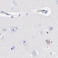
Positive control tissue: Duodenum (Endocrine cells, HPA High)
Negative control tissue: Adipose tissue (HPA Not detected)
ICC-IF cell lines (HPA subcellular resource): HPA ICC-IF images show CPS1 in A-431, U-251MG, U2OS, Sperm, with annotated localisation: Nucleoli (supported), Nucleoli rim (supported) (HPA subcellular).
Technical controls: Include a no-primary, secondary-only control; an isotype control matched to the primary antibody’s rabbit host and clonality; and, if available, a CPS1 knockout specimen or immunizing-peptide block as a biological specificity control (caption: rabbit anti-CPS1; standard IHC practice). For liver sections, block endogenous peroxidase and account for endogenous biotin when using the caption’s biotin-based detection system (caption: biotinylated secondary and streptavidin–biotin complex; standard IHC practice).
⚠️Feasibility: A target-specific fixation window or fixation effect is unreported, and the selected A01320-2 tissue-IHC caption does not state the fixative (caption: fixative not stated). The demonstrated paraffin-section procedure used heat retrieval in EDTA at pH 8.0; this supports trying that retrieval condition but does not establish that retrieval is required (caption: EDTA pH 8.0). The supplied evidence does not establish whether frozen sections or IF are easier; for the demonstrated liver IHC workflow, endogenous biotin can complicate biotin-based detection (caption: liver tissue and biotin-based detection; standard IHC practice).

HPA tissue IHC evidence for CPS1

Comprehensive Human Protein Atlas IHC scoring per tissue (reliability: Enhanced — High consistency between antibody staining and RNA expression data.). Rows are taken directly from the HPA tissue chart — click any row's HPA link to view the source.

Positive expression · recommended positive controls

TissueCell typeLevelEvidenceSource
Duodenum Endocrine cells High Protein (IHC) HPA →
Liver Hepatocytes High Protein (IHC) HPA →
Small intestine Endocrine cells High Protein (IHC) HPA →

Undetected expression · recommended negative controls

TissueCell typeLevelEvidenceSource
Adipose tissue Adipocytes Not detected Protein (IHC) HPA →
Adrenal gland Glandular cells Not detected Protein (IHC) HPA →
Appendix Endocrine cells Not detected Protein (IHC) HPA →
Bone marrow Hematopoietic cells Not detected Protein (IHC) HPA →
Breast Adipocytes Not detected Protein (IHC) HPA →
Section 3

Advanced CPS1 IHC Tips

Use the paraffin-section IHC method as the starting point, then assess staining against the tissue pattern and appropriate controls (datasheet A01320-2; HPA tissue IHC).

Which retrieval conditions should I try first when CPS1 staining is weak?
Start with heat-mediated retrieval in EDTA at pH 8.0 for paraffin sections (datasheet A01320-2). The selected image caption gives no heating time, so compare 10 and 20 minutes on adjacent sections while keeping detection conditions identical (datasheet A01320-2; standard IHC practice). Include liver as a positive control and inspect hepatocyte staining for a granular cytoplasmic pattern before increasing retrieval intensity (HPA tissue IHC; standard IHC practice). If signal remains weak, assess a different retrieval buffer only as a fallback and record any gain alongside changes in tissue integrity or background (standard IHC practice).
Could fixation explain inconsistent CPS1 staining between paraffin blocks?
Target-specific fixation sensitivity is unknown because the selected paraffin-section caption does not state its fixative or fixation duration (datasheet A01320-2). Record the fixative and processing history for each block, and compare adjacent sections from blocks processed similarly before attributing differences to CPS1 expression (standard IHC practice). Keep retrieval at EDTA pH 8.0 and the primary incubation at 2 μg/ml overnight at 4°C during that comparison (datasheet A01320-2). Use hepatocyte staining and section morphology to judge whether a weak block warrants a controlled retrieval comparison; neither pattern alone identifies the cause (HPA tissue IHC; standard IHC practice).
Where should convincing CPS1 chromogenic staining appear?
Look first for selective, granular cytoplasmic staining in hepatocytes and small-intestinal cells, consistent with the tissue IHC profile and mitochondrial localisation (HPA tissue IHC; UniProt P31327). Score the labelled cells and their compartments separately, because a diffuse wash across tissue does not reproduce that reported pattern (HPA tissue IHC; standard IHC practice). UniProt also records nucleolar and hepatocyte cell-surface localisation, although the protein has no transmembrane segment (UniProt P31327). If nuclear or membrane-associated signal appears, compare it with the granular cytoplasmic signal, an appropriate negative control, and cell morphology before assigning it to CPS1 (standard IHC practice).
Could epitope placement or CPS1 processing change the IHC pattern?
CPS1 has 3 listed isoforms, while the antibody epitope is not specified in the supplied caption (UniProt P31327; datasheet A01320-2). The annotated mature chain spans residues 39–1500, so an epitope near the precursor’s beginning could behave differently from one within that chain (UniProt P31327; standard IHC practice). Annotated glycosylation sites include residues 537, 1331, and 1332, alongside multiple modified lysines; their effects on this antibody’s staining are unreported (UniProt P31327; datasheet A01320-2). If sections disagree, obtain the immunogen or epitope information and compare independently validated reagents before claiming isoform-specific staining (standard IHC practice).
How can I investigate a CPS1 IHC pattern with multiplex IF?
Treat IF as a separate validation experiment: the selected antibody evidence describes paraffin-section chromogenic IHC, while HPA reports nucleolar signals in its subcellular IF data (datasheet A01320-2; HPA subcellular). Pair CPS1 with a marker identifying the expected hepatocyte or small-intestinal cell population, and assess whether the signals occur in the same cells (HPA tissue IHC; standard IF practice). Select spectrally separated fluorophores and inspect unstained tissue for autofluorescence before choosing the CPS1 channel (standard IF practice). For a mitochondrial or nucleolar epitope, permeabilise fixed cells sufficiently to reach that intracellular compartment, then check morphology and background against a matched control (UniProt P31327; standard IF practice).
How should I troubleshoot diffuse brown staining around CPS1-positive cells?
Compare the stained section with a no-primary control to locate signal from detection chemistry or tissue rather than antibody binding (standard IHC practice). The selected workflow uses 10% goat serum, a biotinylated secondary antibody, a streptavidin–biotin complex, and DAB; those details identify useful points for control comparisons (datasheet A01320-2). Apply a peroxidase block before DAB detection and consider a biotin-free polymer system if the biotin-based workflow produces persistent background (standard IHC practice). Judge any adjustment against retained granular cytoplasmic staining in hepatocytes and preservation of section morphology, rather than against a lower overall brown intensity alone (HPA tissue IHC; standard IHC practice).
How can I compare CPS1 staining across liver sections reproducibly? ⚠ ANSWER MARKED FOR VERIFICATION
Define the region of interest and exclude folds, necrosis, and section edges before measuring CPS1 staining (standard IHC practice). Record the percentage of viable hepatocytes at each intensity from 0–3, then calculate an H-score from 0–300 using percentage times intensity (standard IHC practice). Normalise the result to the viable hepatocyte population or measured viable tissue area, and report the denominator consistently across sections (standard IHC practice). Keep retrieval and detection settings constant, and document whether the counted signal is granular cytoplasmic staining consistent with the tissue profile (datasheet A01320-2; HPA tissue IHC; standard IHC practice).
When is an unusual CPS1 staining pattern more likely to be artefact?
A credible positive result should align with cellular morphology and the reported selective granular cytoplasmic pattern in hepatocytes or small-intestinal cells (HPA tissue IHC; standard IHC practice). Treat staining concentrated at section edges, necrotic areas, or across unrelated cell types as suspect until a controlled repeat resolves it (standard IHC practice). Nucleolar or cell-surface signal deserves separate assessment because those localisations are annotated, but they do not by themselves establish specificity in chromogenic tissue sections (UniProt P31327; HPA subcellular; standard IHC practice). Compare a no-primary control and, where practical, an independent antibody while checking whether peroxidase or biotin-based detection contributes to the pattern (datasheet A01320-2; standard IHC practice).
Boster reagents

Best CPS1 / Carbamoyl-phosphate synthase [ammonia], mitochondrial IHC Antibodies

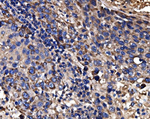
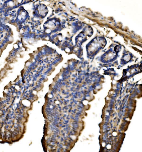
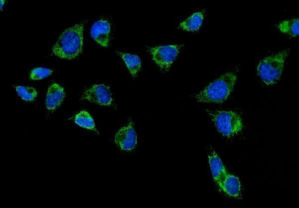
A01320-2 has IHC images from human liver cancer and mouse and rat colon paraffin sections, plus IF data from HeLa cells (A01320-2 image captions).

Real IHC data IHC analysis of CPS1 using anti-CPS1 antibody (A01320-2). CPS1 was detected in a paraffin-embedded section of human liver cancer tissue. Heat mediated antigen retrieval was performed in EDTA buffer (pH 8.0, epitope retrieval solution). The tissue section was blocked with 10% goat serum. The tissue section was then incubated with 2 μg/ml rabbit anti-CPS1 Antibody (A01320-2) overnight at 4°C. Biotinylated goat anti-rabbit IgG was used as secondary antibody and incubated for 30 minutes at 37°C. The tissue section was developed using Strepavidin-Biotin-Complex (SABC) (Catalog # SA1022) with DAB as the chromogen.
Anti-CPS1 Antibody ®
Cat # A01320-2

A01320-2 has pictured paraffin-section IHC in human liver cancer and mouse and rat colon, and IF/ICC in HeLa cells (A01320-2 image captions). M01320-1 lists IHC and IF/ICC for human, mouse and rat, but has no supplied IHC or IF image captions (M01320-1 catalog applications, reactivity and image alts).

Which to pick: Choose A01320-2 for tissue IHC: its paraffin-section captions report EDTA pH 8 retrieval and 2 μg/ml primary antibody; the fixative is unreported (A01320-2 IHC image captions). Choose A01320-2 when an IF/ICC image is useful; its HeLa-cell caption reports 5 μg/ml, while M01320-1 is a monoclonal option listed for IF/ICC without a supplied image (A01320-2 IF image caption; M01320-1 catalog clone, applications and image alts). For cross-species tissue IHC, A01320-2 has human, mouse and rat captions; monkey appears in its catalog reactivity but has no supplied IHC caption (A01320-2 IHC image captions and catalog reactivity).

Each figure is that product's own IHC / IF validation image from its datasheet.

References

  1. UniProt Consortium. UniProt entry P31327 (CPSM_HUMAN, Carbamoyl-phosphate synthase [ammonia], mitochondrial).
  2. Human Protein Atlas. CPS1 tissue IHC expression (reliability: Enhanced).
  3. Human Protein Atlas. CPS1 subcellular location (ICC-IF): Mainly localized to the nucleoli and nucleoli rim. In addition localized to the mid piece and principal piece..
  4. Human Protein Atlas. CPS1 antibody validation summary (2 antibodies).
  5. Aberrant expression and distribution of enzymes of the urea cycle and other ammonia metabolizing pathways in dogs with congenital portosystemic shunts. PloS one 2014 — PMC4063766.
  6. Expression profiling of CPS1 in Correa's cascade and its association with gastric cancer prognosis. Oncology letters 2021 — PMC8045184.
  7. A constitutive knockout of murine carbamoyl phosphate synthetase 1 results in death with marked hyperglutaminemia and hyperammonemia. Journal of inherited metabolic disease 2019 — PMC6728231.
  8. Liver injury in non-alcoholic fatty liver disease is associated with urea cycle enzyme dysregulation. Scientific reports 2022 — PMC8888708.
  9. PubMed PMID:1840546 — UniProt-cited evidence.
  10. PubMed PMID:9711878 — UniProt-cited evidence.
  11. PubMed PMID:12853138 — UniProt-cited evidence.