This website uses cookies to ensure you get the best experience on our website.


- Table of Contents
Plan CPSF6 chromogenic IHC around the ubiquitous nuclear staining observed in tissues (HPA tissue IHC). This guide covers consistent fixation, antibody titration at 2–5 μg/ml (datasheet A04551-1), and interpretation of possible nuclear–cytoplasmic shuttling (UniProt).

Expected localisation, validated protocols, controls and antibodies — the at-a-glance facts below, then the full design guide.
| Expected localisation | Nuclear staining observed; cytoplasmic shuttling reported (HPA tissue IHC; UniProt) | |
| Staining pattern | Ubiquitous nuclear staining across tissue cells (HPA tissue IHC) | |
| Antigen retrieval | EDTA pH 8.0 HIER, heat-mediated (datasheet A04551-1) | |
| Positive control | Adipose tissue+4 more · see all | |
| Negative control | None in HPA (detected in all 45 tissues); use no-primary + isotype controls |
| Fixation | Matched tissue-IHC evidence does not establish the fixation claim. Validate the specimen-specific method before use. (selected-SKU IHC image A04551-1) | |
| Caveat | Nuclear–cytoplasmic shuttling may alter the pattern (UniProt) | |
| Regulation | No expression regulator annotated (UniProt) | |
| Isoform / epitope | 3 isoforms; verify antibody epitope coverage (UniProt) |
The catalog antibody uses heat-mediated EDTA pH 8.0 retrieval (datasheet A04551-1). Published IHC protocols cover HCC and breast cancer tissue microarrays (PMC11234225; PMC5514498).
| Sample | Paraffin-embedded human liver cancer tissue; fixative not specified (datasheet A04551-1) |
| Fixation | Image fixative and duration unreported (datasheet A04551-1); verify before use. |
| Sectioning | 4–5 µm sections on charged slides (standard) |
| Deparaffinisation | Xylene, graded ethanol series to water (standard) |
| Antigen retrieval | Heat retrieval: EDTA pH 8.0 (datasheet A04551-1); 20 min, 95–100 °C (standard) |
| Peroxidase block | 3% H2O2, 10 min, room temperature (standard) |
| Blocking | 10% goat serum (datasheet A04551-1) |
| Primary antibody | Rabbit anti-CPSF6, 2-5 μg/ml (datasheet A04551-1) |
| Primary incubation | Overnight at 4 °C (datasheet A04551-1) |
| Detection | Streptavidin-biotin complex (SABC), DAB chromogen (datasheet A04551-1) |
| Counterstain | Hematoxylin, blue, dehydrate and mount (standard) |
| Expected result | CPSF6-positive staining in adipocytes of adipose tissue (HPA tissue IHC: High). HPA tissue profile: Ubiquitous nuclear expression. No signal in the no-primary control. |
CPSF6 should appear predominantly in nuclei, consistent with its nucleoplasmic and nuclear-speckle localization (UniProt Q16630; HPA: nucleoplasm and nuclear speckles). Expect staining across many cell types rather than a restricted tissue compartment (HPA: ubiquitous nuclear expression; low tissue specificity). HPA rates the tissue IHC pattern Supported, with medium consistency between antibody staining and RNA expression (HPA: tissue IHC). CPSF6 has no transmembrane segment (UniProt Q16630: topology).
| Nuclear staining in multiple cell types, with some variation between nuclei. | This fits the broad tissue pattern (HPA: ubiquitous nuclear expression). Compare identifiable cells within the same section: HPA reports High staining in adipocytes, respiratory epithelial cells, glial cells and bone-marrow hematopoietic cells (HPA: tissue IHC). These examples are useful positive references, not a required intensity for every cell. |
| Nuclei show uneven or punctate signal. | Nucleoplasmic and speckle-associated localization makes nuclear texture plausible (HPA: enhanced nucleoplasm and nuclear speckles in ICC-IF). UniProt also describes paraspeckle-associated puncta whose distribution varies with the cell cycle (UniProt Q16630: subcellular location). Chromogenic IHC may not resolve these structures; judge the overall nuclear compartment first. |
| Strong staining is mainly cytoplasmic or outlines cell membranes. | Predominantly cytoplasmic staining conflicts with the observed tissue IHC pattern (HPA: ubiquitous nuclear expression). CPSF6 can shuttle between nucleus and cytoplasm (UniProt Q16630: subcellular location), so isolated cytoplasmic signal is not automatically false; membrane outlining is unexpected for a protein without a transmembrane segment (UniProt Q16630: topology). |
| An isolated cell population stains while surrounding expected-positive nuclei do not. | Treat the apparent cell restriction cautiously: HPA describes ubiquitous nuclear expression and low tissue specificity (HPA: tissue IHC). Check morphology and controls for cross-reactivity or endogenous detection activity (general IHC practice). Do not label that population CPSF6-positive solely because it contains chromogen. |
| Diffuse color covers nuclei, cytoplasm and empty areas, or no nuclei stain in a positive-reference section. | Diffuse, compartment-independent color limits localization calls (general IHC practice). An absent signal in a section containing a documented High-staining cell population conflicts with the HPA reference pattern (HPA: tissue IHC); inspect controls and processing before interpreting it as biological loss. |
| Tissue reference and confidence | HPA reports ubiquitous nuclear expression and Low tissue specificity, with Supported IHC reliability and medium staining–RNA consistency (HPA: tissue IHC). Use its High-staining examples as references, while allowing biological and assay variation. |
| Subnuclear distribution | Nucleoplasm and nuclear speckles are enhanced in ICC-IF images (HPA: subcellular). UniProt also places CPSF6 in paraspeckle-associated puncta and notes cell-cycle variation (UniProt Q16630: subcellular location); these details may exceed chromogenic IHC resolution. |
| Isoforms and antibody recognition | UniProt lists three CPSF6 isoforms (UniProt Q16630: isoforms). The supplied sources do not map this section's IHC antibody epitope to an isoform; avoid interpreting cell-to-cell intensity differences as isoform changes without separate evidence. |
| Processing and membrane pattern | The annotated chain spans residues 1–551, with no signal peptide or propeptide and no transmembrane segment (UniProt Q16630: processing and topology). Those annotations offer no basis for expecting a shed extracellular or membrane-rim IHC pattern. |
| IF/ICC: what should I expect? | Expect nucleoplasmic signal and nuclear speckles (HPA: subcellular ICC-IF); HPA039973 has Enhanced ICC validation but Supported IHC validation (HPA: antibodies). IF/ICC staining conditions belong to its separate guide; ICC validation does not specify this section's IHC conditions. |
| Situation | Likely cause | Next action |
|---|---|---|
| Nuclear staining is weak throughout a section that contains an HPA High-staining cell population. | The result falls short of the reference pattern (HPA: tissue IHC); antibody performance or a general staining-step failure is possible (general IHC practice). | Check the positive-control section and reagent sequence; optimize retrieval and primary-antibody dilution using the antibody's IHC-P instructions (general IHC practice). No CPSF6-specific retrieval setting or dilution is supplied. |
| Chromogen appears broadly in cytoplasm or extracellular space. | The pattern obscures the reported nuclear distribution (HPA: tissue IHC) and may reflect nonspecific staining or detection background (general IHC practice). | Compare with a no-primary control; review blocking, washes and detection chemistry, then reassess nuclear signal against local background (general IHC practice). |
| A few isolated cells stain strongly while nearby nuclei appear blank. | A sharply restricted pattern conflicts with low tissue specificity and ubiquitous nuclear expression (HPA: tissue IHC); cross-reactivity or endogenous detection activity remains possible (general IHC practice). | Identify the stained cells by morphology, inspect a no-primary control, and compare with a documented High-staining reference cell population (HPA: tissue IHC; general IHC practice). |
| Only membrane rims stain. | A membrane-rim pattern is inconsistent with the lack of a transmembrane segment and the predominantly nuclear reference pattern (UniProt Q16630: topology; HPA: tissue IHC). | Review controls and antibody specificity before scoring; record nuclear and membrane signal separately (general IHC practice). Do not count rim-only staining as the expected CPSF6 result. |
| Nuclear staining is visible but appears smooth rather than punctate. | Chromogenic sections may not resolve nuclear speckles that are enhanced in ICC-IF images (HPA: subcellular); puncta also vary with the cell cycle (UniProt Q16630: subcellular location). | Assess whether signal is chiefly nuclear and above background (HPA: tissue IHC; general IHC practice). Reserve fine subnuclear-pattern questions for the separate IF/ICC guide. |
| The test section is negative and the positive-control section is also negative. | A shared assay problem is more plausible than a tissue-specific CPSF6 absence when the reference is HPA's ubiquitous nuclear pattern (HPA: tissue IHC; general IHC practice). | Check tissue integrity, reagent sequence, retrieval and detection controls before interpreting the test section; use the antibody's IHC-P instructions for assay settings (general IHC practice). |
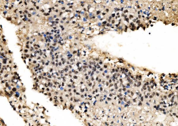
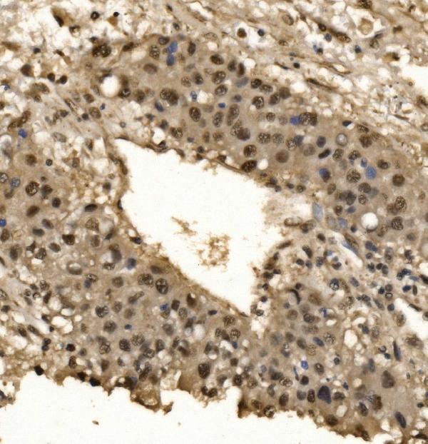
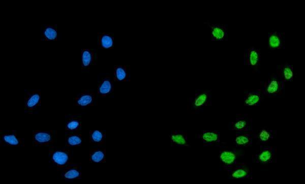
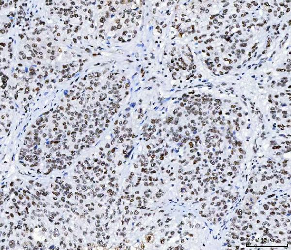
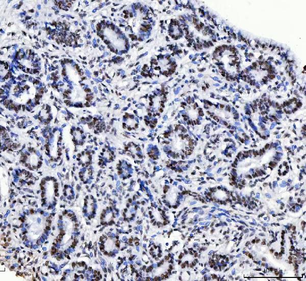
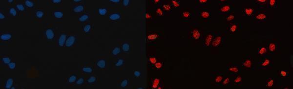


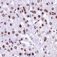


Comprehensive Human Protein Atlas IHC scoring per tissue (reliability: Supported — Medium consistency between antibody staining and RNA expression data.). Rows are taken directly from the HPA tissue chart — click any row's HPA link to view the source.
| Tissue | Cell type | Level | Evidence | Source |
|---|---|---|---|---|
| Adipose tissue | Adipocytes | High | Protein (IHC) | HPA → |
| Adrenal gland | Glandular cells | High | Protein (IHC) | HPA → |
| Appendix | Glandular cells | High | Protein (IHC) | HPA → |
| Bone marrow | Hematopoietic cells | High | Protein (IHC) | HPA → |
| Breast | Adipocytes | High | Protein (IHC) | HPA → |
| Tissue | Cell type | Level | Evidence | Source |
|---|---|---|---|---|
| None in HPA: CPSF6 is detected in all 45 scored tissues. Use a no-primary (secondary-only) and an isotype control instead. | ||||
Troubleshoot CPSF6 staining in paraffin sections by checking retrieval, nuclear localisation, assay controls and scoring before interpreting differences between samples.
Two anti-CPSF6 antibodies have human paraffin-section IHC figures and cell-based IF/ICC figures (catalog image captions). Catalog reactivity includes human, monkey, mouse, and rat (catalog reactivity).
A04551-1 has IHC images from human liver cancer and appendiceal adenocarcinoma paraffin sections, plus an IF/ICC image from MCF-7 cells (A04551-1 image captions). M04551 has IHC images from human squamous cell carcinoma, cervical cancer, colorectal adenocarcinoma, and ovarian cancer paraffin sections, plus an IF/ICC image from U20S cells (M04551 image captions).
Which to pick: For tissue IHC, A04551-1 is a rabbit antibody shown on human liver cancer paraffin sections, while M04551 is a mouse monoclonal, clone 3F11E1, shown on human squamous cell carcinoma paraffin sections; both captions use EDTA retrieval at pH 8.0 and 2 μg/ml primary antibody, and neither reports the fixative (catalog host/clone; A04551-1 and M04551 IHC captions). For IF/ICC, either SKU has a cell-based image and a listed concentration of 5 μg/ml; choose according to the rabbit or mouse secondary antibody available (catalog IF/ICC applications and IF captions). For work across species, both list human, monkey, mouse, and rat reactivity, while their pictured tissue IHC examples are human (catalog reactivity; A04551-1 and M04551 IHC captions).