This website uses cookies to ensure you get the best experience on our website.


- Table of Contents
Plan CPT2 IHC on paraffin sections with catalog antibody M02112 at 1:50 (datasheet: M02112). Assess cytoplasmic staining by cell type, using the reported staining in glandular cells and kidney collecting ducts as references (HPA tissue IHC).

Expected localisation, validated protocols, controls and antibodies — the at-a-glance facts below, then the full design guide.
| Expected localisation | Cytoplasmic staining (HPA tissue IHC); mitochondrial inner membrane (UniProt) | |
| Staining pattern | Cytoplasmic staining in most tissues (HPA tissue IHC) | |
| Antigen retrieval | EDTA pH 8.0 HIER, heat-mediated (datasheet M02112) | |
| Positive control | Adrenal gland+4 more · see all | |
| Negative control | Adipose tissue+4 more · see all |
| Fixation | Matched tissue-IHC evidence does not establish the fixation claim. Validate the specimen-specific method before use. (selected-SKU IHC image M02112) | |
| Caveat | Staining and RNA show medium consistency (HPA tissue IHC) | |
| Regulation | Liver-enhanced RNA expression (HPA tissue RNA) | |
| Isoform / epitope | No isoforms annotated; mature chain spans aa 26–658 (UniProt) |
The catalog antibody protocol is followed by published CPT2 chromogenic IHC protocols for GM tissue and renal cortex (PMC11308078; PMC13297011).
| Sample | Paraffin-embedded human acinic cell carcinoma of parotid tissue; fixative not specified (datasheet M02112) |
| Fixation | Image fixative and duration unreported (datasheet M02112); verify before use. |
| Sectioning | 4–5 µm sections on charged slides (standard) |
| Deparaffinisation | Xylene, graded ethanol series to water (standard) |
| Antigen retrieval | Heat retrieval: EDTA pH 8.0 (datasheet M02112); 20 min, 95–100 °C (standard) |
| Peroxidase block | 3% H2O2, 10 min, room temperature (standard) |
| Blocking | 10% goat serum (datasheet M02112) |
| Primary antibody | Rabbit monoclonal (clone FGB-3) anti-CPT2, 1:50 (datasheet M02112) |
| Primary incubation | Overnight at 4 °C (datasheet M02112) |
| Detection | HRP-conjugated secondary, DAB chromogen (datasheet M02112) |
| Counterstain | Hematoxylin, blue, dehydrate and mount (standard) |
| Expected result | CPT2-positive staining in glandular cells of adrenal gland (HPA tissue IHC: High). HPA tissue profile: Cytoplasmic expression in most tissues. No signal in the no-primary control. |
CPT2 is associated with the mitochondrial inner membrane, with annotated matrix-facing regions and no transmembrane segment (UniProt P23786 topology). In paraffin-section IHC, expect cytoplasmic staining in appropriate cells, including glandular cells of colon and stomach and kidney collecting ducts, all rated High by HPA (HPA tissue IHC). HPA describes cytoplasmic expression in most tissues and rates the tissue IHC profile Enhanced, with medium consistency between staining and RNA data (HPA tissue IHC).
| Distinct cytoplasmic chromogen in colon or stomach glandular cells, or kidney collecting ducts, with comparatively clear nuclei. | This fits the High cell-specific staining reported in these sites (HPA tissue IHC). The cytoplasmic appearance is compatible with mitochondrial CPT2 (UniProt P23786 topology); conventional chromogenic IHC alone cannot establish that each stained structure is a mitochondrion. Compare the cells within the section as well as the overall tissue signal. |
| Predominantly nuclear or nucleolar chromogen in tissue IHC, with little convincing cytoplasmic signal. | Treat this as a compartment mismatch against the HPA cytoplasmic tissue profile and UniProt mitochondrial location (HPA tissue IHC; UniProt P23786 topology). Investigate antibody specificity and detection background before calling it CPT2. HPA reports uncertain nucleoplasmic and nucleolar signals in ICC-IF, so those observations do not establish an expected nuclear IHC pattern (HPA subcellular ICC-IF). |
| Strong staining in HPA-listed negative cells, such as adipocytes or lymph-node germinal-center cells. | HPA reports CPT2 as Not detected in those particular cell populations (HPA tissue IHC). Unexpected signal may reflect cross-reactivity or endogenous detection activity (standard IHC practice). Check the identity of the stained cells and the no-primary control; a negative rating for one cell population does not make every cell in that tissue a negative control. |
| Diffuse chromogen across stroma, nuclei, and empty spaces, obscuring cell boundaries. | A field-wide haze does not match the reported cytoplasmic, cell-dependent profile (HPA tissue IHC). Nonspecific primary binding, incomplete blocking, or detection background are possible general IHC causes (standard IHC practice). Assess a no-primary control and optimize blocking and washes before interpreting weak cellular color as a positive result. |
| No convincing signal in an otherwise evaluable section containing colon or stomach glandular cells, or kidney collecting ducts. | Those cell types are rated High by HPA, making absent signal a reason to check the assay (HPA tissue IHC). Confirm tissue identity and section quality, then review the catalog antibody's IHC-P dilution and detection controls (standard IHC practice). HPA's Enhanced rating describes evidence for the staining profile; it does not guarantee a positive result in every preparation (HPA tissue IHC). |
| Subcellular interpretation | UniProt places CPT2 at the mitochondrial inner membrane and annotates matrix regions at residues 26–178 and 209–658, without a transmembrane segment (UniProt P23786 topology). Use this to judge compartment plausibility, while recognizing that routine chromogenic IHC usually reads as cytoplasmic signal rather than resolvable membrane topology (standard IHC practice). |
| Choice of comparison cells | HPA rates glandular cells in adrenal gland, colon, duodenum, gallbladder, rectum, and stomach; kidney collecting ducts; and placental syncytiotrophoblasts High (HPA tissue IHC). Its Not detected calls apply to named cells, including adipocytes and lymph-node germinal-center cells, rather than entire organs (HPA tissue IHC). Select controls by cell population. |
| Antibody validation | HPA lists IHC as Enhanced for HPA028201 and HPA028202, and Supported for HPA028214 (HPA antibodies). Enhanced reflects reproduction by independent antibodies or orthogonal data under the supplied definition. These ratings support the tissue pattern; they do not validate an unrelated catalog antibody or establish its working dilution (HPA antibodies; standard IHC practice). |
| Processing and antigen retrieval | UniProt annotates a mature CPT2 chain spanning residues 26–658 and no glycosylation sites (UniProt P23786 processing). The supplied sources do not map the catalog antibody epitope or report CPT2-specific retrieval or fixation sensitivity. Follow the catalog antibody's IHC-P instructions for retrieval, then optimize against appropriate cell controls (standard IHC practice). |
| Situation | Likely cause | Next action |
|---|---|---|
| Positive-control glandular cells are blank. | A failed staining run, unsuitable antibody dilution, or ineffective detection are possible general IHC causes; the listed glandular cells are High in HPA (HPA tissue IHC; standard IHC practice). | Verify the section and control identity, follow the catalog antibody's IHC-P instructions, and check that the detection system worked on a separate assay control (standard IHC practice). |
| Color fills the section or appears in the no-primary control. | Endogenous enzyme activity or detection-reagent background can produce chromogen without specific primary binding (standard IHC practice). | Apply the appropriate endogenous-activity block for the detection chemistry, inspect the no-primary control, and review washes and detection-reagent exposure (standard IHC practice). |
| Negative-control cells stain strongly, but the no-primary control is clear. | Primary-antibody cross-reactivity or nonspecific binding is possible; adipocytes and lymph-node germinal-center cells are Not detected in HPA tissue IHC (HPA tissue IHC; standard IHC practice). | Confirm cell identity, review the IHC-validated antibody's dilution and blocking conditions, and compare with an independently validated antibody when available (standard IHC practice). |
| Only nuclei or nucleoli show a strong IHC signal. | That pattern conflicts with HPA's cytoplasmic tissue profile and UniProt's mitochondrial assignment; HPA's nuclear ICC-IF locations are uncertain (HPA tissue IHC; HPA subcellular ICC-IF; UniProt P23786 topology). | Check cytoplasmic positive-control cells and no-primary background before scoring. Seek independent antibody confirmation if nuclear staining persists (standard IHC practice). |
| Weak cytoplasmic staining is hard to separate from background. | HPA includes Low calls for thyroid glandular cells, cortical neurons, and several other populations; weak signal can also be obscured by routine IHC background (HPA tissue IHC; standard IHC practice). | Anchor interpretation to an HPA High cell population, compare matched controls, and optimize blocking, washes, and catalog-recommended dilution (HPA tissue IHC; standard IHC practice). |
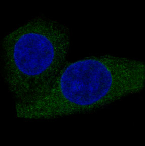
| Q: Should IF/ICC show the same pattern as tissue IHC? | HPA supports a mitochondrial ICC-IF location but marks nucleoplasm and nucleoli uncertain; these are separate observations from the cytoplasmic tissue IHC profile (HPA subcellular ICC-IF; HPA tissue IHC). | A: Expect mitochondrial signal in IF/ICC, and treat nuclear signal cautiously. Use the separate IF/ICC guide for assay design; do not transfer an IHC-P condition to IF/ICC without validation (HPA subcellular ICC-IF; standard IF practice). |
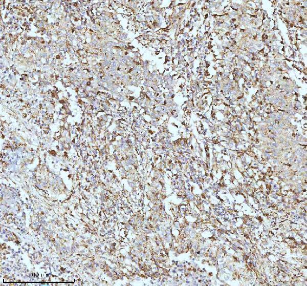
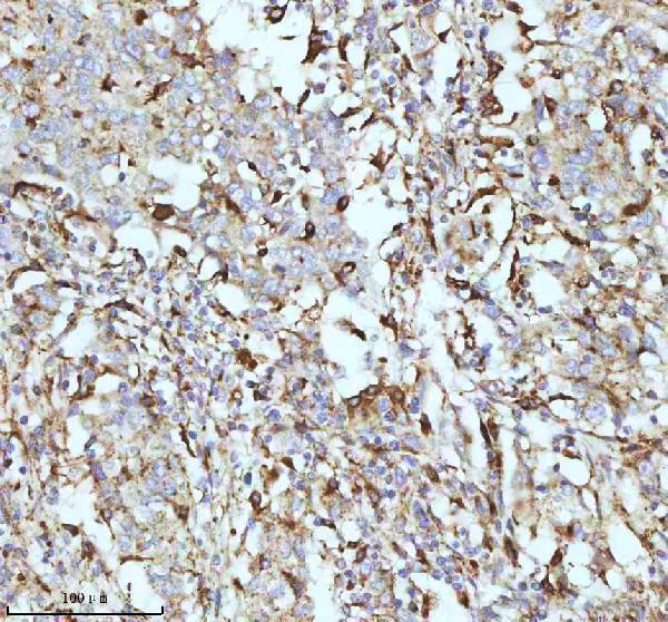
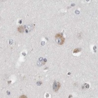
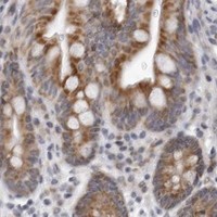
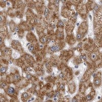
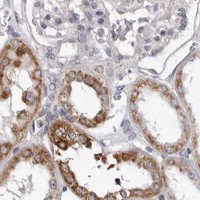


Comprehensive Human Protein Atlas IHC scoring per tissue (reliability: Enhanced — Medium consistency between antibody staining and RNA expression data.). Rows are taken directly from the HPA tissue chart — click any row's HPA link to view the source.
| Tissue | Cell type | Level | Evidence | Source |
|---|---|---|---|---|
| Adrenal gland | Glandular cells | High | Protein (IHC) | HPA → |
| Colon | Glandular cells | High | Protein (IHC) | HPA → |
| Duodenum | Glandular cells | High | Protein (IHC) | HPA → |
| Gallbladder | Glandular cells | High | Protein (IHC) | HPA → |
| Kidney | Collecting ducts | High | Protein (IHC) | HPA → |
| Tissue | Cell type | Level | Evidence | Source |
|---|---|---|---|---|
| Adipose tissue | Adipocytes | Not detected | Protein (IHC) | HPA → |
| Bone marrow | Hematopoietic cells | Not detected | Protein (IHC) | HPA → |
| Cerebellum | Cells in granular layer | Not detected | Protein (IHC) | HPA → |
| Lymph node | Germinal center cells | Not detected | Protein (IHC) | HPA → |
| Ovary | Ovarian stroma cells | Not detected | Protein (IHC) | HPA → |
Troubleshoot CPT2 staining in paraffin-section chromogenic IHC, with one entry for planning a separate IF experiment.
Catalog antibody M02112 has human paraffin-section IHC images and an MCF-7 IF image (M02112 image captions); both antibodies list Human, Mouse, and Rat reactivity (catalog applications/reactivity).
M02112 lists IHC and ICC/IF, with IHC images from human parotid acinic cell carcinoma and cervical cancer paraffin sections and an IF image from MCF-7 cells (M02112 applications/image captions). A02112-1 lists IHC and Human, Mouse, and Rat reactivity, but has no IHC image in the supplied catalog (A02112-1 applications/reactivity/image alts).
Which to pick: For tissue IHC, choose M02112: its own image captions document paraffin sections, EDTA retrieval at pH 8.0, and 1:50 primary dilution; the fixative is unreported (M02112 IHC image captions). For IF/ICC, choose M02112 because ICC/IF is listed and its IF caption identifies MCF-7 cells (M02112 applications/IF image caption). For cross-species planning, both SKUs list Human, Mouse, and Rat reactivity; M02112 is monoclonal, while A02112-1 is an IHC-listed rabbit antibody without a supplied IHC image (catalog reactivity/host/clone/applications/image alts).