This website uses cookies to ensure you get the best experience on our website.


- Table of Contents
Plan CPXM2 paraffin IHC with the catalog antibody’s 1:100–1:300 dilution range (datasheet A14237) and epididymal glandular cells as a medium-staining reference (HPA tissue IHC). Interpret cytoplasmic or membranous staining cautiously because secreted CPXM2 may differ from its tissue RNA distribution (HPA tissue IHC; UniProt).

Expected localisation, validated protocols, controls and antibodies — the at-a-glance facts below, then the full design guide.
| Expected localisation | Cytoplasmic or membranous in selected tissues (HPA tissue IHC) | |
| Staining pattern | Selected tissues show cytoplasmic or membranous staining (HPA tissue IHC) | |
| Antigen retrieval | Tris-EDTA pH 9.0 HIER, heat-mediated (datasheet A14237) | |
| Positive control | Epididymis | |
| Negative control | Adipose tissue+4 more · see all |
| Fixation | Keep fixation consistent across sections (standard IHC practice; not target-specific) | |
| Caveat | Secretion may separate tissue RNA and protein patterns (HPA tissue IHC) | |
| Regulation | Expression regulators unannotated (UniProt) | |
| Isoform / epitope | No isoforms; no cytoplasmic domain; mature chain 26–756 (UniProt) |
The catalog antibody protocol uses Tris-EDTA pH 9.0 retrieval (datasheet A14237). The published CPXM2 IHC protocols below provide four additional study-specific workflows (PMC6718098; PMC6138928; PMC7127851; PMC9293992).
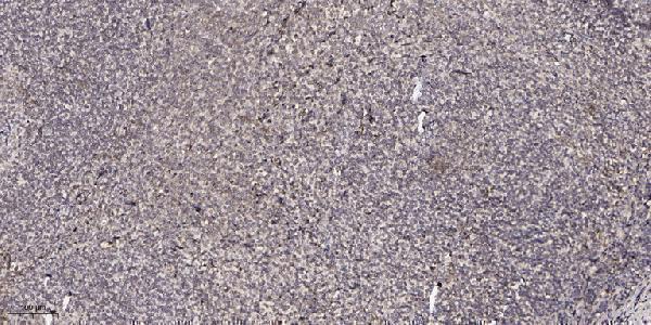
| Sample | Paraffin-embedded human tonsil tissue; fixative not specified (datasheet A14237) |
| Fixation | Image fixative and duration unreported (datasheet A14237); verify before use. |
| Sectioning | 4–5 µm sections on charged slides (standard) |
| Deparaffinisation | Xylene, graded ethanol series to water (standard) |
| Antigen retrieval | Heat retrieval: Tris-EDTA pH 9.0 (datasheet A14237); 20 min, 95–100 °C (standard) |
| Peroxidase block | 3% H2O2, 10 min, room temperature (standard) |
| Blocking | 10% normal serum of the secondary host, 30 min, room temperature (standard) |
| Primary antibody | Rabbit anti-CPXM2, 1:100-1:300 (datasheet A14237) |
| Primary incubation | Overnight at 4 °C (standard) |
| Detection | HRP-polymer secondary, DAB chromogen 5–10 min (standard) |
| Counterstain | Hematoxylin, blue, dehydrate and mount (standard) |
| Expected result | CPXM2-positive staining in glandular cells of epididymis (HPA tissue IHC: Medium). HPA tissue profile: Cytoplasmic or membranous expression in selected tissues. No signal in the no-primary control. |
CPXM2 is a secreted protein with a signal peptide and no transmembrane segment (UniProt Q8N436). In tissue IHC, HPA describes cytoplasmic or membranous staining in selected tissues, including medium staining of epididymal glandular cells (HPA tissue IHC). Treat that appearance as a reference pattern, not a definitive compartment assignment: HPA rates the tissue staining Uncertain and cautions that secreted protein and RNA locations may differ (HPA tissue IHC).
| Epididymal glandular cells show moderate cytoplasmic or cell-edge chromogen, with discernible unstained areas. | This matches the reported medium glandular-cell signal and selected-tissue cytoplasmic or membranous pattern (HPA tissue IHC). Because CPXM2 is secreted, a cell-edge appearance alone does not establish membrane anchoring (UniProt Q8N436). |
| Strong, sharply nuclear staining dominates the section. | Nuclear staining falls outside HPA’s reported cytoplasmic or membranous tissue pattern (HPA tissue IHC). Check counterstain overlap, detection background, and antibody specificity before scoring it as CPXM2; the available record does not validate a nuclear pattern. |
| Adipocytes or bone-marrow hematopoietic cells stain prominently. | HPA reports CPXM2 as not detected in those sampled cells (HPA tissue IHC). An unexpected signal raises concern for cross-reactivity or endogenous chromogen activity; confirm the cell identity and compare antibody-omission and detection controls (general IHC practice). |
| Brown signal coats most cells, extracellular spaces, and blank regions without a clear cell pattern. | That distribution is difficult to reconcile with HPA’s selected-tissue pattern (HPA tissue IHC). Diffuse chromogen may reflect background from detection chemistry or inadequate blocking; assess a matched negative control before interpreting extracellular signal (general IHC practice). |
| Epididymal glandular cells have no visible signal. | A missing signal conflicts with HPA’s medium reference staining, but its reliability is Uncertain (HPA tissue IHC). First check tissue preservation, antibody and detection controls, and whether the sampled section contains the relevant glandular cells (general IHC practice). |
| Secretion and apparent cell-edge signal | CPXM2 has a cleaved signal peptide, residues 1–25, and a secreted chain beginning at residue 26, with no transmembrane segment (UniProt Q8N436). Interpret cell-edge chromogen in that context; morphology alone cannot prove surface retention. |
| Reported tissue distribution | Epididymal glandular cells are medium; respiratory and several gastrointestinal epithelial or glandular cells are low (HPA tissue IHC). Use the reported level for the exact sampled cell type, rather than expecting equal staining across tissues. |
| RNA versus tissue protein | HPA lists RNA as tissue enhanced in blood vessel and choroid plexus, while warning that secreted variants can make RNA and protein locations differ (HPA tissue IHC). Do not treat RNA enrichment as an IHC positive-control result. |
| Antibody-validation limit | The listed rabbit polyclonal antibody HPA048375 has Uncertain IHC validation (HPA antibodies). Use staining morphology and appropriate controls together; the supplied record does not provide an independently confirmed IHC pattern. |
| Processing and glycosylation | UniProt annotates a signal peptide and five glycosylation sites (UniProt Q8N436). These features describe the protein, but the supplied sources do not establish their effect on antigen retrieval, staining intensity, or antibody epitope access. |
| IF/ICC Q&A: Is a cellular IF pattern established? | No ICC-IF image-bearing cell line or main subcellular location is supplied; HPA’s subcellular summary says Secreted (HPA subcellular). This IHC tissue pattern cannot by itself validate an IF/ICC compartment. |
| Situation | Likely cause | Next action |
|---|---|---|
| No staining in epididymal glandular cells | The result differs from HPA’s medium observation, although its reliability is Uncertain (HPA tissue IHC). A failed detection step or missing target cells is also possible (general IHC practice). | Verify glandular-cell presence and section quality; run antibody and detection controls, then review the chosen retrieval and antibody conditions as general IHC variables. No CPXM2-specific retrieval condition is established by the supplied sources. |
| Weak staining in bronchus, colon, or stomach | HPA reports low staining in the listed respiratory or gastrointestinal cells (HPA tissue IHC). Weak signal can therefore fit the reference pattern; judge it against background and the exact cell type. | Compare cells within the section and a matched negative control; record intensity and distribution without upgrading faint, nonspecific chromogen to a strong positive (general IHC practice). |
| Prominent nuclear signal | HPA describes cytoplasmic or membranous expression in selected tissues, not a nuclear pattern (HPA tissue IHC). Counterstain overlap or nonspecific detection can complicate visual assignment (general IHC practice). | Inspect the chromogen separately from the counterstain, compare antibody-omission controls, and avoid scoring nuclear signal as CPXM2 until independently supported (general IHC practice). |
| Widespread staining in reported negative cells | HPA reports adipocytes and bone-marrow hematopoietic cells as not detected in its sampled tissues (HPA tissue IHC). Cross-reactivity or endogenous detection activity could explain unexpected chromogen (general IHC practice). | Confirm cell identity and compare a no-primary control; check endogenous activity blocking and detection reagents before interpreting the signal (general IHC practice). |
| Diffuse brown haze obscures cell boundaries | A broad haze prevents comparison with HPA’s cell-specific reference pattern (HPA tissue IHC). In IHC, inadequate blocking, residual detection reagent, or excessive chromogen development may contribute to background (general IHC practice). | Review blocking, wash, and chromogen-development steps against a matched negative control; score only signal that remains spatially interpretable (general IHC practice). |
| An IF/ICC image seems to show a precise organelle location | HPA supplies no ICC-IF image-bearing cell line or main subcellular location for CPXM2; its summary is Secreted (HPA subcellular). | Treat the organelle assignment as unverified and seek independent localisation evidence before using it to interpret this tissue IHC result (HPA subcellular; general IF practice). |


Comprehensive Human Protein Atlas IHC scoring per tissue (reliability: Uncertain — At least one protein variant secreted, tissue location of RNA and protein might differ and correlation is complex.). Rows are taken directly from the HPA tissue chart — click any row's HPA link to view the source.
| Tissue | Cell type | Level | Evidence | Source |
|---|---|---|---|---|
| Epididymis | Glandular cells | Medium | Protein (IHC) | HPA → |
| Tissue | Cell type | Level | Evidence | Source |
|---|---|---|---|---|
| Adipose tissue | Adipocytes | Not detected | Protein (IHC) | HPA → |
| Adrenal gland | Glandular cells | Not detected | Protein (IHC) | HPA → |
| Bone marrow | Hematopoietic cells | Not detected | Protein (IHC) | HPA → |
| Breast | Adipocytes | Not detected | Protein (IHC) | HPA → |
| Caudate | Glial cells | Not detected | Protein (IHC) | HPA → |
Troubleshoot CPXM2 chromogenic IHC in paraffin sections using the catalog antibody’s tonsil image and the reported localisation evidence.
A14237 has IHC image data from paraffin-embedded human tonsil (catalog IHC image caption). IF is listed without an image (catalog applications; catalog IF image alts); human and mouse reactivity is listed (catalog reactivity).
A14237 is listed for IHC and IF, with human and mouse reactivity (catalog applications; catalog reactivity). Its supplied image shows paraffin-embedded human tonsil IHC at 1:200 with Tris-EDTA pH 9.0 retrieval; no IF image is supplied (catalog IHC image caption; catalog IF image alts).
Which to pick: For tissue IHC, choose A14237 based on its human paraffin-section image; the fixative is unreported (catalog IHC image caption). For IF, A14237 is listed at 1:50, but ICC validation and an IF image are unreported, and clonality is unspecified (catalog applications; catalog IF dilution; catalog IF image alts; catalog clone field). For mouse work, A14237 lists mouse reactivity, though its supplied IHC image shows human tonsil (catalog reactivity; catalog IHC image caption).