CR2 / Complement receptor type 2 · IHC design guide

Design Immunohistochemistry for CR2

Plan chromogenic CR2 IHC on paraffin sections with tonsil or spleen as positive controls (HPA tissue IHC). Interpret cytoplasmic lymphoid staining (HPA tissue IHC) alongside CR2’s membrane topology (UniProt).

Evidence assembled Oct 2026 · For research use; verify linked source records and product datasheet before use
Immunohistochemistry protocol sheet for CR2 (IHC for CR2): expected localisation Cytoplasmic lymphoid stain (HPA tissue IHC); cell membrane (UniProt), antibody M01632-2, validated IHC image, and IHC protocol steps
Printable CR2 IHC protocol sheet — expected localisation Cytoplasmic lymphoid stain (HPA tissue IHC); cell membrane (UniProt), antibody M01632-2, controls and protocol steps. Open the full CR2 IHC guide →

CR2 Immunohistochemistry Experimental Design Guide

Expected localisation, validated protocols, controls and antibodies — the at-a-glance facts below, then the full design guide.

Must know before staining
Expected localisation Cytoplasmic lymphoid stain (HPA tissue IHC); cell membrane (UniProt)
Staining pattern Cytoplasmic staining in lymphoid tissue, including germinal centers (HPA tissue IHC)
Antigen retrieval Citrate pH 6.0 HIER, 95–98 °C, 20 min (rule: cytoplasmic / membrane antigen)
Positive control ⓘ Appendix+3 more · see all
Negative control ⓘ Adipose tissue+4 more · see all
Important caveats
Reasons your staining may differ from the expected pattern.
Fixation Matched tissue-IHC evidence does not establish the fixation claim. Validate the specimen-specific method before use. (selected-SKU IHC image M01632-2)
Caveat Tissue IHC appears cytoplasmic despite membrane topology (HPA tissue IHC; UniProt)
Regulation Expressed in mature B cells (UniProt)
Isoform / epitope 4 isoforms; check extracellular versus cytoplasmic epitope (UniProt)
Section 1

Recommended CR2 IHC & IF Protocols

The catalog antibody’s IHC-P protocol is paired with a published CR2 immunohistochemistry example in human tissues (PMC12328216 methods).

Recommended immunohistochemistry (IHC-P) protocol parameters
SampleTissue sections; selected-image fixative not specified (standard IHC workflow)
FixationImage fixative and duration unreported (datasheet M01632-2); verify before use.
Sectioning4–5 µm sections on charged slides (standard)
DeparaffinisationXylene, graded ethanol series to water (standard)
Antigen retrievalHeat-induced epitope retrieval in citrate buffer, pH 6.0, 20 min at 95–98 °C (standard rule: cytoplasmic / membrane antigen)
Peroxidase block3% H2O2, 10 min, room temperature (standard)
Blocking10% normal serum of the secondary host, 30 min, room temperature (standard)
Primary antibodyRabbit anti-CR2, 1:1000-1:4000 (datasheet M01632-2)
Primary incubationOvernight at 4 °C (standard)
DetectionHRP-polymer secondary, DAB chromogen 5–10 min (standard)
CounterstainHematoxylin, blue, dehydrate and mount (standard)
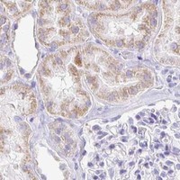
Expected resultCR2-positive staining in lymphoid tissue of appendix (HPA tissue IHC: High). HPA tissue profile: Cytoplasmic expression in lymphoid tissues. No signal in the no-primary control.
💡Decision noteStart with citrate pH 6.0 retrieval at 95–98 °C for 20 min (page antigen retrieval); assess cell-membrane staining (UniProt P20023 topology).
Section 2

What Is the Expected CR2 Staining Pattern?

CR2 is a cell membrane protein with an extracellular region at residues 21–971 and a transmembrane segment at 972–999 (UniProt P20023 topology). In tissue IHC, expect staining in lymphoid structures, especially germinal center cells and splenic white pulp cells (HPA: High). HPA describes its tissue staining profile as cytoplasmic expression in lymphoid tissues and rates the tissue evidence Enhanced, reflecting consistency between staining and RNA expression (HPA: tissue IHC).

What am I looking at on my slide?
Strong staining in lymph node or tonsil germinal centers, or in splenic white pulp.This matches the reported high signal in those specific tissue compartments (HPA: tissue IHC). Score the relevant cells and structures; a high tissue rating does not mean every cell on the section must stain.
Cytoplasmic staining in lymphoid structures, with or without a clear membrane rim.HPA reports a cytoplasmic tissue IHC profile, while UniProt assigns CR2 to the cell membrane (HPA: tissue IHC; UniProt P20023 subcellular location). Record the visible pattern rather than rejecting cytoplasmic signal solely because it lacks a sharp rim.
Predominantly nuclear staining in the expected lymphoid cells.A nuclear-dominant pattern does not match the reported cytoplasmic tissue profile or membrane assignment (HPA: tissue IHC; UniProt P20023 subcellular location). Treat it as suspect until a control and an independently validated stain support it.
Strong staining in cell populations reported as undetected, such as adipocytes or adrenal glandular cells.That distribution conflicts with the named negative populations (HPA: Not detected in adipocytes and adrenal glandular cells). Consider cross-reactivity or endogenous detection activity; an HPA negative call applies to the listed cells, not necessarily every cell in that tissue.
No staining in germinal centers of a lymph node or tonsil positive control.This fails to reproduce a reported high-signal setting (HPA: High in lymph node and tonsil germinal center cells). Check the control section and staining run before calling the experimental sample CR2 negative (general IHC practice).
💡Expected CR2 appearanceCall the result positive when lymphoid cells show convincing signal in germinal centers or splenic white pulp, where HPA reports High staining; cytoplasmic tissue staining is documented even though CR2 is membrane assigned (HPA: tissue IHC; UniProt P20023). Nuclear-dominant or widespread uniform staining outside the expected cells warrants investigation (HPA: tissue IHC; general IHC practice).
How each factor affects the staining
Cellular locationCR2 has an extracellular region at residues 21–971, one transmembrane segment, and a cytoplasmic tail at 1000–1033 (UniProt P20023 topology). Epitope location matters when interpreting a membrane rim, but no antibody epitope is supplied.
Tissue and cell selectionAppendix lymphoid tissue, lymph node and tonsil germinal centers, and splenic white pulp are reported High (HPA: tissue IHC). Choose one of these structures for a positive control and assess its named cells, not the whole tissue indiscriminately.
Reported negative populationsHPA reports Not detected in adipocytes, adrenal glandular cells, bone marrow hematopoietic cells, bronchial respiratory epithelial cells, and the specified sampled brain cell populations (HPA: tissue IHC). Use each call only for its named cell population.
Tissue IHC versus subcellular summaryThe cytoplasmic tissue IHC profile and membrane subcellular summary describe different observations (HPA: tissue IHC; HPA: subcellular). The supplied subcellular record has no ICC-IF image-bearing cell lines, so it cannot resolve this appearance difference in a chosen cell model.
Antibody validationThree listed antibodies, HPA052942, HPA060715, and CAB002659, have Enhanced IHC status (HPA: antibodies). This supports comparing IHC patterns, but the payload gives no ICC validation status for them (HPA: antibodies).
Processing and variantsUniProt lists a cleaved signal peptide at residues 1–20, four isoforms, and 11 glycosylation sites (UniProt P20023 processing, isoforms, glycosylation). Without the antibody epitope, these facts do not predict a different staining pattern or a retrieval requirement.
Why is my staining missing, weak or wrong?
SituationLikely causeNext action
No signal in the expected lymphoid positive control.The observed result conflicts with High staining in lymph node or tonsil germinal center cells (HPA: tissue IHC); the supplied sources do not identify a CR2-specific fixation or retrieval failure.Confirm the relevant germinal centers are present, then review antibody dilution, antigen retrieval, detection reagents, and the run control using the antibody's IHC instructions (general IHC practice).
Weak signal appears throughout the section without clear lymphoid enrichment.Diffuse staining lacks the reported lymphoid pattern (HPA: tissue IHC). Background from nonspecific binding or detection chemistry is possible (general IHC practice).Compare with a no-primary control, inspect blocking and wash steps, and reduce excess primary or detection reagent according to the assay instructions (general IHC practice).
Nuclei stain more strongly than the lymphoid cell bodies.Nuclear-dominant signal is discordant with the cytoplasmic tissue profile and membrane assignment (HPA: tissue IHC; UniProt P20023 subcellular location).Check the no-primary control and counterstain appearance, then compare with an independently validated IHC antibody if available (general IHC practice; HPA: Enhanced IHC antibodies).
Unexpected strong staining appears in a reported negative cell population.A strong signal in named undetected cells conflicts with their HPA tissue IHC calls (HPA: Not detected). Cross-reactivity or endogenous detection activity may explain it (general IHC practice).Verify cell identity and compare positive and no-primary controls; review the blocking and detection chemistry before assigning the signal to CR2 (general IHC practice).
The slide shows cytoplasmic signal but no crisp membrane outline.HPA reports cytoplasmic tissue IHC staining, despite a membrane subcellular assignment (HPA: tissue IHC; UniProt P20023). A missing rim alone cannot establish a false positive.Judge whether staining is confined to the reported lymphoid structures, and document the observed compartment; use an independently validated IHC antibody to investigate a consequential discrepancy (HPA: tissue IHC; general IHC practice).
Can the tissue IHC pattern establish an IF/ICC result?The supplied subcellular summary says Membrane but lists no ICC-IF image-bearing cell lines, and the listed antibodies have no ICC status (HPA: subcellular; HPA: antibodies).Use the separate IF/ICC guide for that application; assess its own positive and negative controls before interpreting fluorescence (general IF practice).

Sample controls for CR2 IHC & IF

🧪Run lymph node first and score germinal center cells, where CR2 staining is High (HPA: High in lymph-node germinal center cells). Use adrenal gland glandular cells as the biological tissue negative (HPA: Not detected in adrenal gland glandular cells); on the lymph-node slide, adjacent cells outside germinal centers should be assessed for background against the control slides, rather than assumed to be CR2-negative.
Positive control tissue: Appendix (Lymphoid tissue, HPA High)
Negative control tissue: Adipose tissue (HPA Not detected)
ICC-IF cell lines (HPA subcellular resource): HPA carries no ICC-IF cell line for CR2; derive a cell-line control from the positive tissue's cell type (Lymphoid tissue) and confirm it by RNA or western blot first.
Technical controls: Include a no-primary, secondary-only control; a nonimmune IgG or isotype control matched to the primary antibody’s host species and clonality; and a matched CR2-knockout specimen if available (standard IHC practice). Block endogenous peroxidase in lymph node, and account for endogenous biotin if using avidin–biotin detection (standard IHC practice).
⚠️Feasibility: A CR2-specific fixation window or fixation effect is unreported in the supplied evidence, and the selected M01632-2 rat-spleen IHC caption does not state a fixative (caption: fixative unreported). Antigen-retrieval dependence is unreported; compare retrieved and unretrieved paraffin sections during optimization (standard IHC practice). The supplied evidence does not establish frozen-section or IF/ICC staining as easier (HPA: no ICC-IF image cell lines); Fc-receptor binding in lymph-node immune cells can create background, so interpret staining alongside the controls (standard IHC practice).

HPA tissue IHC evidence for CR2

Comprehensive Human Protein Atlas IHC scoring per tissue (reliability: Enhanced — High consistency between antibody staining and RNA expression data.). Rows are taken directly from the HPA tissue chart — click any row's HPA link to view the source.

Positive expression · recommended positive controls

TissueCell typeLevelEvidenceSource
Appendix Lymphoid tissue High Protein (IHC) HPA →
Lymph node Germinal center cells High Protein (IHC) HPA →
Spleen Cells in white pulp High Protein (IHC) HPA →
Tonsil Germinal center cells High Protein (IHC) HPA →

Undetected expression · recommended negative controls

TissueCell typeLevelEvidenceSource
Adipose tissue Adipocytes Not detected Protein (IHC) HPA →
Adrenal gland Glandular cells Not detected Protein (IHC) HPA →
Bone marrow Hematopoietic cells Not detected Protein (IHC) HPA →
Breast Adipocytes Not detected Protein (IHC) HPA →
Bronchus Respiratory epithelial cells Not detected Protein (IHC) HPA →
Section 3

Advanced CR2 IHC Tips

Troubleshoot CR2 staining in paraffin sections by checking retrieval, tissue context, membrane localisation, and controls before interpreting chromogenic signal.

How should I retrieve CR2 in paraffin sections when staining is weak?
Use citrate pH 6.0 heat-induced epitope retrieval at 95–98 °C for 20 min (page retrieval setting). Keep section thickness, cooling time, and antibody incubation consistent while comparing retrieval runs (standard IHC practice). CR2 has a large extracellular region spanning residues 21–971 and 11 listed glycosylation sites, but the catalog antibody’s epitope is unspecified (UniProt P20023 topology; UniProt P20023 glycosylation; supplied product evidence). Compare signal in germinal center cells or splenic white pulp with a matched negative control (HPA: high in lymph node germinal center cells and splenic white pulp; standard IHC practice). If retrieval increases diffuse staining without improving that expected pattern, reassess antibody concentration and detection background (standard IHC practice).
How can I assess whether fixation is causing weak CR2 staining?
Target-specific fixation sensitivity is unknown because the supplied CR2 tissue caption does not report a fixative (catalog antibody M01632-2 caption). Record fixation duration and processing conditions for each specimen, then compare sections processed alike before changing antibody conditions (standard IHC practice). Run the same retrieval setting—citrate pH 6.0, 95–98 °C, 20 min—across that comparison (page retrieval setting; standard IHC practice). Include lymphoid tissue with an expected positive cell population and a negative reagent control in each run (HPA: high in lymph node germinal center cells; standard IHC practice). Interpret any association with fixation as an experiment-specific observation until independently reproduced (standard IHC practice).
Should CR2 staining appear on the membrane or in the cytoplasm?
Prioritise membrane-associated staining: CR2 is a cell-membrane protein with an extracellular region at residues 21–971 and a transmembrane segment at 972–999 (UniProt P20023 topology). HPA also describes cytoplasmic expression in lymphoid tissues, so record cytoplasmic and membrane patterns separately rather than combining them into one score (HPA tissue IHC profile; standard IHC practice). Examine germinal center cells or splenic white pulp first, where HPA reports high staining (HPA: high in lymph node germinal center cells and splenic white pulp). For diffuse staining outside those compartments, inspect morphology, retrieval consistency, and negative controls before calling it CR2 (standard IHC practice).
Can this antibody distinguish CR2 isoforms or detect an extracellular epitope?
CR2 has 4 listed isoforms, A through D, but no isoform-specific antibody epitope is supplied (UniProt P20023 isoforms; supplied product evidence). The reference protein has 15 extracellular Sushi domains across residues 21–970 and a cytoplasmic tail at 1000–1033 (UniProt P20023 domains and topology). Do not infer which side of the membrane the catalog antibody binds from a positive chromogenic stain alone (supplied product evidence; standard IHC interpretation). Compare staining across identically processed positive and negative tissues, and request an epitope map before making isoform-specific claims (HPA tissue IHC; standard IHC practice). If the map becomes available, check its sequence against each isoform before interpreting differences (standard IHC practice).
How should I investigate CR2 by IF alongside this chromogenic IHC guide?
Treat IF as a separate application because the supplied catalog evidence shows tissue IHC in rat spleen and provides no IF validation (catalog antibody M01632-2 caption; supplied product evidence). For multiplex IF, pair CR2 with a marker identifying the expected B-cell population, then assess colocalisation at cell boundaries (UniProt P20023 tissue specificity and membrane localisation; standard IF practice). Choose fluorophores after measuring tissue autofluorescence in unstained sections, reserving a spectrally clear channel for weaker signal (standard IF practice). If an epitope map places binding on the extracellular side, first assess staining without permeabilisation; a cytoplasmic epitope requires access across the membrane (UniProt P20023 topology; standard IF practice). Keep IF optimisation separate from chromogenic IHC scoring (standard IHC/IF practice).
What should I check when CR2 DAB staining looks diffuse or widespread?
Check a negative reagent control and inspect whether color follows tissue edges, damaged areas, or deposits rather than intact cells (standard chromogenic IHC practice). Quench endogenous peroxidase before horseradish peroxidase detection and assess nonspecific secondary-antibody binding with an appropriate control (standard chromogenic IHC practice). CR2 is reported at the cell membrane, while HPA describes cytoplasmic expression in lymphoid tissues; neither supports treating uniform tissue-wide color as sufficient evidence (UniProt P20023 localisation; HPA tissue IHC profile). Compare suspected signal with germinal center cells or splenic white pulp and with an HPA-negative compartment such as adipocytes (HPA: high in lymph node germinal center cells and splenic white pulp; HPA: not detected in adipose tissue adipocytes).
How should I quantify CR2 staining across lymphoid sections? ⚠ ANSWER MARKED FOR VERIFICATION
Define the anatomical region and cell population before scoring, such as germinal center cells or splenic white pulp cells (HPA: high in lymph node germinal center cells and splenic white pulp; standard IHC practice). Report the percentage of positive intact cells and an intensity-based H-score, or count positive cells per mm² when cell density varies (standard quantitative IHC practice). Normalise cell counts to the measured viable region area, and compare H-scores within the same defined cell population across specimens (standard quantitative IHC practice). Record membrane and cytoplasmic signal separately because UniProt lists membrane localisation while HPA reports cytoplasmic expression in lymphoid tissues (UniProt P20023 localisation; HPA tissue IHC profile). Apply one fixed scoring threshold to matched controls and study sections (standard quantitative IHC practice).
When is CR2 staining convincing rather than an artefact?
A convincing result places signal in intact cells of an expected lymphoid compartment, especially germinal center cells or splenic white pulp (HPA: high in lymph node germinal center cells and splenic white pulp; standard IHC interpretation). Membrane-associated staining fits CR2 topology, although HPA also reports cytoplasmic expression in lymphoid tissues (UniProt P20023 topology; HPA tissue IHC profile). Recheck staining restricted to section edges, necrotic areas, or cells outside the expected population against negative reagent controls (standard chromogenic IHC practice; UniProt P20023 tissue specificity). Endogenous peroxidase can create DAB signal, so assess the peroxidase-block control before interpreting brown deposits (standard chromogenic IHC practice). Compare staining with a low-expression reference compartment, such as adipocytes in adipose tissue, on a comparable run (HPA: not detected in adipose tissue adipocytes; standard IHC practice).
Boster reagents

Best CR2 / Complement receptor type 2 IHC Antibodies

Catalog anti-CR2 antibodies have IHC images from spleen and tonsil (catalog IHC image captions), list IF (catalog applications), and cover human, mouse and rat samples (catalog reactivity).

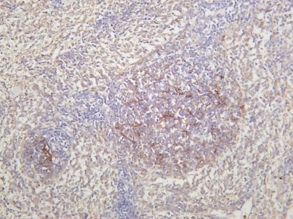
Real IHC data Rat spleen was stained with anti-CD21 rabbit antibody
Anti-CD21 Rabbit Monoclonal Antibody
Cat # M01632-2
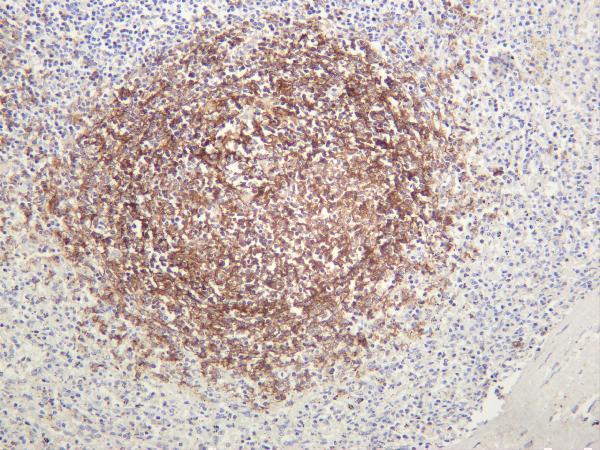
Real IHC data Immunohistochemical analysis of paraffin-embedded human spleen, using CD21 Antibody.
Anti-CD21 CR2 Rabbit Monoclonal Antibody
Cat # M01632

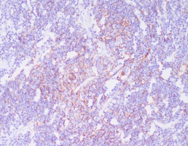
M01632 has an IHC image from paraffin-embedded human spleen and lists IHC, ICC and IF with human and mouse reactivity (catalog IHC image caption; applications; reactivity). M01632-2 has IHC images from rat, human and mouse spleen and human tonsil, and lists IHC and IF with reactivity across those three species (catalog IHC image captions; applications; reactivity).

Which to pick: For human tissue IHC on paraffin sections, choose M01632: its own image shows paraffin-embedded human spleen, but the fixative is unreported (catalog IHC image caption). For IF/ICC, choose monoclonal M01632 when ICC is required; IF is listed, but no IF image is supplied (catalog title; applications; IF image alts). For work across human, mouse and rat, choose monoclonal M01632-2 for IHC or IF; its IHC images show spleen staining in all three species, but their processing and fixative are unreported (catalog title; applications; reactivity; IHC image captions).

Each figure is that product's own IHC / IF validation image from its datasheet.

References

  1. UniProt Consortium. UniProt entry P20023 (CR2_HUMAN, Complement receptor type 2).
  2. Human Protein Atlas. CR2 tissue IHC expression (reliability: Enhanced).
  3. Human Protein Atlas. CR2 subcellular location (ICC-IF): Membrane.
  4. Human Protein Atlas. CR2 antibody validation summary (3 antibodies).
  5. Encapsulated Cell Technology-Based Delivery of a Complement Inhibitor Reduces Choroidal Neovascularization in a Mouse Model. Translational vision science & technology 2018 — PMC5846441.
  6. The use of Matrigel combined with encapsulated cell technology to deliver a complement inhibitor in a mouse model of choroidal neovascularization. Molecular vision 2020 — PMC7245607.
  7. R9AP is a common receptor for EBV infection in epithelial cells and B cells. Nature 2025 — PMC12328216.
  8. Dissecting the complement pathway in hepatic injury and regeneration with a novel protective strategy. The Journal of experimental medicine 2014 — PMC4144741.
  9. PubMed PMID:2563370 — UniProt-cited evidence.
  10. PubMed PMID:2832506 — UniProt-cited evidence.
  11. PubMed PMID:2827171 — UniProt-cited evidence.