CREBBP / CREB-binding protein · IHC design guide

Design Immunohistochemistry for CREBBP

This guide sets nuclear staining expectations for CREBBP in paraffin-section IHC (HPA tissue IHC). Use colon glandular cells as a positive reference (HPA tissue IHC), then score nuclear and cytoplasmic signal separately (UniProt).

Evidence assembled Oct 2026 · For research use; verify linked source records and product datasheet before use
Immunohistochemistry protocol sheet for CREBBP (IHC for CREBBP): expected localisation Ubiquitous nuclear staining in tissue (HPA tissue IHC), antibody A00205-3, validated IHC image, and IHC protocol steps
Printable CREBBP IHC protocol sheet — expected localisation Ubiquitous nuclear staining in tissue (HPA tissue IHC), antibody A00205-3, controls and protocol steps. Open the full CREBBP IHC guide →

CREBBP Immunohistochemistry Experimental Design Guide

Expected localisation, validated protocols, controls and antibodies — the at-a-glance facts below, then the full design guide.

Must know before staining
Expected localisation Ubiquitous nuclear staining in tissue (HPA tissue IHC)
Staining pattern Nuclei across tissues; strong in colon glandular cells (HPA tissue IHC)
Antigen retrieval Tris-EDTA pH 9.0 HIER, 95–98 °C, 20 min (rule: nuclear antigen)
Positive control ⓘ Cerebellum+4 more · see all
Negative control ⓘ None in HPA (detected in all 45 tissues); use no-primary + isotype controls
Important caveats
Reasons your staining may differ from the expected pattern.
Fixation Keep fixation consistent across sections (standard IHC practice; not target-specific)
Caveat UV damage can shift CREBBP from nucleus to cytoplasm (UniProt)
Regulation ALX1 promotes nuclear relocalization (UniProt)
Isoform / epitope 2 isoforms; epitope differences are unspecified (UniProt)
Section 1

Recommended CREBBP IHC & IF Protocols

Compare the catalog antibody’s IHC-P protocol (datasheet) with published CREBBP staining in valve tissue (PMC7411470), mouse molars (PMC11967410), and human bladder cancer (PMC4411067).

Recommended immunohistochemistry (IHC-P) protocol parameters
SampleParaffin-embedded human tonsil carcinoma tissue; fixative not specified (datasheet A00205-3)
FixationImage fixative and duration unreported (datasheet A00205-3); verify before use.
Sectioning4–5 µm sections on charged slides (standard)
DeparaffinisationXylene, graded ethanol series to water (standard)
Antigen retrievalHeat-induced epitope retrieval in Tris-EDTA buffer, pH 9.0, 20 min at 95–98 °C (standard rule: nuclear antigen)
Peroxidase block3% H2O2, 10 min, room temperature (standard)
Blocking10% normal serum of the secondary host, 30 min, room temperature (standard)
Primary antibodyRabbit anti-CREBBP, 1:50-1:200 (datasheet A00205-3)
Primary incubationOvernight at 4 °C (standard)
DetectionHRP-polymer secondary, DAB chromogen 5–10 min (standard)
CounterstainHematoxylin, blue, dehydrate and mount (standard)
Expected resultCREBBP-positive staining in cells in granular layer of cerebellum (HPA tissue IHC: High). HPA tissue profile: Ubiquitous nuclear expression. No signal in the no-primary control.
💡Decision noteStart with Tris-EDTA HIER at pH 9.0, 95–98 °C for 20 min (page retrieval rule: nuclear antigen); compare citrate/EDTA at pH 6.2 (PMC11967410).
Section 2

What Is the Expected CREBBP Staining Pattern?

CREBBP should stain mainly nuclei across many tissue cell types (HPA: ubiquitous nuclear expression; IHC reliability Supported). High staining is reported in cerebellar granular-layer cells, cervical squamous epithelium and colonic glands (HPA: tissue IHC). UniProt also lists cytoplasmic localization under specified conditions; CREBBP has no transmembrane segment (UniProt Q92793: localization and topology).

What am I looking at on my slide?
Nuclear chromogen in the expected cells, with clearly visible counterstained tissue architecture.This fits the reported ubiquitous nuclear IHC pattern (HPA: tissue IHC, Supported). Compare nuclei within the same section and a known-positive control before judging relative intensity; HPA levels describe observed staining, not a universal cutoff for each run (HPA: tissue IHC; standard IHC practice).
Predominantly cytoplasmic or surface staining, with little nuclear signal in an otherwise intact section.Check specificity and detection background because routine tissue IHC is reported as nuclear and CREBBP has no membrane-spanning segment (HPA: tissue IHC; UniProt Q92793: topology). Cytoplasmic localization is possible in specified contexts, including after UV damage, so compartment alone cannot prove an artefact (UniProt Q92793: localization).
Strong staining in a cell population expected to stain at a low level, while the expected nuclei are weak.For example, HPA reports low staining in adipocytes and high staining in colonic glandular cells (HPA: tissue IHC). Consider cross-reactivity or endogenous chromogenic activity and compare a control section; a low HPA level is not evidence that a cell type must be negative (standard IHC practice; HPA: tissue IHC).
Diffuse color over nuclei, cytoplasm and empty spaces, obscuring cell boundaries.Treat this as background until controls distinguish specific nuclear staining from nonspecific antibody binding or detection activity (standard IHC practice). It does not resemble the reported ubiquitous nuclear pattern (HPA: tissue IHC); avoid scoring faint nuclei through a similarly colored background.
No nuclear signal in a known-positive tissue on the same run.A negative test section cannot be interpreted confidently if the positive control also fails (standard IHC practice). Cerebellar granular-layer cells, cervical squamous epithelium and colonic glands are reported high-staining examples (HPA: tissue IHC); troubleshoot the run before calling the sample CREBBP-negative.
💡Expected CREBBP appearanceCall a result positive when cellular nuclei show discernible staining in expected populations, including the HPA high-staining examples; widespread extranuclear color without convincing nuclear signal is suspect (HPA: tissue IHC; standard IHC practice).
How each factor affects the staining
Tissue and cell populationHPA reports ubiquitous nuclear expression but different staining levels: high in colonic glandular cells and low in skeletal-muscle myocytes, among other examples (HPA: tissue IHC). Select a documented high-staining population for the positive control; do not turn the low group into a negative control (standard IHC practice).
Compartment and contextThe main tissue IHC expectation is nuclear (HPA: tissue IHC). UniProt lists both nucleus and cytoplasm, with relocalization after UV damage or in the presence of ALX1 (UniProt Q92793: localization). A contextual cytoplasmic signal needs corroboration; routine cytoplasmic color alone does not establish relocalization (standard IHC practice).
Antibody validationHPA lists Supported IHC validation for HPA055861 and CAB004212 (HPA: antibodies). This supports interpretation of the reported pattern for those antibodies; it does not validate a different catalog antibody or supply its dilution, retrieval conditions or performance in every tissue (HPA: antibodies; standard IHC practice).
Isoforms and epitopeUniProt records two CREBBP isoforms and KIX, bromodomain and HAT regions (UniProt Q92793: isoforms and domains). The payload gives no IHC antibody epitope, so isoform coverage or domain-dependent staining cannot be inferred for the catalog antibody; consult its own epitope information if available (standard IHC practice).
IF/ICC Q: What pattern should be expected?A: Mainly nucleoplasm and nuclear bodies, with both listed as enhanced locations (HPA: subcellular ICC-IF). This supports an IF/ICC localization expectation only; the separate IF/ICC guide should handle its protocol, and the ICC image pattern does not define chromogenic tissue intensity (HPA: subcellular ICC-IF; HPA: tissue IHC).
Why is my staining missing, weak or wrong?
SituationLikely causeNext action
The high-staining positive control has no nuclear chromogen.The run may have failed at antibody binding, antigen retrieval or chromogenic detection; this pattern alone does not identify which step failed (standard IHC practice).Check the antibody's IHC-P instructions, control-slide handling and detection reagents, then repeat with a documented high-staining population such as colonic glands (standard IHC practice; HPA: tissue IHC, High in colonic glandular cells).
Color is mainly cytoplasmic or follows cell outlines.Nonspecific binding or detection background is possible; routine HPA tissue staining is nuclear (standard IHC practice; HPA: tissue IHC). Context-dependent cytoplasmic CREBBP remains possible (UniProt Q92793: localization).Compare the positive control and a matched detection control, then assess whether nuclei carry independent signal before interpreting relocalization (standard IHC practice).
Low-staining populations appear stronger than expected.Cross-reactivity or endogenous detection activity may contribute; HPA's low category does not mean absent expression (standard IHC practice; HPA: tissue IHC).Compare cell morphology and nuclear distribution with a high-staining control; include an appropriate antibody-omission detection control when evaluating chromogenic background (standard IHC practice).
Diffuse chromogen obscures the nuclear pattern.Nonspecific binding, insufficient washing or endogenous chromogenic activity can raise background in IHC (standard IHC practice).Review blocking, washing and detection controls, and titrate the IHC-validated antibody using its instructions; score only nuclei distinguishable from surrounding background (standard IHC practice).
Only scattered nuclei stain in a tissue expected to show broad expression.Uneven section processing or detection may be involved, while cell-to-cell intensity can also vary; HPA describes ubiquitous nuclear expression without requiring identical intensity in every cell (standard IHC practice; HPA: tissue IHC).Inspect section quality and within-slide staining uniformity, then compare the same run's positive control before assigning a biological distribution (standard IHC practice).
A sample is called negative despite a weak counterstain or damaged morphology.Cell identity and nuclear boundaries may be unreliable, limiting interpretation of the reported nuclear pattern (standard IHC practice; HPA: tissue IHC).Review an intact section with interpretable counterstain and a successful positive control; report the sample as indeterminate if those requirements are unmet (standard IHC practice).

Sample controls for CREBBP IHC & IF

🧪Run colon first and expect staining in glandular cells (HPA: High in colon glandular cells). HPA detects CREBBP in all 45 scored tissues, so use no-primary and isotype controls for the negative; any unstained cells on the positive slide should show only background, but cannot be designated true CREBBP-negative cells from these data (HPA: no negative tissue rows).
Positive control tissue: Cerebellum (Cells in granular layer, HPA High)
Negative control tissue: None in HPA: CREBBP is detected in all 45 scored tissues. Use a no-primary (secondary-only) and an isotype control instead.
ICC-IF cell lines (HPA subcellular resource): HPA ICC-IF images show CREBBP in A-431, U-251MG, U2OS, MCF-7, ASC52telo, hTERT-RPE1 (serum starved), KOLF2.1J, U2OS, siRNA 1 (10x), U2OS, siRNA 1 (40x), U2OS, siRNA 2 (10x), U2OS, siRNA 2 (40x), U2OS, scrambled (10x), U2OS, scrambled (40x), NIH 3T3, with annotated localisation: Nucleoplasm (enhanced), Nuclear bodies (enhanced) (HPA subcellular).
Technical controls: Include a no-primary, secondary-only control and an isotype control matched to the primary antibody’s host species, immunoglobulin class and concentration; a CREBBP knockout sample processed alongside the tissue provides a biological negative (standard IHC practice). Quench endogenous peroxidase and, if using avidin–biotin detection, assess endogenous biotin in colon (selected-SKU caption: avidin–peroxidase detection).
⚠️Feasibility: A target-specific fixation window or fixation effect is unreported, and the selected A00205-3 paraffin-section caption does not state a fixative (selected-SKU caption: fixative unreported). Retrieval dependence is unreported; optimize antigen retrieval on sections processed the same way, and do not infer that frozen sections or IF are easier from the available evidence (HPA: ICC-IF images; selected-SKU caption: paraffin section). In colon, exclude luminal debris or trapped chromogen from glandular-cell scoring (HPA: High in colon glandular cells; standard IHC practice).

HPA tissue IHC evidence for CREBBP

Comprehensive Human Protein Atlas IHC scoring per tissue (reliability: Supported — High consistency between antibody staining and RNA expression data.). Rows are taken directly from the HPA tissue chart — click any row's HPA link to view the source.

Positive expression · recommended positive controls

TissueCell typeLevelEvidenceSource
Cerebellum Cells in granular layer High Protein (IHC) HPA →
Cervix Squamous epithelial cells High Protein (IHC) HPA →
Colon Glandular cells High Protein (IHC) HPA →
Endometrium Cells in endometrial stroma High Protein (IHC) HPA →
Esophagus Squamous epithelial cells High Protein (IHC) HPA →

Undetected expression · recommended negative controls

TissueCell typeLevelEvidenceSource
None in HPA: CREBBP is detected in all 45 scored tissues. Use a no-primary (secondary-only) and an isotype control instead.
Section 3

Advanced CREBBP IHC Tips

Troubleshoot CREBBP staining in paraffin sections by checking retrieval, nuclear localisation, controls and scoring before interpreting changes in signal.

How should I retrieve CREBBP in paraffin sections when nuclear staining is weak?
Use Tris-EDTA at pH 9.0 for heat-induced epitope retrieval at 95–98 °C for 20 min (page retrieval setting). Keep heating, cooling, section thickness and antibody incubation consistent across comparison slides so a change in nuclear signal can be attributed to the retrieval trial (standard IHC practice). Score intact nuclei separately from diffuse cytoplasmic colour: CREBBP is mainly nucleoplasmic and present in nuclear bodies, although cytoplasmic localisation is documented (HPA subcellular; UniProt Q92793). If nuclear staining remains weak, trial another retrieval condition on matched sections with the same controls, recording its buffer and pH as a fallback experiment (standard IHC practice).
Can fixation explain weak or patchy CREBBP staining in paraffin sections?
Target-specific fixation sensitivity for CREBBP is unknown from the supplied evidence; do not assign weak staining to a particular fixative or fixation duration (supplied evidence). The catalog image documents paraffin-embedded human tonsil carcinoma stained at 1:50, but its caption does not state the fixative (caption A00205-3). Record each specimen’s fixative, duration, processing history and time before fixation, then compare sections processed together with the same retrieval and detection conditions (standard IHC practice). Check tissue preservation and nuclear counterstain before scoring: damaged or poorly preserved areas can make staining difficult to interpret, regardless of the target (standard IHC practice).
Should CREBBP staining be nuclear, cytoplasmic, or both?
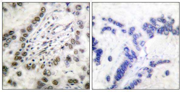
Expect a predominantly nuclear readout: tissue IHC reports ubiquitous nuclear expression, while cellular imaging places CREBBP mainly in nucleoplasm and nuclear bodies (HPA tissue IHC; HPA subcellular). Cytoplasmic signal is biologically possible because UniProt lists both compartments and reports relocalisation under defined conditions, including after UV damage (UniProt Q92793). The catalog paraffin-section image describes nuclear and cytoplasmic staining in human tonsil carcinoma at 1:50 (caption A00205-3). Score nuclear and cytoplasmic compartments separately, compare each with the PBS-for-primary control shown for that image, and avoid treating diffuse cytoplasmic colour alone as proof of CREBBP (caption A00205-3; standard IHC practice).
How can isoforms or epitope placement affect CREBBP IHC interpretation?
CREBBP has 2 annotated isoforms and spans 2,442 amino acids; the record identifies KIX, bromodomain and HAT regions (UniProt Q92793). Establish which sequence the antibody recognises and whether that sequence is shared by both isoforms before calling an absent stain an absent protein (UniProt Q92793; standard IHC practice). The supplied caption does not identify this antibody’s epitope, so its 1:50 tissue staining cannot establish isoform coverage (caption A00205-3). CREBBP also has annotated modified residues, including phosphorylation and arginine methylation; test retrieval and antibody specificity empirically rather than assigning an epitope-accessibility effect to a particular modification without evidence (UniProt Q92793; standard IHC practice).
How should an IF follow-up check the cell source of CREBBP staining?
On the separate IF/ICC workflow, multiplex CREBBP with a marker that identifies the cell population being evaluated, and inspect whether CREBBP signal falls within those cells’ nuclei (standard IF practice; HPA subcellular). Choose fluorophores after measuring tissue autofluorescence in the intended channels, and include single-label controls to assess bleed-through (standard IF practice). For an intracellular nuclear epitope, provide permeabilisation that allows antibody access across the plasma membrane and nuclear envelope; CREBBP has no annotated transmembrane segment (UniProt Q92793; standard IF practice). Compare nuclear patterns with HPA’s nucleoplasm and nuclear-body localisation, but optimise IF fixation independently because the supplied paraffin IHC caption reports no fixative (HPA subcellular; caption A00205-3).
What controls help distinguish CREBBP signal from chromogenic background?
Run a matched no-primary control: the catalog paraffin-section image used PBS instead of primary antibody, followed by biotinylated secondary antibody and avidin–peroxidase detection (caption A00205-3). For a peroxidase and DAB workflow, include a peroxidase block and inspect the no-primary slide for enzyme-related colour; these are general IHC controls, not CREBBP-specific validation (standard IHC practice). Compare intact nuclear staining with diffuse pigment, section folds, damaged edges and necrotic areas before raising antibody concentration (HPA tissue IHC; standard IHC practice). If background persists, assess blocking, washing and detection chemistry on adjacent sections, keeping retrieval and exposure conditions matched (standard IHC practice).
How should I quantify CREBBP staining across tissue sections? ⚠ ANSWER MARKED FOR VERIFICATION
Define the cell population and compartment before scoring, since HPA describes widespread nuclear expression and the catalog image also describes cytoplasmic colour (HPA tissue IHC; caption A00205-3). For nuclear chromogenic staining, report the percentage of positive intact nuclei and an H-score based on staining intensity; record cytoplasmic staining separately (standard IHC practice). Normalise counts to the number of evaluable cells, or report positive-cell density per mm² of viable tissue when cell counts are impractical (standard IHC practice). Apply the same threshold, counterstain assessment, region selection and control-based background correction across sections, excluding folds, edges and necrotic areas (standard IHC practice).
When is an apparent CREBBP-positive area likely to be artefact?
Give greatest weight to reproducible staining in intact nuclei: HPA reports ubiquitous nuclear tissue expression and enhanced nucleoplasmic and nuclear-body localisation (HPA tissue IHC; HPA subcellular). Examine the cells actually stained; high signal is reported in cerebellar granular-layer cells and several epithelial populations, whereas skeletal-muscle myocytes and adipocytes are listed as low (HPA tissue IHC). Treat colour concentrated at cut edges, in necrosis, or in the no-primary control as suspect, and check endogenous peroxidase before interpreting DAB deposits (standard IHC practice; caption A00205-3). Cytoplasmic staining warrants compartment-specific reporting and control comparison, because UniProt documents cytoplasmic CREBBP and the catalog image describes both compartments (UniProt Q92793; caption A00205-3).
Boster reagents

Best CREBBP / CREB-binding protein IHC Antibodies

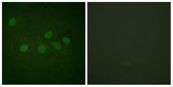
Anti-CREBBP antibodies have IHC images from human and mouse tissues and an IF image from HeLa cells (catalog image captions); all three list human, mouse and rat reactivity (catalog reactivity).

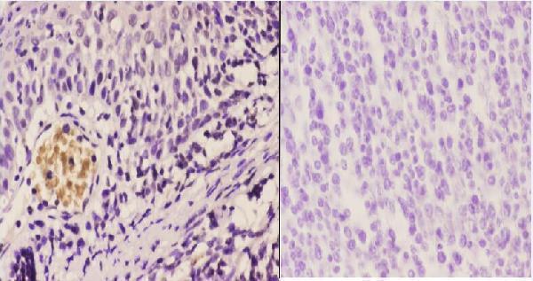
Real IHC data Immunohistochemistry (IHC) analyzes of CBP (E1528) pAb in paraffin-embedded human tonsil carcinoma tissue at 1:50.showing cytoplasmic and nucleus staining. Negative control (the right)Using PBS instead of primary antibody, secondary antibody is Goat Anti-Rabbit IgG-biotin followed by avidin-peroxidase.
Anti-CBP (E1528) CREBBP Antibody
Cat # A00205-3
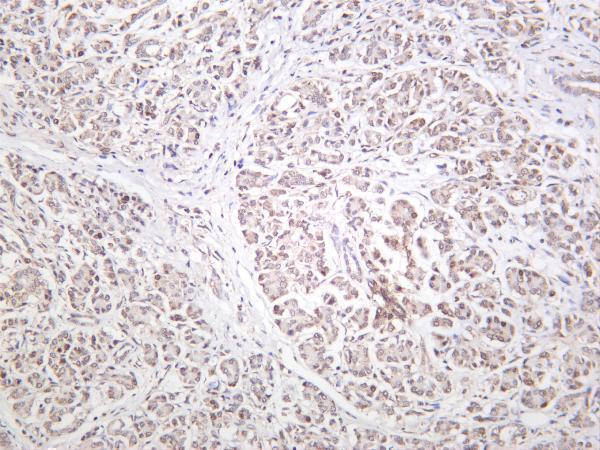
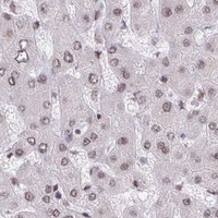
Real IHC data Human pancreas was stained with anti-CREBBP rabbit antibody
Anti-CREBBP Rabbit Monoclonal Antibody
Cat # M00205-1
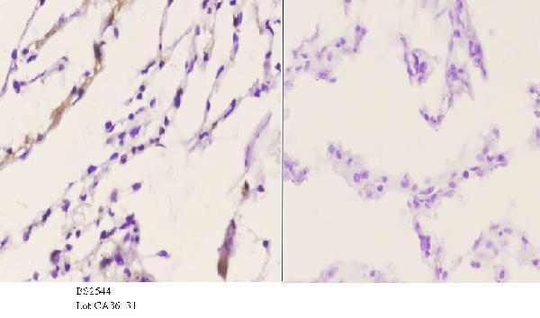
Real IHC data Immunohistochemistry analysis of paraffin-embedded human lung carcinoma tissue, using CBP Antibody. The picture on the right is blocked with the synthesized peptide.
Anti-CBP CREBBP Antibody
Cat # A00205-1

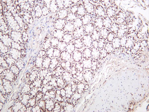
A00205-3 has IHC images from paraffin-embedded human tonsil and lung carcinoma (catalog IHC captions); M00205-1 has IHC images from human pancreas and mouse colon (catalog IHC captions). A00205-1 has an IHC image from paraffin-embedded human lung carcinoma and an IF image from HeLa cells, each with a peptide-blocked comparison (catalog IHC and IF captions).

Which to pick: For paraffin-section IHC, choose A00205-3 for its two tissue images with primary-omission controls; its listed IHC dilution is 1:50–1:200, and the captions do not report a fixative (catalog IHC captions and dilution). For cell IF/ICC, start with A00205-1 because its IF image shows HeLa cells with a peptide-blocked comparison; its listed IF dilution is 1:200–1:1000 (catalog IF caption and dilution). For work spanning species, M00205-1 offers a recombinant rabbit monoclonal antibody with IHC images from human and mouse tissue and listed rat reactivity; all three SKUs list human, mouse and rat reactivity (catalog description, IHC captions and reactivity).

Each figure is that product's own IHC / IF validation image from its datasheet.

References

  1. UniProt Consortium. UniProt entry Q92793 (CBP_HUMAN, CREB-binding protein).
  2. Human Protein Atlas. CREBBP tissue IHC expression (reliability: Supported).
  3. Human Protein Atlas. CREBBP subcellular location (ICC-IF): Mainly localized to the nucleoplasm and nuclear bodies..
  4. Human Protein Atlas. CREBBP antibody validation summary (2 antibodies).
  5. Upregulated microRNA‑330‑3p promotes calcification in the bicuspid aortic valve via targeting CREBBP. Molecular medicine reports 2020 — PMC7411470.
  6. Temporal and spatial distribution of histone acetylation in mouse molar development. PeerJ 2025 — PMC11967410.
  7. Prognostic Significance of CREB-Binding Protein and CD81 Expression in Primary High Grade Non-Muscle Invasive Bladder Cancer: Identification of Novel Biomarkers for Bladder Cancer Using Antibody Microarray. PloS one 2015 — PMC4411067.
  8. Abexinostat, a histone deacetylases inhibitor, for patients with relapsed or refractory follicular lymphoma: a multi-center, single-arm phase 2 study. Signal transduction and targeted therapy 2026 — PMC13111648.
  9. PubMed PMID:9238046 — UniProt-cited evidence.
  10. PubMed PMID:9177780 — UniProt-cited evidence.
  11. PubMed PMID:8782817 — UniProt-cited evidence.