This website uses cookies to ensure you get the best experience on our website.


- Table of Contents
Plan chromogenic CREBZF IHC in paraffin sections with the catalog antibody at 1:100–1:300 (datasheet A07952-2). Compare cytoplasmic tissue staining (HPA tissue IHC) with its nuclear molecular annotation (UniProt), and interpret the pattern cautiously because tissue IHC reliability is uncertain (HPA tissue IHC).

Expected localisation, validated protocols, controls and antibodies — the at-a-glance facts below, then the full design guide.
| Expected localisation | Cytoplasmic in tissue (HPA tissue IHC); nuclear location (UniProt) | |
| Staining pattern | Cytoplasmic staining in most tissues (HPA tissue IHC) | |
| Antigen retrieval | Tris-EDTA pH 9.0 HIER, heat-mediated (datasheet A07952-2) | |
| Positive control | Caudate+4 more · see all | |
| Negative control | Adrenal gland+4 more · see all |
| Fixation | Keep fixation consistent across sections. (standard IHC practice; not target-specific) | |
| Caveat | Tissue staining has low concordance with RNA expression (HPA tissue IHC) | |
| Regulation | Adult tissue abundance varies (UniProt) | |
| Isoform / epitope | No annotated isoforms; one 1–354 chain (UniProt) |
Compare the catalog antibody’s IHC-P protocol with four published CREBZF IHC methods (PMC5915772; PMC6274897; PMC7085260; PMC3878900).
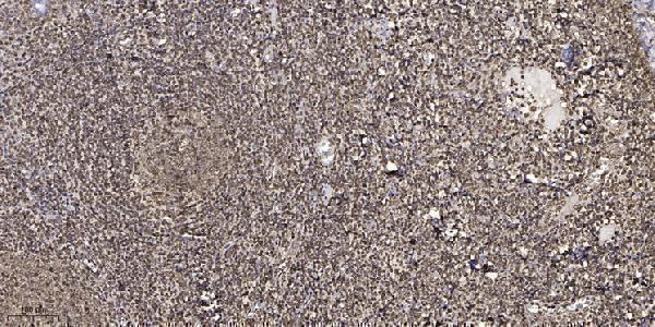
| Sample | Paraffin-embedded human tonsil tissue; fixative not specified (datasheet A07952-2) |
| Fixation | Image fixative and duration unreported (datasheet A07952-2); verify before use. |
| Sectioning | 4–5 µm sections on charged slides (standard) |
| Deparaffinisation | Xylene, graded ethanol series to water (standard) |
| Antigen retrieval | Heat retrieval: Tris-EDTA pH 9.0 (datasheet A07952-2); 20 min, 95–100 °C (standard) |
| Peroxidase block | 3% H2O2, 10 min, room temperature (standard) |
| Blocking | 10% normal serum of the secondary host, 30 min, room temperature (standard) |
| Primary antibody | Rabbit anti-CREBZF, 1:100-1:300 (datasheet A07952-2) |
| Primary incubation | Overnight at 4 °C (standard) |
| Detection | HRP-polymer secondary, DAB chromogen 5–10 min (standard) |
| Counterstain | Hematoxylin, blue, dehydrate and mount (standard) |
| Expected result | CREBZF-positive staining in neuronal cells of caudate (HPA tissue IHC: Medium). HPA tissue profile: Cytoplasmic expression in most tissues. No signal in the no-primary control. |
CREBZF is a nuclear transcription factor with no transmembrane segment (UniProt Q9NS37). In tissue IHC, HPA instead reports mainly cytoplasmic staining, including medium signal in cardiomyocytes and kidney glomerular cells (HPA tissue IHC). Treat that pattern as provisional: HPA rates its tissue IHC reliability Uncertain because antibody staining and RNA expression show low consistency (HPA tissue IHC).
| Medium cytoplasmic staining in cardiomyocytes or kidney glomerular cells. | This matches the reported tissue IHC pattern (HPA tissue IHC: Medium in both). Record the stained cell type and compartment; the pattern is provisional because tissue IHC reliability is Uncertain (HPA tissue IHC). |
| Nuclear signal accompanies cytoplasmic staining in cells of interest. | Nuclear localisation is biologically plausible (UniProt Q9NS37: nucleus), but HPA tissue IHC describes chiefly cytoplasmic staining (HPA tissue IHC). Score the compartments separately and seek independent confirmation before treating either as definitive. |
| Staining is confined to extracellular material or appears as granular deposit outside cells. | That distribution does not match the reported cellular IHC pattern (HPA tissue IHC) or nuclear localisation (UniProt Q9NS37). Check the no-primary control and chromogen distribution; deposition or background is a more useful first interpretation than target localisation (general IHC practice). |
| Strong signal appears in cholangiocytes or adrenal glandular cells. | HPA reports these specific cell populations as Not detected (HPA tissue IHC). Review cell identification and no-primary controls; cross-reactivity or endogenous detection activity is possible (general IHC practice), although HPA's Uncertain rating prevents a definitive false-positive call (HPA tissue IHC). |
| No staining appears in cardiomyocytes or kidney glomerular cells. | These are reported Medium examples (HPA tissue IHC), so inspect positive-control performance, retrieval and detection conditions (general IHC practice). An absent signal alone cannot establish absent CREBZF, given the Uncertain tissue IHC reliability (HPA tissue IHC). |
| How should the compartment mismatch affect scoring? | UniProt places CREBZF in the nucleus, including PML nuclear bodies (UniProt Q9NS37); HPA tissue IHC describes cytoplasmic expression in most tissues (HPA tissue IHC). Record nuclear and cytoplasmic scores separately, without assuming the IHC pattern resolves the disagreement. |
| Which tissue examples are useful for comparison? | Heart cardiomyocytes and kidney glomerular cells are Medium examples; adrenal glandular cells and liver cholangiocytes are Not detected examples (HPA tissue IHC). Compare the named cell populations, rather than treating an entire organ as uniformly positive or negative. |
| How strong is the antibody evidence? | The tissue IHC profile is Uncertain, with low consistency between staining and RNA expression (HPA tissue IHC). HPA021526 has Uncertain IHC status; HPA063652 has Supported ICC status, which does not validate the tissue IHC pattern (HPA antibodies). |
| What does IF/ICC show? | HPA reports approved nucleoplasm and mitochondria localisation in ICC-IF, with images from HaCaT, HeLa and U2OS (HPA subcellular). This is a separate assay and does not turn mitochondrial staining into an established paraffin-section IHC expectation (HPA subcellular; HPA tissue IHC). |
| Can processing or topology explain membrane staining? | CREBZF has no transmembrane segment, signal peptide, propeptide or reported isoforms, and its annotated chain spans residues 1–354 (UniProt Q9NS37). A membrane-only pattern has no support from those annotations; check controls and antibody specificity (general IHC practice). |
| What is known about fixation sensitivity? | No target-specific fixation effect is reported in the supplied UniProt or HPA evidence. If signal is weak, review the laboratory's general retrieval and detection controls (general IHC practice); do not infer that fixation selectively altered CREBZF staining. |
| Situation | Likely cause | Next action |
|---|---|---|
| Signal covers nuclei, cytoplasm and empty slide areas uniformly. | Nonspecific background, residual reagent or excessive detection can produce widespread colour (general IHC practice). | Compare with the no-primary control; review blocking, washing and detection conditions (general IHC practice). Do not score diffuse slide-wide colour as the HPA cellular pattern (HPA tissue IHC). |
| A tissue expected to be positive has no visible signal. | HPA reports Medium cardiomyocyte and glomerular-cell staining, but rates tissue IHC Uncertain (HPA tissue IHC); technical failure and biological variability remain possible. | Check a same-run positive control, section quality, retrieval and detection steps (general IHC practice). Document the absent signal without converting HPA's example into a guaranteed positive. |
| Only nuclear staining is present in an IHC section. | Nuclear localisation fits UniProt, while HPA tissue IHC chiefly reports cytoplasmic staining (UniProt Q9NS37; HPA tissue IHC). | Score nuclear staining separately; compare cell distribution with controls and, if needed, an independent antibody (general IHC practice). Report the source disagreement rather than silently combining compartments. |
| Signal appears in an HPA Not detected cell population. | Unexpected staining may reflect cell misidentification, cross-reactivity or endogenous detection activity (general IHC practice); the HPA IHC assessment is Uncertain (HPA tissue IHC). | Verify the cell population and inspect no-primary and detection controls (general IHC practice). Treat HPA's Not detected label as a comparison point, not proof that every signal is artefactual. |
| Brown precipitate seems to outline vessels or sit outside cells. | Extracellular deposits are inconsistent with the cellular IHC profile (HPA tissue IHC) and nuclear annotation (UniProt Q9NS37); chromogen artefact is possible (general IHC practice). | Inspect an unstained or no-primary section and check wash and chromogen handling (general IHC practice). Exclude deposits from cellular scoring. |
| ICC-IF shows mitochondrial signal, but IHC scoring expects cytoplasm. | Approved ICC-IF localisation includes mitochondria and nucleoplasm (HPA subcellular); tissue IHC describes broad cytoplasmic expression with Uncertain reliability (HPA tissue IHC). | Keep assay-specific annotations separate. Record the observed IHC compartment and cell type, and use independent validation if a precise mitochondrial claim is needed (general IHC practice). |


Comprehensive Human Protein Atlas IHC scoring per tissue (reliability: Uncertain — Low consistency between antibody staining and RNA expression data. Pending external verification.). Rows are taken directly from the HPA tissue chart — click any row's HPA link to view the source.
| Tissue | Cell type | Level | Evidence | Source |
|---|---|---|---|---|
| Caudate | Neuronal cells | Medium | Protein (IHC) | HPA → |
| Cerebellum | Purkinje cells | Medium | Protein (IHC) | HPA → |
| Cerebral cortex | Neuropil | Medium | Protein (IHC) | HPA → |
| Esophagus | Squamous epithelial cells | Medium | Protein (IHC) | HPA → |
| Gallbladder | Glandular cells | Medium | Protein (IHC) | HPA → |
| Tissue | Cell type | Level | Evidence | Source |
|---|---|---|---|---|
| Adrenal gland | Glandular cells | Not detected | Protein (IHC) | HPA → |
| Bone marrow | Hematopoietic cells | Not detected | Protein (IHC) | HPA → |
| Breast | Adipocytes | Not detected | Protein (IHC) | HPA → |
| Cervix | Glandular cells | Not detected | Protein (IHC) | HPA → |
| Duodenum | Glandular cells | Not detected | Protein (IHC) | HPA → |
Use compartment-aware controls when evaluating CREBZF in chromogenic paraffin-section IHC; reported nuclear and tissue-staining patterns require careful interpretation (UniProt Q9NS37; HPA tissue IHC).
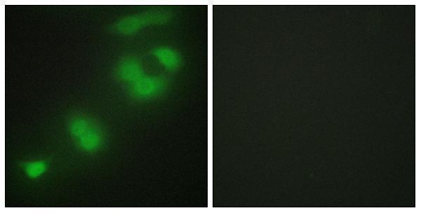
A07952-2 has IHC data from paraffin-embedded human tonsil and IF data from HepG2 cells (catalog image captions). Listed reactivity covers Human and Mouse (catalog reactivity).
A07952-2 will render with an IHC image of paraffin-embedded human tonsil (catalog IHC image caption). Its IF image shows HepG2 cells with peptide blocking; IHC and IF are listed applications (catalog IF image caption; catalog applications).
Which to pick: For tissue IHC, choose A07952-2 for paraffin sections: its image caption reports Tris-EDTA pH 9 retrieval and a 1:200 primary dilution; the fixative is unreported (catalog IHC image caption). For IF, A07952-2 has a HepG2 image; ICC validation is unreported, and the antibody is rabbit-hosted with no clone specified (catalog IF image caption; catalog host/clone). For cross-species work, A07952-2 lists Human and Mouse reactivity, while its supplied IHC image documents human tissue only (catalog reactivity; catalog IHC image caption).