This website uses cookies to ensure you get the best experience on our website.


- Table of Contents
Plan chromogenic CREM IHC in paraffin sections using nuclear staining in spermatocytes and spermatids as the tissue benchmark (HPA tissue IHC). This guide covers fixation consistency, antibody titration, and interpretation across 29 isoforms (UniProt).

Expected localisation, validated protocols, controls and antibodies — the at-a-glance facts below, then the full design guide.
| Expected localisation | Nuclear in spermatocytes and spermatids (HPA tissue IHC) | |
| Staining pattern | Nuclear staining in spermatocytes and spermatids (HPA tissue IHC) | |
| Antigen retrieval | Tris-EDTA pH 8.0 HIER, heat-mediated (datasheet A04379-1) | |
| Positive control | Testis+4 more · see all | |
| Negative control | Adipose tissue+4 more · see all |
| Fixation | Keep fixation consistent across paraffin sections (standard IHC practice; not target-specific) | |
| Caveat | Isoform variation may affect antibody recognition (UniProt) | |
| Regulation | High in round or early spermatids (HPA tissue IHC) | |
| Isoform / epitope | 29 isoforms; check epitope coverage (UniProt) |
Compare the catalog antibody’s IHC-P protocol (datasheet A04379-1) with four published CREM IHC protocols (PMC8329441; PMC5013872; PMC6389866; PMC3572472).
| Sample | Paraffin-embedded Human brain tissue; fixative not specified (datasheet A04379-1) |
| Fixation | Image fixative and duration unreported (datasheet A04379-1); verify before use. |
| Sectioning | 4–5 µm sections on charged slides (standard) |
| Deparaffinisation | Xylene, graded ethanol series to water (standard) |
| Antigen retrieval | Heat retrieval: Tris-EDTA pH 8.0 (datasheet A04379-1); 20 min, 95–100 °C (standard) |
| Peroxidase block | 3% H2O2, 10 min, room temperature (standard) |
| Blocking | 10% normal serum of the secondary host, 30 min, room temperature (standard) |
| Primary antibody | Rabbit anti-CREM, 1:100-1:300 (datasheet A04379-1) |
| Primary incubation | Overnight at 4 °C (standard) |
| Detection | HRP-polymer secondary, DAB chromogen 5–10 min (standard) |
| Counterstain | Hematoxylin, blue, dehydrate and mount (standard) |
| Expected result | CREM-positive staining in round or early spermatids of testis (HPA tissue IHC: High). HPA tissue profile: Nuclear expression in spermatocytes and spermatids. No signal in the no-primary control. |
CREM is a nuclear and cytoplasmic transcriptional regulator with no transmembrane segment (UniProt Q03060). In paraffin sections, the clearest expected IHC signal is nuclear staining in testicular spermatocytes and spermatids, strongest in round or early spermatids (HPA tissue IHC: Enhanced; High in round or early spermatids). Interpret staining by cell type and compartment together: HPA reports additional, generally medium staining in selected epithelial cells (HPA tissue IHC).
| Strong nuclear chromogen in round or early spermatids, with nuclear staining in spermatocytes. | This matches the reference pattern. HPA rates round or early spermatids High and describes nuclear expression in spermatocytes and spermatids (HPA tissue IHC: Enhanced). |
| Testicular signal is predominantly diffuse cytoplasmic, with little nuclear staining. | Treat this as a localisation mismatch and possible staining artefact against the HPA tissue pattern (HPA tissue IHC: nuclear expression). Cytoplasmic CREM remains biologically plausible, so compartment alone cannot establish antibody specificity (UniProt Q03060: nucleus; cytoplasm). |
| Strong staining appears in adipocytes or lymph node germinal center cells. | These cell types are listed as Not detected (HPA tissue IHC). Cross-reactivity or endogenous detection activity is possible; a positive result there needs controls before it is attributed to CREM (general IHC practice). |
| Brown haze covers tissue, including cells expected to have little signal. | A widespread, poorly localised deposit cannot be scored as the expected cell-specific nuclear pattern (HPA tissue IHC). Excess background from staining or detection is a possible cause (general IHC practice). |
| No signal appears in round or early spermatids on a testis section. | That conflicts with the High reference staining (HPA tissue IHC). Check section quality and the staining run before calling the sample CREM-negative; absence of chromogen alone does not identify the failed step (general IHC practice). |
| Reference tissue and antibody validation | Testis round or early spermatids provide a High-staining reference, while HPA lists both HPA001818 and CAB018352 as IHC Enhanced (HPA tissue IHC; HPA antibodies). Those ratings support the reported pattern; they do not validate every antibody or staining run. |
| Cell type and compartment | HPA describes nuclear staining in spermatocytes and spermatids, but also Medium staining in goblet and selected glandular cells (HPA tissue IHC). UniProt lists both nucleus and cytoplasm; do not require every positive tissue to resemble testis. |
| Isoforms and antibody epitope | UniProt lists 29 CREM isoforms and identifies isoform 14 as the major testis activator (UniProt Q03060). No epitope is supplied here, so isoform coverage and any isoform-specific staining difference cannot be predicted for an antibody. |
| Protein features and target-specific limits | CREM has no signal peptide or transmembrane segment; its recorded chain spans residues 1–345 and includes several phosphorylated serines (UniProt Q03060). These facts do not establish antigen-retrieval conditions or fixation sensitivity; neither is reported in the supplied sources. |
| IF/ICC Q: What compartment should be checked? | A: Expect mainly nucleoplasmic signal, with additional vesicle localisation (HPA subcellular ICC-IF: nucleoplasm supported; vesicles approved). This ICC-IF observation informs localisation checks; it is not an IHC-P staining protocol or a substitute for the tissue IHC pattern. |
| Situation | Likely cause | Next action |
|---|---|---|
| Testis positive control has weak or absent staining. | Possible section, retrieval, primary-antibody, or detection failure (general IHC practice); round or early spermatids are expected to stain High (HPA tissue IHC). | Review tissue morphology and run controls, then optimise retrieval and primary concentration within the IHC-validated antibody's instructions; compare nuclear signal in round or early spermatids (general IHC practice; HPA tissue IHC). |
| Testis staining is mostly cytoplasmic rather than nuclear. | The distribution conflicts with the HPA testis pattern, although UniProt also lists cytoplasm as a CREM location (HPA tissue IHC; UniProt Q03060). | Check nuclear counterstain and cell identity, reduce background, and compare an independently validated IHC antibody if available; avoid scoring cytoplasmic colour alone as the reference pattern (general IHC practice; HPA tissue IHC). |
| Strong colour appears in an HPA Not detected cell type. | Cross-reactivity or endogenous detection activity could explain the discrepancy; HPA lists adipocytes and lymph node germinal center cells as Not detected (HPA tissue IHC; general IHC practice). | Inspect a no-primary control and evaluate endogenous-enzyme blocking for the chromogenic system; repeat with a validated antibody before assigning the colour to CREM (general IHC practice). |
| Diffuse brown background obscures nuclei. | Excess reagent, inadequate blocking, or insufficient washing can produce diffuse signal (general IHC practice). The expected testis pattern is cell-specific and nuclear (HPA tissue IHC). | Titrate the primary antibody, review blocking and wash steps, and compare with a no-primary control; score only cells whose nuclear signal remains distinguishable (general IHC practice). |
| The no-primary control develops brown deposits. | Primary-antibody-independent colour points to the detection system or tissue activity, not a demonstrated CREM signal (general IHC practice). | Check reagent controls and apply the appropriate endogenous-activity block for the chromogen system, then rerun the control alongside the testis reference section (general IHC practice; HPA tissue IHC: High in round or early spermatids). |
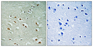
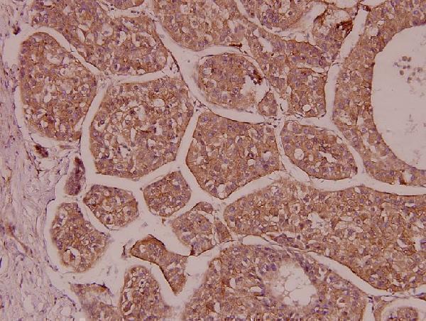


Comprehensive Human Protein Atlas IHC scoring per tissue (reliability: Enhanced — High consistency between antibody staining and RNA expression data.). Rows are taken directly from the HPA tissue chart — click any row's HPA link to view the source.
| Tissue | Cell type | Level | Evidence | Source |
|---|---|---|---|---|
| Testis | Round or early spermatids | High | Protein (IHC) | HPA → |
| Appendix | Goblet cells | Medium | Protein (IHC) | HPA → |
| Colon | Goblet cells | Medium | Protein (IHC) | HPA → |
| Duodenum | Goblet cells | Medium | Protein (IHC) | HPA → |
| Endometrium | Glandular cells | Medium | Protein (IHC) | HPA → |
| Tissue | Cell type | Level | Evidence | Source |
|---|---|---|---|---|
| Adipose tissue | Adipocytes | Not detected | Protein (IHC) | HPA → |
| Caudate | Glial cells | Not detected | Protein (IHC) | HPA → |
| Cerebral cortex | Endothelial cells | Not detected | Protein (IHC) | HPA → |
| Cervix | Glandular cells | Not detected | Protein (IHC) | HPA → |
| Esophagus | Squamous epithelial cells | Not detected | Protein (IHC) | HPA → |
Troubleshoot CREM staining in paraffin sections by checking retrieval, nuclear localisation and cell type before interpreting chromogenic signal (datasheet A04379-1; HPA tissue IHC).
Two anti-CREM antibodies have IHC images from paraffin-embedded human brain and liver carcinoma (A04379-1 and A04379 image captions); both list human, mouse and rat reactivity, and A04379-1 lists IF (catalog applications and reactivity).
A04379 will render with IHC of paraffin-embedded human liver carcinoma at 1:100 (A04379 image caption). A04379-1 will render with IHC of paraffin-embedded human brain at 1:100 after Tris-EDTA pH 8.0 retrieval (A04379-1 image caption); IF is also listed for this SKU (catalog applications).
Which to pick: For tissue IHC, choose A04379 for the shown paraffin-embedded human liver carcinoma sample (A04379 image caption), or A04379-1 for the shown paraffin-embedded human brain sample (A04379-1 image caption); neither caption reports the fixative (image captions). For IF/ICC planning, A04379-1 is the relevant starting point because it lists IF at 1:50 (catalog applications and IF dilution); ICC testing and an IF image are unreported (catalog applications and image captions). For mouse or rat tissue, either SKU lists reactivity with those species (catalog reactivity), though both IHC images show human samples (image captions); both antibodies have a rabbit host, and clone status is unreported (catalog host and clone).