CRHBP / Corticotropin-releasing hormone-binding protein · IHC design guide

Design Immunohistochemistry for CRHBP

Plan CRHBP paraffin IHC around cytoplasmic staining and plasma positivity (HPA tissue IHC). Use testis round or early spermatids, reported as high, as a reference for scoring (HPA tissue IHC).

Evidence assembled Oct 2026 · For research use; verify linked source records and product datasheet before use
Immunohistochemistry protocol sheet for CRHBP (IHC for CRHBP): expected localisation Cytoplasmic staining (HPA tissue IHC), antibody A06764, validated IHC image, and IHC protocol steps
Printable CRHBP IHC protocol sheet — expected localisation Cytoplasmic staining (HPA tissue IHC), antibody A06764, controls and protocol steps. Open the full CRHBP IHC guide →

CRHBP Immunohistochemistry Experimental Design Guide

Expected localisation, validated protocols, controls and antibodies — the at-a-glance facts below, then the full design guide.

Must know before staining
Expected localisation Cytoplasmic staining (HPA tissue IHC)
Staining pattern Cytoplasmic staining in most tissues, with plasma positivity (HPA tissue IHC)
Antigen retrieval Citrate pH 6.0 HIER, 95–98 °C, 20 min (rule: cytoplasmic / membrane antigen)
Positive control ⓘ Testis
Negative control ⓘ Adipose tissue+4 more · see all
Important caveats
Reasons your staining may differ from the expected pattern.
Fixation Keep formalin fixation consistent across sections (standard IHC practice; not target-specific)
Caveat Secretion can separate tissue staining from RNA location (HPA tissue IHC)
Regulation No expression regulator annotated (UniProt)
Isoform / epitope No isoforms annotated; mature chain starts at aa 25 (UniProt)
Section 1

Recommended CRHBP IHC & IF Protocols

The catalog antibody’s IHC-P protocol is accompanied by 3 published CRHBP IHC protocols covering HCC sections and rat hippocampus (PMC6096283; PMC4777610; PMC11633302).

Recommended immunohistochemistry (IHC-P) protocol parameters
SampleParaffin-embedded mouse kidney tissue; fixative not specified (datasheet A06764)
FixationImage fixative and duration unreported (datasheet A06764); verify before use.
Sectioning4–5 µm sections on charged slides (standard)
DeparaffinisationXylene, graded ethanol series to water (standard)
Antigen retrievalHeat-induced epitope retrieval in citrate buffer, pH 6.0, 20 min at 95–98 °C (standard rule: cytoplasmic / membrane antigen)
Peroxidase block3% H2O2, 10 min, room temperature (standard)
Blocking10% normal serum of the secondary host, 30 min, room temperature (standard)
Primary antibodyRabbit anti-CRHBP, 1:100–1:500 starting range (standard)
Primary incubationOvernight at 4 °C (standard)
DetectionHRP-polymer secondary, DAB chromogen 5–10 min (standard)
CounterstainHematoxylin, blue, dehydrate and mount (standard)
Expected resultCRHBP-positive staining in round or early spermatids of testis (HPA tissue IHC: High). HPA tissue profile: Cytoplasmic expression in most tissues, including plasma positivity. No signal in the no-primary control.
💡Decision noteStart with citrate pH 6.0 retrieval at 95–98 °C for 20 min (page retrieval). Compare the published high-pressure conditions if staining needs optimization (PMC6096283; PMC11633302).
Section 2

What Is the Expected CRHBP Staining Pattern?

CRHBP is secreted and has no transmembrane segment (UniProt P24387: subcellular location and topology). In tissue IHC, expect predominantly cytoplasmic staining, with possible plasma positivity (HPA tissue IHC: Approved; profile). Round or early spermatids in testis are a documented high-staining population (HPA tissue IHC: High). Because CRHBP is secreted, its tissue protein location need not match its RNA location (HPA tissue IHC: reliability description).

What am I looking at on my slide?
Strong cytoplasmic staining in round or early spermatids, with restrained background in adjacent structures.This matches the documented high-staining testis population (HPA tissue IHC: High in round or early spermatids). Plasma positivity can also fit the reported tissue profile (HPA tissue IHC: plasma positivity). Judge the cellular pattern alongside the specimen's morphology (general IHC practice).
Predominantly nuclear staining, or a crisp membrane rim presented as the principal CRHBP pattern.Treat this as a localisation mismatch: the reported tissue pattern is cytoplasmic (HPA tissue IHC: profile), and CRHBP has no transmembrane segment (UniProt P24387: topology). Review staining controls and antibody specificity before assigning such signal to CRHBP (general IHC practice).
Strong staining in a population listed as not detected, such as adrenal glandular cells or adipocytes.This conflicts with those sampled HPA observations (HPA tissue IHC: Not detected in adrenal glandular cells and adipocytes). Consider cross-reactivity or endogenous detection activity, then check a reagent control (general IHC practice). An HPA negative call is a reference observation, not proof that every specimen must be negative.
Broad, diffuse colour covers cells and surrounding tissue, obscuring cellular boundaries.CRHBP can be present in plasma (HPA tissue IHC: profile), so extracellular colour alone is not decisive. If colour is widespread without a discernible cell-specific pattern, investigate nonspecific background, blocking, washes and detection controls (general IHC practice). Compare with the high-staining spermatid pattern (HPA tissue IHC: High).
No detectable staining in round or early spermatids in a testis section.The expected high-staining reference population is missing (HPA tissue IHC: High in round or early spermatids). Check tissue preservation and section quality, antibody and detection steps, and a positive control (general IHC practice). A failed run cannot establish CRHBP absence in the specimen.
💡Expected CRHBP appearanceA convincing positive shows high cytoplasmic staining in round or early spermatids (HPA tissue IHC: High; cytoplasmic profile); dominant nuclear or crisp membrane-rim staining is a localisation mismatch (HPA tissue IHC: profile; UniProt P24387: topology).
How each factor affects the staining
Secretion and compartmentCRHBP is secreted and lacks a transmembrane segment (UniProt P24387: subcellular location and topology). HPA nevertheless reports cytoplasmic tissue staining and plasma positivity (HPA tissue IHC: profile). Interpret cellular signal and plasma signal separately; neither alone defines an ICC organelle location.
Protein processingUniProt annotates a signal peptide at residues 1–24 and a mature chain at 25–322 (UniProt P24387: processing). The supplied sources give no antibody epitope, so they cannot establish whether either region is recognised after processing.
GlycosylationOne glycosylation site is annotated at residue 204 (UniProt P24387: glycosylation). Its effect on recognition by a particular IHC antibody is unreported in the supplied sources; do not infer a need for antigen retrieval or altered staining from this annotation alone.
RNA versus protein locationRNA is tissue enhanced in brain and liver, while the IHC profile describes cytoplasmic staining in most tissues (HPA: RNA specificity and tissue IHC profile). HPA cautions that locations of secreted RNA and protein may differ (HPA tissue IHC: reliability description); RNA enrichment alone does not establish a positive control cell.
Antibody evidenceTwo listed antibodies have different IHC status: HPA036234 is Supported and HPA046120 is Approved (HPA: antibody validation). These labels support reviewing antibody-specific evidence; they do not supply the catalog antibody's epitope, dilution, antigen retrieval method or fixation sensitivity.
IF/ICC: what location is established?Only a secreted summary is available; HPA gives no main ICC location and no cell-line ICC-IF images (HPA subcellular: summary and image fields). UniProt also calls CRHBP secreted (UniProt P24387: subcellular location). Thus this record cannot specify an IF organelle pattern; consult the separate IF/ICC guide for experimental design.
Why is my staining missing, weak or wrong?
SituationLikely causeNext action
Testis positive control is blank.The documented high-staining round or early spermatid signal is absent (HPA tissue IHC: High); a staining step or specimen issue may be responsible (general IHC practice).Confirm the expected cells are present; review section quality, primary antibody, detection reagent and counterstain, then repeat with a working control (general IHC practice).
A not-detected reference population stains strongly.Cross-reactivity or endogenous detection activity can mimic target staining (general IHC practice); compare the specific population with its HPA call (HPA tissue IHC: Not detected).Use an appropriate primary-omission or detection control, review antibody validation, and score the cell type rather than the tissue name alone (general IHC practice; HPA: antibody validation).
Diffuse background makes cells hard to score.Nonspecific reagent signal or insufficient washing is possible (general IHC practice). Plasma positivity is also reported for CRHBP (HPA tissue IHC: profile).Inspect a detection control, blocking and washes; compare extracellular colour with cellular staining in the high-staining spermatid reference (general IHC practice; HPA tissue IHC: High).
Signal is mainly nuclear or outlines membranes.That pattern conflicts with the cytoplasmic IHC profile (HPA tissue IHC: profile) and the lack of a transmembrane segment (UniProt P24387: topology).Check morphology and detection controls, then compare another validated antibody's IHC evidence where available (general IHC practice; HPA: antibody validation).
Brain or liver RNA enrichment is used to declare an IHC result positive.The RNA designation is tissue enhanced, while HPA warns that secreted protein and RNA locations may differ (HPA: RNA specificity and reliability description).Score observed protein staining by cell type and compartment; use the documented high-staining testis population as the supplied positive reference (HPA tissue IHC: High).
An IF image is assigned a specific intracellular organelle.HPA provides no main ICC location or cell-line ICC-IF images for CRHBP (HPA subcellular: location and image fields).Report the observed IF pattern without naming an established CRHBP organelle location; follow the separate IF/ICC guide for controls and imaging choices (HPA subcellular: Secreted; general IF practice).

Sample controls for CRHBP IHC & IF

🧪Run testis first and expect staining in round or early spermatids (High; HPA: testis). Run adipose tissue as the negative tissue, focusing on adipocytes (HPA: Not detected in adipocytes); on the testis slide, use cells outside the round or early spermatid compartment as background comparators without assuming they are target-negative.
Positive control tissue: Testis (Round or early spermatids, HPA High)
Negative control tissue: Adipose tissue (HPA Not detected)
ICC-IF cell lines (HPA subcellular resource): HPA carries no ICC-IF cell line for CRHBP; derive a cell-line control from the positive tissue's cell type (Round or early spermatids) and confirm it by RNA or western blot first.
Technical controls: Include a no-primary (secondary-only) slide, an isotype control matched to the primary antibody’s host species, immunoglobulin class and clonality, and CRHBP-knockout tissue or a validated peptide-block control (standard IHC practice). Check testis sections for endogenous peroxidase signal and nonspecific chromogen deposition (standard chromogenic IHC practice).
⚠️Feasibility: A target-specific fixation window and retrieval dependency are unreported in the supplied evidence; the paraffin-section mouse kidney caption gives a 1:300 dilution but does not report a fixative (A06764 caption). The supplied evidence does not establish whether frozen sections or IF are easier, and HPA lists no ICC-IF cell-line images (HPA subcellular). No target-specific testis artefact is reported in the supplied evidence.

HPA tissue IHC evidence for CRHBP

Comprehensive Human Protein Atlas IHC scoring per tissue (reliability: Approved — Secreted protein, tissue location of RNA and protein is expected to differ.). Rows are taken directly from the HPA tissue chart — click any row's HPA link to view the source.

Positive expression · recommended positive controls

TissueCell typeLevelEvidenceSource
Testis Round or early spermatids High Protein (IHC) HPA →

Undetected expression · recommended negative controls

TissueCell typeLevelEvidenceSource
Adipose tissue Adipocytes Not detected Protein (IHC) HPA →
Adrenal gland Glandular cells Not detected Protein (IHC) HPA →
Appendix Glandular cells Not detected Protein (IHC) HPA →
Bone marrow Hematopoietic cells Not detected Protein (IHC) HPA →
Breast Adipocytes Not detected Protein (IHC) HPA →
Section 3

Advanced CRHBP IHC Tips

Troubleshoot CRHBP staining in paraffin sections by checking retrieval, cell context, extracellular signal, and controls (UniProt P24387; HPA tissue IHC).

What retrieval conditions should I start with for CRHBP in paraffin sections?
Start with citrate buffer at pH 6.0, heated to 95–98 °C for 20 min (page retrieval rule). Apply the same cooling and wash sequence to control and test sections so retrieval differences do not masquerade as expression differences (standard IHC practice). The selected A06764 image documents CRHBP staining in paraffin-embedded mouse kidney at 1:300, but its caption does not report retrieval conditions (A06764 caption). If staining is weak, compare a modestly longer citrate retrieval with the starting condition on serial sections, while checking tissue morphology and background (standard IHC practice). Do not treat stronger staining alone as proof of specificity (standard IHC practice).
How should I handle fixation when CRHBP staining varies between sections?
Target-specific fixation sensitivity is unknown because the supplied CRHBP evidence gives no fixation comparison, and the A06764 paraffin-section caption does not state a fixative (A06764 caption). Record the fixative, fixation duration, tissue thickness, and processing history for each specimen before comparing staining (standard IHC practice). Keep those variables consistent across matched sections, then run a small fixation or retrieval comparison only if suitable specimens are available (standard IHC practice). Inspect morphology alongside signal: damaged tissue can make a staining difference difficult to interpret (standard IHC practice). The HPA staining profile and UniProt protein annotations do not establish an optimal fixative (HPA tissue IHC; UniProt P24387).
Where should convincing CRHBP staining appear in tissue?
Interpret CRHBP as a secreted protein with no annotated transmembrane segment, so extracellular or plasma-associated signal can be biologically plausible (UniProt P24387 topology; HPA tissue IHC). HPA also reports cytoplasmic staining in most tissues and describes plasma positivity (HPA tissue IHC). For cellular interpretation, compare signal with cell boundaries and adjacent extracellular space on the same section, using a counterstain to identify the relevant cells (standard IHC practice). HPA reports high staining in round or early spermatids in testis, offering a documented cellular reference for comparison (HPA tissue IHC). Avoid assigning diffuse signal to a particular producing cell solely from its location, because tissue RNA and protein locations may differ for a secreted protein (HPA tissue IHC).
Could processing or glycosylation alter which CRHBP epitope is visible?
Check the antibody's stated immunogen before interpreting a missing signal: the CRHBP precursor has a 1–24 signal peptide and a 25–322 mature chain (UniProt P24387 processing). An epitope confined to the cleaved signal peptide would have a different interpretation from one in the mature chain (UniProt P24387 processing; standard epitope interpretation). UniProt annotates one glycosylation site at residue 204 and 0 isoforms; these annotations do not identify the catalog antibody's epitope (UniProt P24387). Compare retrieval conditions and controls before attributing weak staining to epitope masking (standard IHC practice). Do not claim isoform-selective staining without an epitope map and supporting validation (UniProt P24387 isoform annotation; standard IHC practice).
How can I cross-check chromogenic CRHBP IHC with multiplex IF?
Use IF as a complementary localisation check and multiplex CRHBP with a validated marker for the expected cell type; HPA identifies round or early spermatids as a high-staining testis population (HPA tissue IHC; standard IF practice). Image an unstained section first, then choose fluorophores and filter sets that separate specific signal from that tissue's autofluorescence (standard IF practice). CRHBP is secreted and has no transmembrane segment, so an intracellular epitope may require mild permeabilisation, whereas extracellular staining should be assessed before permeabilisation (UniProt P24387 topology; standard IF practice). Keep antibody omission and single-color controls to identify background and channel bleed-through (standard IF practice). Do not transfer the A06764 paraffin-section conditions directly to IF, because its caption describes IHC only (A06764 caption).
How do I reduce diffuse or enzyme-related background without losing CRHBP signal?
First inspect a no-primary control beside the test section to distinguish detection-system staining from antibody-dependent staining (standard IHC practice). For chromogenic IHC, block endogenous peroxidase before an HRP and DAB detection step, and match blocking and wash conditions across sections (standard IHC practice). Titrate the primary antibody around the documented 1:300 A06764 mouse-kidney image condition if using that exact antibody, checking whether cellular contrast improves (A06764 caption; standard IHC practice). CRHBP is secreted and HPA reports plasma positivity, so diffuse signal is not automatically nonspecific (UniProt P24387; HPA tissue IHC). Judge it against section morphology, controls, and the expected cell context rather than intensity alone (standard IHC practice).
How should I score CRHBP staining across paraffin sections? ⚠ ANSWER MARKED FOR VERIFICATION
Define the compartment and cell population before scoring, because HPA describes both cytoplasmic expression and plasma positivity for secreted CRHBP (HPA tissue IHC; UniProt P24387). For a defined cellular population, report the percentage of positive cells or an H-score using fixed intensity categories and the same threshold across specimens (standard IHC practice). For extracellular signal, quantify stained area or optical density per mm² of viable tissue with a consistent background subtraction rule (standard IHC practice). Normalize cellular counts to eligible cells and extracellular measurements to analyzed viable area, excluding folds and necrotic regions (standard IHC practice). Keep acquisition, counterstain, detection development, and region selection consistent, and report cellular and extracellular results separately (standard IHC practice).
What makes a CRHBP-positive region credible rather than an artefact?
A credible result has reproducible staining in an anatomically defined region and survives comparison with no-primary and detection controls (standard IHC practice). HPA reports high staining in testis round or early spermatids and no detected staining in several listed cell populations, which can inform context but does not validate every specimen or antibody (HPA tissue IHC). Check whether signal follows plausible cytoplasmic or extracellular distribution for secreted CRHBP, rather than appearing only at cut edges or within necrotic tissue (UniProt P24387; HPA tissue IHC; standard IHC practice). Persistent brown signal in a no-primary control raises concern for endogenous enzyme or detection background (standard IHC practice). Treat an unexpected cell population or exclusively nuclear pattern as requiring independent validation before calling it CRHBP (UniProt P24387 topology; standard IHC practice).
Boster reagents

Best CRHBP / Corticotropin-releasing hormone-binding protein IHC Antibodies

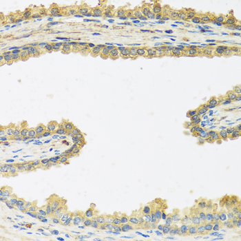
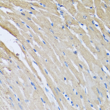
A06764 is listed for IHC-P and IF/ICC in human, mouse, and rat; supplied images show paraffin-section IHC in mouse and rat kidney (catalog applications/reactivity; IHC image captions).

Real IHC data Immunohistochemistry analysis of CRHBP in paraffin-embedded mouse kidney tissue using CRHBP pAb at a dilution of 1:300 (40x lens).
Anti-CRHBP Antibody
Cat # A06764

A06764 will render with a mouse kidney paraffin-section IHC figure at 1:300 (mouse IHC image caption). A second caption documents rat kidney paraffin-section IHC at 1:300; IF/ICC is listed without a supplied IF figure (rat IHC image caption; catalog applications; IF image alts).

Which to pick: Choose A06764 for paraffin-section tissue IHC based on its mouse and rat kidney figures; the fixative is unreported (IHC image captions). For IF/ICC, A06764 is the listed rabbit antibody, with an IF dilution range of 1:50–1:200 (catalog host, applications and IF dilution). For cross-species work, A06764 lists human, mouse and rat reactivity, with direct IHC image evidence for mouse and rat only (catalog reactivity; IHC image captions).

Each figure is that product's own IHC / IF validation image from its datasheet.

References

  1. UniProt Consortium. UniProt entry P24387 (CRHBP_HUMAN, Corticotropin-releasing hormone-binding protein).
  2. Human Protein Atlas. CRHBP tissue IHC expression (reliability: Approved).
  3. Human Protein Atlas. CRHBP subcellular location (ICC-IF): Secreted.
  4. Human Protein Atlas. CRHBP antibody validation summary (2 antibodies).
  5. Decreased CRHBP expression is predictive of poor prognosis in patients with hepatocellular carcinoma. Oncology letters 2018 — PMC6096283.
  6. Repeated exposure to neurotoxic levels of chlorpyrifos alters hippocampal expression of neurotrophins and neuropeptides. Toxicology 2016 — PMC4777610.
  7. Expression of genes related to the hypothalamic-pituitary-adrenal axis in murine fetal lungs in late gestation. Reproductive biology and endocrinology : RB&E 2010 — PMC2989976.
  8. Innovative Dual mRNA-Lipid Nanoparticle Therapy Targeting CRHBP and CFHR3 for Enhanced Treatment of Hepatocellular Carcinoma. International journal of nanomedicine 2024 — PMC11633302.
  9. PubMed PMID:1846945 — UniProt-cited evidence.
  10. PubMed PMID:8307998 — UniProt-cited evidence.
  11. PubMed PMID:15489334 — UniProt-cited evidence.