This website uses cookies to ensure you get the best experience on our website.


- Table of Contents
Plan chromogenic CRK IHC in paraffin sections using the reported cytoplasmic and membranous tissue pattern (HPA tissue IHC). Select positive control cells from the reported high-staining tissues (HPA tissue IHC), and check isoform coverage before scoring (UniProt).

Expected localisation, validated protocols, controls and antibodies — the at-a-glance facts below, then the full design guide.
| Expected localisation | Cytoplasmic and membranous staining (HPA tissue IHC) | |
| Staining pattern | Membranous and cytoplasmic staining in several tissues (HPA tissue IHC) | |
| Antigen retrieval | EDTA pH 8.0 HIER, heat-mediated (datasheet PB9101) | |
| Positive control | Duodenum+4 more · see all | |
| Negative control | Adipose tissue+4 more · see all |
| Fixation | Keep fixation conditions consistent across sections. (standard IHC practice; not target-specific) | |
| Caveat | Presumed off-target staining can confound scoring (HPA tissue IHC) | |
| Regulation | Adhesion recruits CRK to the plasma membrane (UniProt) | |
| Isoform / epitope | Two isoforms; verify antibody epitope coverage (UniProt) |
The catalog antibody protocol (datasheet: PB9101) is accompanied by four published CRK IHC protocols (PMC11896132; PMC4477307; PMC4471787; PMC3695334).
| Sample | Paraffin-embedded human lung cancer tissue; fixative not specified (datasheet PB9101) |
| Fixation | Image fixative and duration unreported (datasheet PB9101); verify before use. |
| Sectioning | 4–5 µm sections on charged slides (standard) |
| Deparaffinisation | Xylene, graded ethanol series to water (standard) |
| Antigen retrieval | Heat retrieval: EDTA pH 8.0 (datasheet PB9101); 20 min, 95–100 °C (standard) |
| Peroxidase block | 3% H2O2, 10 min, room temperature (standard) |
| Blocking | 10% goat serum (datasheet PB9101) |
| Primary antibody | Rabbit anti-CRK, 2-5μg/ml (datasheet PB9101) |
| Primary incubation | Overnight at 4 °C (datasheet PB9101) |
| Detection | HRP-conjugated secondary, DAB chromogen (datasheet PB9101) |
| Counterstain | Hematoxylin, blue, dehydrate and mount (standard) |
| Expected result | CRK-positive staining in glandular cells of duodenum (HPA tissue IHC: High). HPA tissue profile: Membranous and cytoplasmic expression in several tissues. No signal in the no-primary control. |
CRK should show membranous and cytoplasmic staining in several tissues (HPA tissue IHC). Expect strong staining in duodenal glandular cells, kidney tubular cells and hepatocytes (HPA: High). CRK has no transmembrane segment and can move to the plasma membrane during cell adhesion (UniProt P46108 topology and localisation). HPA rates its tissue IHC profile Approved, with medium consistency between staining and RNA data (HPA tissue IHC).
| Membranous and cytoplasmic staining in duodenal glandular cells or kidney tubular cells. | This matches the reported compartments and High staining in those cells (HPA tissue IHC). Judge the named cell population, since adjacent cells need not have the same intensity. |
| Predominantly nuclear staining in a tissue section, with little membranous or cytoplasmic signal. | This departs from the reported tissue IHC profile (HPA tissue IHC). Check specificity before calling it CRK; nucleoplasmic localisation is uncertain in ICC-IF and does not establish a nuclear IHC pattern (HPA subcellular). |
| Strong staining of adipocytes in adipose tissue or hematopoietic cells in bone marrow. | Those specific cell populations are reported as Not detected (HPA tissue IHC). Consider cross-reactivity or endogenous detection activity, and inspect a no-primary control (general IHC practice). |
| Diffuse chromogen over cells and tissue spaces, obscuring cell boundaries. | That distribution cannot be scored reliably against the membranous and cytoplasmic profile (HPA tissue IHC). It may reflect nonspecific detection or background; compare with a no-primary control (general IHC practice). |
| No convincing signal in duodenal glandular cells or hepatocytes. | Both are reported High (HPA tissue IHC). First assess whether the run detected antigen in a known-positive section; a blank section alone cannot establish absent CRK expression (general IHC practice). |
| Compartment and membrane association | CRK is cytoplasmic and can move to the plasma membrane during adhesion (UniProt P46108); tissue IHC reports both membranous and cytoplasmic staining (HPA tissue IHC). Membrane signal need not imply a transmembrane protein (UniProt P46108 topology). |
| Cell population and staining level | Hepatocytes and salivary glandular cells are High, while colon glandular cells are Medium (HPA tissue IHC). Compare the identified cells rather than assigning one intensity to an entire organ. |
| Tissue IHC evidence limit | The profile is Approved, with medium staining–RNA consistency; presumed off-target binding was observed and disregarded (HPA tissue IHC). Treat an unexpected pattern as a finding to validate, not as established CRK localisation. |
| Antibody-specific validation | CAB010485 is Approved for IHC, while HPA068087 is Approved for ICC and has no listed IHC rating (HPA antibodies). Validation in one application does not supply an IHC rating for the other antibody. |
| Isoforms and antibody recognition | Crk-II and Crk-I are listed isoforms (UniProt P46108). The supplied record gives no antibody epitope or isoform recognition data, so a difference between preparations cannot be assigned to an isoform from staining alone. |
| IF/ICC Q: Should its pattern match tissue IHC exactly? | No exact match is established: plasma membrane is the main approved ICC-IF location; actin filaments and basal body are additional approved locations, while nucleoplasm is uncertain (HPA subcellular). Interpret those images within ICC-IF, separately from the tissue IHC profile (HPA tissue IHC). |
| Situation | Likely cause | Next action |
|---|---|---|
| Known-positive cells are blank. | The section or detection run may have failed; High staining is reported in duodenal glandular cells and hepatocytes (HPA tissue IHC). | Run a known-positive section alongside the sample and confirm that the detection reagents and counterstain worked (general IHC practice). |
| Only nuclei stain strongly. | A nuclear-dominant tissue pattern does not match the reported membranous and cytoplasmic IHC profile (HPA tissue IHC). | Inspect a no-primary control and repeat with an IHC-validated antibody if available (general IHC practice; HPA antibodies). |
| An HPA Not detected cell population stains strongly. | Adipose-tissue adipocytes and bone-marrow hematopoietic cells are reported Not detected (HPA tissue IHC); cross-reactivity or endogenous activity is possible (general IHC practice). | Check a no-primary control and whether background follows the cells or the detection chemistry (general IHC practice). |
| Chromogen is diffuse across the section. | Background can obscure the cell boundaries needed to judge CRK's tissue pattern (HPA tissue IHC; general IHC practice). | Compare with a no-primary control, then review blocking, washes and detection development using the assay controls (general IHC practice). |
| Colon glandular cells look weaker than kidney tubular cells. | Colon glandular cells are Medium and kidney tubular cells are High in the HPA profile (HPA tissue IHC). | Score each named population against its reported level before treating the difference as an assay failure (HPA tissue IHC). |
| An ICC-IF image shows nucleoplasmic signal but the IHC section does not. | Nucleoplasmic ICC-IF localisation is uncertain, while tissue IHC reports membranous and cytoplasmic staining (HPA subcellular; HPA tissue IHC). | Evaluate each application with its own validated antibody and controls; do not use the uncertain ICC-IF location as an IHC acceptance criterion (HPA antibodies; HPA subcellular). |




Comprehensive Human Protein Atlas IHC scoring per tissue (reliability: Approved — Medium consistency between antibody staining and RNA expression data. Presumed off target binding observed and disregarded.). Rows are taken directly from the HPA tissue chart — click any row's HPA link to view the source.
| Tissue | Cell type | Level | Evidence | Source |
|---|---|---|---|---|
| Duodenum | Glandular cells | High | Protein (IHC) | HPA → |
| Kidney | Cells in tubules | High | Protein (IHC) | HPA → |
| Liver | Hepatocytes | High | Protein (IHC) | HPA → |
| Salivary gland | Glandular cells | High | Protein (IHC) | HPA → |
| Small intestine | Glandular cells | High | Protein (IHC) | HPA → |
| Tissue | Cell type | Level | Evidence | Source |
|---|---|---|---|---|
| Adipose tissue | Adipocytes | Not detected | Protein (IHC) | HPA → |
| Adrenal gland | Glandular cells | Not detected | Protein (IHC) | HPA → |
| Bone marrow | Hematopoietic cells | Not detected | Protein (IHC) | HPA → |
| Breast | Adipocytes | Not detected | Protein (IHC) | HPA → |
| Caudate | Glial cells | Not detected | Protein (IHC) | HPA → |
Troubleshoot CRK staining in paraffin sections by checking retrieval, compartment, cell identity and controls before comparing staining intensity.
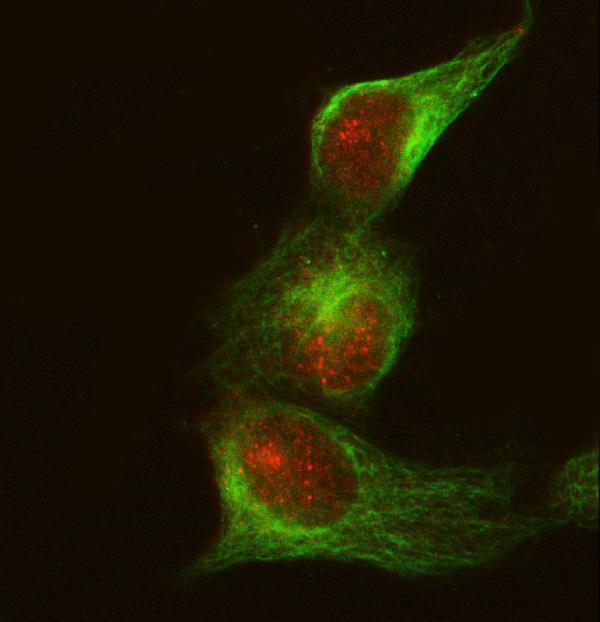
Anti-CRK antibodies have IHC images from paraffin sections of human lung cancer and mouse and rat brain (PB9101 image captions), plus an IF image from HeLa cells (M02533-3 image caption).
PB9101 has IHC images from paraffin sections of human lung cancer and mouse and rat brain (PB9101 image captions). M02533-3 has an IF image from HeLa cells (M02533-3 image caption) and is listed for ICC/IF (catalog applications).
Which to pick: Choose PB9101 for tissue IHC: it is listed for IHC in human, mouse and rat (catalog applications and reactivity), and its images show paraffin sections from all three species (PB9101 image captions). Choose M02533-3, a rabbit monoclonal, for IF/ICC (catalog: clone 24C53; catalog applications); its reactivity lists human, mouse and rat, while the supplied IF image shows HeLa cells (catalog reactivity; M02533-3 image caption). PB9101's captions support paraffin sections; the fixative is unreported (PB9101 image captions).