This website uses cookies to ensure you get the best experience on our website.


- Table of Contents
Plan CRKL staining in paraffin sections with the IHC-validated antibody at 1:50–1:200 (datasheet: M02100-2). Assess cytoplasmic staining in glandular cells while accounting for the uncertain tissue-IHC reliability (HPA tissue IHC).

Expected localisation, validated protocols, controls and antibodies — the at-a-glance facts below, then the full design guide.
| Expected localisation | Cytoplasmic staining in several tissues (HPA tissue IHC) | |
| Staining pattern | Cytoplasmic staining in glandular cells (HPA tissue IHC) | |
| Antigen retrieval | EDTA pH 8.0 HIER, heat-mediated (datasheet M02100-2) | |
| Positive control | Colon+4 more · see all | |
| Negative control | Adipose tissue+4 more · see all |
| Fixation | Keep fixation consistent across sections. (standard IHC practice; not target-specific) | |
| Caveat | Staining has low consistency with RNA expression (HPA tissue IHC) | |
| Regulation | Expression regulation not annotated (UniProt) | |
| Isoform / epitope | 0 isoforms; no processing variants annotated (UniProt) |
The catalog antibody uses EDTA pH 8.0 retrieval (datasheet M02100-2). Four published IHC protocols describe gastric, lung and breast tissue sections (PMC3388458; PMC3778295; PMC6679112; PMC5112787).
| Sample | Paraffin-embedded mouse brain tissue; fixative not specified (datasheet M02100-2) |
| Fixation | Image fixative and duration unreported (datasheet M02100-2); verify before use. |
| Sectioning | 4–5 µm sections on charged slides (standard) |
| Deparaffinisation | Xylene, graded ethanol series to water (standard) |
| Antigen retrieval | Heat retrieval: EDTA pH 8.0 (datasheet M02100-2); 20 min, 95–100 °C (standard) |
| Peroxidase block | 3% H2O2, 10 min, room temperature (standard) |
| Blocking | 10% goat serum (datasheet M02100-2) |
| Primary antibody | Rabbit monoclonal (clone 32C63) anti-CRKL, 1:50-1:200 (datasheet M02100-2) |
| Primary incubation | Overnight at 4 °C (datasheet M02100-2) |
| Detection | HRP-conjugated secondary, DAB chromogen (datasheet M02100-2) |
| Counterstain | Hematoxylin, blue, dehydrate and mount (standard) |
| Expected result | CRKL-positive staining in glandular cells of colon (HPA tissue IHC: High). HPA tissue profile: Cytoplasmic expression in several tissues. No signal in the no-primary control. |
In paraffin-section IHC, expect predominantly cytoplasmic CRKL staining in glandular cells of colon, fallopian tube, rectum, seminal vesicle, small intestine, and stomach (HPA tissue IHC: High; cytoplasmic expression in several tissues). CRKL has no transmembrane segment (UniProt P46109 topology). Treat this as a provisional pattern: HPA rates its tissue IHC reliability Uncertain because staining and RNA expression show low consistency (HPA tissue IHC: Uncertain).
| Cytoplasmic staining in glandular cells of colon or small intestine. | This matches a reported high-staining cell type and compartment (HPA tissue IHC: High; cytoplasmic expression). Score the glandular cells themselves, then compare staining with a negative control. The match supports interpretation but cannot establish antibody specificity alone because the tissue IHC profile is rated Uncertain (HPA tissue IHC: reliability). |
| Predominantly nuclear staining with little cytoplasmic staining in paraffin-section IHC. | This differs from the reported tissue IHC pattern (HPA tissue IHC: cytoplasmic expression). Check whether the apparent nuclear signal also occurs in a negative control, and review counterstain and detection background (general IHC practice). Nuclear staining is not inherently impossible: HPA reports nucleoplasm as the main location in ICC-IF, an observation from a different application (HPA subcellular: nucleoplasm, enhanced). |
| Strong staining in adipocytes or esophageal squamous epithelial cells. | Those cell types are reported as Not detected in the cited tissue IHC profile (HPA tissue IHC: adipose tissue; esophagus). Investigate cross-reactivity and endogenous chromogenic activity with appropriate negative controls (general IHC practice). Because the HPA profile is Uncertain, discordance is a reason to investigate, not proof that an individual stained cell is false (HPA tissue IHC: reliability). |
| Diffuse color covers cells and surrounding section without a clear cellular pattern. | A diffuse deposit cannot be confidently scored as CRKL localisation. Examine negative controls and distinguish localized stain from nonspecific background or endogenous detection activity (general chromogenic IHC practice). The expected tissue IHC observation is cellular, predominantly cytoplasmic expression, rather than uniform color across the section (HPA tissue IHC: profile). |
| No glandular-cell signal in colon despite an otherwise readable section. | Colon glandular cells are reported High, so absence warrants a run-quality check (HPA tissue IHC: colon). Confirm that the IHC-validated antibody and detection system work on a known-positive control; review retrieval and primary-antibody steps using the validated procedure (general IHC practice). A single negative result does not overturn an Uncertain tissue profile (HPA tissue IHC: reliability). |
| Tissue and cell-type context | Colon, fallopian tube, rectum, seminal vesicle, small intestine, and stomach glandular cells are reported High; bronchial respiratory epithelial cells are Medium (HPA tissue IHC). Use the named cell type when interpreting a section, since an organ-level positive label does not describe every cell in that organ (general IHC interpretation). |
| Evidence strength | The tissue IHC profile is Uncertain because antibody staining and RNA expression have low consistency (HPA tissue IHC: reliability). The listed IHC antibody HPA001100 is also marked Uncertain, whereas HPA000532 is marked Enhanced for ICC, a separate application (HPA antibodies). Keep these validation labels attached to their respective assays. |
| Topology and protein forms | CRKL has no transmembrane segment, signal peptide, or propeptide, and its annotated chain spans residues 1–303 (UniProt P46109 topology and processing). These annotations offer no basis for expecting a membrane-restricted or shed-protein pattern; they do not independently establish the observed tissue compartment (UniProt P46109; HPA tissue IHC: cytoplasmic expression). |
| Phosphorylation and epitope interpretation | UniProt lists phosphotyrosines at residues 127 and 207 (UniProt P46109 modified residues). The payload gives no antibody epitope or phospho-specific validation, so a stain cannot be read as phosphorylation status or used to infer that either modification changes antigen retrieval (UniProt P46109; supplied HPA antibody data). |
| IF/ICC Q&A: should the same compartment be expected? | HPA ICC-IF reports mainly nucleoplasmic CRKL, with additional cytosolic localisation (HPA subcellular: nucleoplasm enhanced; cytosol approved). This explains why an IF image may differ from the cytoplasmic tissue IHC profile (HPA tissue IHC: profile). Interpret each application with its own evidence; the ICC validation label does not validate a paraffin-section IHC result (HPA antibodies). |
| Situation | Likely cause | Next action |
|---|---|---|
| No signal in colon glandular cells. | A reported High cell type is negative (HPA tissue IHC: colon); a failed staining run or an unsupported biological difference remains possible. | Check a known-positive section, primary-antibody application, retrieval, and detection controls against the validated IHC procedure (general IHC practice). Record the discordance without calling colon universally negative because HPA rates the profile Uncertain (HPA tissue IHC: reliability). |
| Color appears mainly in nuclei on a paraffin section. | The distribution conflicts with the cytoplasmic tissue IHC profile, although ICC-IF reports a nucleoplasmic main location (HPA tissue IHC: profile; HPA subcellular). | Inspect negative controls and cellular boundaries, then document the nuclear pattern separately from the tissue IHC expectation (general IHC interpretation). Do not transfer the ICC-IF localisation or its Enhanced antibody label directly to IHC (HPA subcellular; HPA antibodies). |
| Adipocytes or esophageal squamous epithelial cells stain strongly. | Both cell types are reported Not detected in tissue IHC (HPA tissue IHC: adipose tissue; esophagus); cross-reactivity or endogenous detection activity may contribute (general IHC practice). | Compare with a negative control and assess whether color follows cells or background (general chromogenic IHC practice). Treat disagreement as a specificity question because the reference tissue profile is Uncertain (HPA tissue IHC: reliability). |
| Diffuse stain obscures glandular-cell boundaries. | Background or overdeveloped chromogen can prevent reliable localisation (general chromogenic IHC practice). | Review negative controls, blocking, detection, and development conditions using the validated procedure; score only interpretable cells (general IHC practice). A uniform deposit does not reproduce HPA's cytoplasmic cellular pattern (HPA tissue IHC: profile). |
| A reported High tissue and a reported Not detected cell type look equally stained. | The expected cell-type contrast is absent (HPA tissue IHC: High and Not detected entries); nonspecific signal or assay variation may explain it (general IHC practice). | Compare matched controls and cell-level staining before assigning positivity (general IHC interpretation). Preserve the HPA uncertainty when reporting the result, since its tissue IHC staining and RNA data have low consistency (HPA tissue IHC: reliability). |


Comprehensive Human Protein Atlas IHC scoring per tissue (reliability: Uncertain — Low consistency between antibody staining and RNA expression data.). Rows are taken directly from the HPA tissue chart — click any row's HPA link to view the source.
| Tissue | Cell type | Level | Evidence | Source |
|---|---|---|---|---|
| Colon | Glandular cells | High | Protein (IHC) | HPA → |
| Fallopian tube | Glandular cells | High | Protein (IHC) | HPA → |
| Rectum | Glandular cells | High | Protein (IHC) | HPA → |
| Seminal vesicle | Glandular cells | High | Protein (IHC) | HPA → |
| Small intestine | Glandular cells | High | Protein (IHC) | HPA → |
| Tissue | Cell type | Level | Evidence | Source |
|---|---|---|---|---|
| Adipose tissue | Adipocytes | Not detected | Protein (IHC) | HPA → |
| Adrenal gland | Glandular cells | Not detected | Protein (IHC) | HPA → |
| Breast | Adipocytes | Not detected | Protein (IHC) | HPA → |
| Caudate | Glial cells | Not detected | Protein (IHC) | HPA → |
| Esophagus | Squamous epithelial cells | Not detected | Protein (IHC) | HPA → |
Use the catalog antibody’s paraffin section result as a starting point, then assess compartment, cell type and controls before scoring CRKL staining.
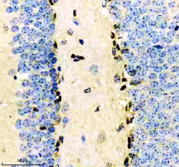
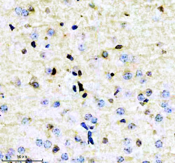
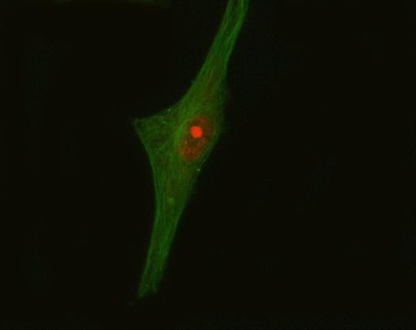
Anti-CRKL antibodies have IHC images from paraffin-embedded mouse and rat brain sections (M02100-2 image captions) and an IF/ICC image from human U87 cells (A02100-1 image caption).
M02100-2 has IHC images from paraffin-embedded mouse and rat brain sections and lists human, mouse and rat reactivity (M02100-2 image captions; catalog reactivity). A02100-1 has an IF/ICC image from U87 cells and lists human reactivity (A02100-1 image caption; catalog reactivity).
Which to pick: Choose M02100-2 for tissue IHC: it is a rabbit monoclonal listed for IHC, with paraffin-section images from mouse and rat brain (catalog: clone 32C63; M02100-2 image captions). It also lists human, mouse and rat reactivity for cross-species planning, although its supplied IHC images show mouse and rat tissue (catalog reactivity; M02100-2 image captions). Choose A02100-1 for human-cell IF/ICC because its image shows CRKL staining in U87 cells; the fixative is unreported in both products’ image captions (A02100-1 image caption; M02100-2 image captions).