CRYAA / Alpha-crystallin A chain · IHC design guide

Design Immunohistochemistry for CRYAA

Plan chromogenic CRYAA IHC in paraffin sections using cytoplasmic staining in lens fiber cells as the reference pattern (HPA tissue IHC). Account for heat shock–associated nuclear translocation when interpreting localisation (UniProt).

Evidence assembled Oct 2026 · For research use; verify linked source records and product datasheet before use
Immunohistochemistry protocol sheet for CRYAA (IHC for CRYAA): expected localisation Lens fiber cell cytoplasm (HPA tissue IHC), antibody PB9940, validated IHC image, and IHC protocol steps
Printable CRYAA IHC protocol sheet — expected localisation Lens fiber cell cytoplasm (HPA tissue IHC), antibody PB9940, controls and protocol steps. Open the full CRYAA IHC guide →

CRYAA Immunohistochemistry Experimental Design Guide

Expected localisation, validated protocols, controls and antibodies — the at-a-glance facts below, then the full design guide.

Must know before staining
Expected localisation Lens fiber cell cytoplasm (HPA tissue IHC)
Staining pattern Lens fiber cells show cytoplasmic staining (HPA tissue IHC)
Antigen retrieval Citrate pH 6 HIER, heat-mediated (datasheet PB9940)
Positive control ⓘ Eye
Negative control ⓘ Adipose tissue+4 more · see all
Important caveats
Reasons your staining may differ from the expected pattern.
Fixation Keep fixation consistent across sections (standard IHC practice; not target-specific). Selected-image fixative and duration unreported (datasheet PB9940); verify before use.
Caveat Heat shock can shift CRYAA into nuclei (UniProt)
Regulation Heat shock triggers nuclear translocation (UniProt)
Isoform / epitope 0 isoforms; chains end at aa 162–173, affecting C-terminal epitope coverage (UniProt)
Section 1

Recommended CRYAA IHC & IF Protocols

The catalog antibody’s IHC-P protocol (datasheet: PB9940) is accompanied by a published human lens capsule protocol (PMC6334984).

Recommended immunohistochemistry (IHC-P) protocol parameters
SampleParaffin-embedded mouse spleen tissues; fixative not specified (datasheet PB9940)
FixationImage fixative and duration unreported (datasheet PB9940); verify before use.
Sectioning4–5 µm sections on charged slides (standard)
DeparaffinisationXylene, graded ethanol series to water (standard)
Antigen retrievalHeat retrieval: Citrate pH 6, 20 min (datasheet PB9940)
Peroxidase block3% H2O2, 10 min, room temperature (standard)
Blocking10% goat serum (datasheet PB9940)
Primary antibodyRabbit anti-CRYAA, 0.5-1μg/ml (datasheet PB9940)
Primary incubationOvernight at 4 °C (datasheet PB9940)
DetectionStreptavidin-biotin complex (SABC), DAB chromogen (datasheet PB9940)
CounterstainHematoxylin, blue, dehydrate and mount (standard)
Expected resultCRYAA-positive staining in lens fiber cells of eye (HPA tissue IHC: Medium). HPA tissue profile: Cytoplasmic expression in lens. No signal in the no-primary control.
💡Decision noteStart with heat-mediated citrate retrieval at pH 6 (datasheet: PB9940); the published capsule excerpt does not specify retrieval (PMC6334984).
Section 2

What Is the Expected CRYAA Staining Pattern?

CRYAA is expected in the cytoplasm of eye lens fiber cells, where HPA reports medium IHC staining (HPA: tissue IHC, Supported). UniProt also lists cytoplasmic and nuclear localization, including movement into nuclear speckles during heat shock (UniProt P02489: subcellular location). It has no transmembrane segment, so a membrane-restricted pattern is unexpected (UniProt P02489: topology). HPA’s tissue rating has external support but no internal RNA correlation (HPA: tissue IHC reliability).

What am I looking at on my slide?
Lens fiber cells show cytoplasmic chromogenic staining, with signal clearly distinguishable from the counterstain.This matches the reported positive cell type, compartment, and medium staining level (HPA: eye tissue IHC). Judge the distribution across recognizable lens fiber cells as well as signal strength; HPA’s medium level is an observation, not a required intensity for every section (HPA: eye tissue IHC; general IHC practice).
A membrane outline dominates the lens fiber cell staining, with little cytoplasmic signal.A membrane-restricted pattern conflicts with the reported cytoplasmic tissue pattern and absence of a transmembrane segment (HPA: eye tissue IHC; UniProt P02489: topology). Review morphology and controls before interpreting it as CRYAA; a compartment mismatch alone does not identify the artifact’s cause (general IHC practice).
Strong staining appears in a cell population listed as not detected, such as adipocytes in adipose tissue.That pattern is discordant with the HPA tissue observation (HPA: adipose tissue IHC, not detected). Cross-reactivity or endogenous chromogenic detection activity are possible technical explanations; test them with controls before assigning a cause (general IHC practice).
Color is widespread across tissue and background, obscuring cell boundaries.A diffuse field cannot establish the lens fiber cytoplasmic pattern reported for CRYAA (HPA: eye tissue IHC; general IHC practice). Background can arise from nonspecific antibody binding or detection chemistry; inspect a no-primary control and the staining distribution before scoring cells (general IHC practice).
No convincing signal is visible in an adequately preserved eye lens section.This disagrees with the HPA observation of medium staining in lens fiber cells (HPA: eye tissue IHC). Check that lens fiber cells are present, then review antibody, retrieval, and detection performance using appropriate run controls; the result alone does not prove CRYAA absence (general IHC practice).
💡Expected CRYAA appearanceCall a section positive when lens fiber cells show discernible cytoplasmic staining consistent with HPA’s medium level; isolated membrane staining or strong signal in HPA-listed negative cell types is suspect (HPA: eye and negative-tissue IHC; UniProt P02489: topology).
How each factor affects the staining
Tissue and cell contextThe protein-level positive reference is eye lens fiber cells; HPA reports cytoplasmic, medium IHC staining there (HPA: eye tissue IHC). Several sampled non-lens cell types are listed as not detected, so their signal needs separate scrutiny (HPA: tissue IHC).
Compartment and topologyCRYAA has no transmembrane segment, while HPA tissue IHC places its observed lens signal in the cytoplasm (UniProt P02489: topology; HPA: eye tissue IHC). A membrane-only outline therefore does not match this reference pattern.
Nuclear interpretationUniProt reports nuclear translocation during heat shock and localization to nuclear splicing speckles (UniProt P02489: subcellular location). HPA ICC-IF also supports additional nucleoplasmic localization (HPA: subcellular ICC-IF). Nuclear signal needs context; it is not automatically an IHC artifact.
IHC antibody evidenceHPA lists IHC support for HPA038430 and ICC support for HPA037737 as separate antibody records (HPA: antibody validation). The tissue pattern and rating support interpretation of the reported IHC result; they do not validate every antibody or staining condition (HPA: tissue IHC and antibody validation).
IF/ICC?For IF/ICC interpretation, HPA reports mainly cytosolic and additionally nucleoplasmic localization, with images in Hep-G2, U-251MG, and U2OS (HPA: subcellular ICC-IF). Those images inform localization; they do not constitute an IHC-P protocol or establish a lens section intensity (HPA: subcellular ICC-IF; general IHC practice).
RNA versus proteinHPA lists group-enriched RNA in kidney and retina, while its supplied protein IHC positive is the eye lens (HPA: tissue RNA specificity and tissue IHC). Do not infer positive kidney or retina IHC from that RNA category alone (general IHC interpretation).
Why is my staining missing, weak or wrong?
SituationLikely causeNext action
Lens fiber cells are present, but the section has no visible CRYAA signal.The result conflicts with medium lens fiber staining reported by HPA; the specific failed step cannot be inferred from a blank section (HPA: eye tissue IHC; general IHC practice).Confirm lens fiber morphology, review the antibody’s IHC-P instructions, and check positive run controls, retrieval execution, and chromogenic detection (general IHC practice).
The entire slide has diffuse brown color, including regions without a clear cellular pattern.Nonspecific binding or chromogenic detection background can obscure the expected cytoplasmic lens pattern (general IHC practice; HPA: eye tissue IHC).Compare a no-primary control, inspect washing and blocking steps, and adjust antibody concentration or detection time using the validated IHC-P procedure (general IHC practice).
A HPA-listed negative cell type stains strongly.For example, adipocytes in adipose tissue are listed as not detected; cross-reactivity or endogenous enzyme activity may explain discordant chromogenic staining (HPA: adipose tissue IHC; general IHC practice).Verify cell identity and compare no-primary and detection controls. If using a peroxidase system, review the endogenous peroxidase blocking step (general IHC practice).
Lens staining is concentrated at cell borders rather than in the cytoplasm.The distribution differs from HPA’s cytoplasmic lens observation and from the non-transmembrane topology (HPA: eye tissue IHC; UniProt P02489: topology).Check section morphology and focus, then compare positive and no-primary controls before scoring border staining as specific (general IHC practice).
Nuclear staining accompanies cytoplasmic staining.Nuclear localization is biologically plausible: UniProt reports heat-shock-related nuclear movement, and HPA ICC-IF reports additional nucleoplasmic signal (UniProt P02489: subcellular location; HPA: subcellular ICC-IF).Record the nuclear and cytoplasmic patterns separately and compare controls and experimental context; do not classify nuclear signal as artifact solely by compartment (general IHC practice).
A researcher expects kidney or retina staining because of the RNA summary.The group-enriched kidney and retina result is RNA evidence; the supplied HPA protein IHC positive is eye lens fiber cells (HPA: tissue RNA specificity and tissue IHC).Use the lens fiber IHC observation as the positive protein reference. Treat any kidney or retina staining as a separate observation requiring its own controls (HPA: eye tissue IHC; general IHC practice).

Sample controls for CRYAA IHC & IF

🧪Run eye tissue first and assess lens fiber cells for staining (HPA: Medium in eye lens fiber cells). Use adipose tissue adipocytes as a negative comparison (HPA: Not detected in adipocytes); treat other cells on the eye section as internal background checks, since their CRYAA status is not specified in the supplied HPA row.
Positive control tissue: Eye (Lens fiber cells, HPA Medium)
Negative control tissue: Adipose tissue (HPA Not detected)
ICC-IF cell lines (HPA subcellular resource): HPA ICC-IF images show CRYAA in Hep-G2, U-251MG, U2OS, with annotated localisation: Cytosol (supported) (HPA subcellular).
Technical controls: Include a no-primary, secondary-only control and a host-matched rabbit IgG isotype control alongside a CRYAA knockout tissue control, if available (PB9940 caption: rabbit primary; standard IHC practice). Because the caption uses biotin-based detection with DAB, check endogenous biotin and peroxidase background on eye sections (PB9940 caption: biotinylated secondary, SABC and DAB; standard IHC practice).
⚠️Feasibility: No target-specific fixation window or fixation effect is reported, and the fixative in the PB9940 paraffin-section caption is unreported; fixation therefore cannot be assumed (PB9940 caption). Heat retrieval in citrate buffer at pH 6 for 20 minutes was used for mouse spleen, but retrieval dependence in eye tissue is unreported (PB9940 caption). Paraffin IHC has the supplied workflow, while frozen-section or IF ease is unestablished; if using IF, assess lens autofluorescence with an unstained section (PB9940 caption; standard IF practice). The selected PB9940 tissue-IHC caption documents paraffin sections, but does not specify the fixative (selected-SKU IHC image PB9940).

HPA tissue IHC evidence for CRYAA

Comprehensive Human Protein Atlas IHC scoring per tissue (reliability: Supported — External characterization data supports antibody staining but no internal RNA data available for correlation.). Rows are taken directly from the HPA tissue chart — click any row's HPA link to view the source.

Positive expression · recommended positive controls

TissueCell typeLevelEvidenceSource
Eye Lens fiber cells Medium Protein (IHC) HPA →

Undetected expression · recommended negative controls

TissueCell typeLevelEvidenceSource
Adipose tissue Adipocytes Not detected Protein (IHC) HPA →
Adrenal gland Glandular cells Not detected Protein (IHC) HPA →
Appendix Glandular cells Not detected Protein (IHC) HPA →
Bone marrow Hematopoietic cells Not detected Protein (IHC) HPA →
Breast Adipocytes Not detected Protein (IHC) HPA →
Section 3

Advanced CRYAA IHC Tips

Use the PB9940 paraffin-section workflow as the starting point, and interpret CRYAA staining against its lens expression and expected cellular location.

How should I retrieve CRYAA in paraffin sections when staining is weak?
Start with heat-mediated citrate retrieval at pH 6 for 20 minutes on paraffin sections (datasheet PB9940). The reported spleen staining used this retrieval condition, followed by 1 μg/mL primary antibody overnight at 4°C (PB9940 caption). If signal is weak, check that sections were fully deparaffinised and that the retrieval bath reached a consistent temperature before changing antibody concentration (standard IHC practice). Compare any adjusted retrieval time with the original condition on adjacent sections, because stronger staining can also increase tissue background (standard IHC practice). Include a lens section to judge whether the change preserves the expected cytoplasmic pattern (HPA: cytoplasmic expression in lens).
Can I infer the best fixative for CRYAA from the available IHC evidence?
The PB9940 spleen caption identifies paraffin sections but does not report a fixative, so CRYAA-specific fixation sensitivity is unknown (PB9940 caption). Record the fixative and fixation duration for each specimen, then compare sections processed with the same citrate pH 6 retrieval and antibody conditions (standard IHC practice; datasheet PB9940). If fixation varies across a series, assess morphology and staining intensity together before attributing a difference to CRYAA abundance (standard IHC practice). Use a lens control in each run to monitor the expected cytoplasmic signal, while recognising that this control cannot establish which fixative is optimal (HPA: cytoplasmic expression in lens; standard IHC practice).
Where should CRYAA staining appear, and how should I assess nuclear signal?
Expect prominent cytoplasmic staining in lens fiber cells; the tissue IHC profile reports medium staining there and describes cytoplasmic lens expression (HPA: Eye, lens fiber cells, Medium; HPA: cytoplasmic expression in lens). CRYAA is also reported in the nucleus and can move into nuclear speckles during heat shock, so nuclear staining is biologically plausible in a suitable context (UniProt P02489: subcellular location). Assess nuclear signal against the counterstain and nearby cytoplasm, rather than calling every dark nucleus positive (standard IHC practice). Document whether staining is diffuse or confined to discrete structures, and use controls before assigning a stress-related explanation (standard IHC practice; UniProt P02489: heat-shock translocation).
Could CRYAA processing or modifications change what this antibody detects?
The record lists 0 isoforms but identifies chains ending at residues 173, 172, 168 and 162 (UniProt P02489: isoforms and processing). It also reports a glycosylation site at residue 162 and modified residues including phosphoserine 45 (UniProt P02489: glycosylation and modified residues). Because the supplied evidence does not map the catalog antibody’s epitope, staining differences cannot be assigned to one processed chain or modification (supplied PB9940 evidence: epitope unspecified). If a sample stains unexpectedly, compare the same retrieval and detection conditions across controls before proposing epitope masking (standard IHC practice).
How should I adapt CRYAA localisation checks to a separate IF experiment?
In a separate IF experiment, pair CRYAA with a validated lens fiber cell marker so the expected cell type can be identified independently (HPA: lens fiber cell staining; standard IF practice). Choose spectrally separated fluorophores and inspect an unstained section in each channel; a far-red CRYAA channel may help if the specimen has shorter-wavelength autofluorescence (standard IF practice). CRYAA is cytoplasmic and can also be nuclear, with no transmembrane segment reported, so permeabilisation should provide access to intracellular epitopes (UniProt P02489: subcellular location and topology; standard IF practice). Optimise permeabilisation on matched samples and verify nuclear calls against the nuclear counterstain and single-label controls (standard IF practice).
How can I distinguish CRYAA signal from chromogenic background?
The PB9940 example used 10% goat serum blocking, 1 μg/mL primary antibody overnight at 4°C, and a biotin-based detection system with DAB (PB9940 caption). For diffuse brown staining, inspect no-primary and detection-only controls, then assess blocking, secondary binding and development time on matched sections (standard IHC practice). Include a peroxidase block when using a peroxidase and DAB workflow, and check whether endogenous biotin contributes signal with the chosen detection system (standard IHC practice; PB9940 caption: biotin-based detection and DAB). Compare background with the expected cytoplasmic lens pattern before raising the primary concentration (HPA: cytoplasmic expression in lens; standard IHC practice).
What is a defensible way to score CRYAA in chromogenic IHC? ⚠ ANSWER MARKED FOR VERIFICATION
Define the tissue compartment and eligible cells before scoring, using lens fiber cells as the reference population when lens is studied (HPA: Eye, lens fiber cells, Medium; standard IHC practice). Record the percentage of positive cells and staining intensity separately, or calculate an H-score from those observations using one threshold across the comparison (standard IHC practice). For spatial analyses, report positive-cell density per mm² of viable tissue and exclude folds, tears and necrotic regions by a prespecified rule (standard IHC practice). Normalise comparisons to the same cell population, sampled area, staining run and exposure to retrieval, rather than comparing raw brown area alone (standard IHC practice; datasheet PB9940: citrate pH 6 retrieval).
What would make an apparent CRYAA-positive area convincing or suspect?
A convincing result has cellular staining in an expected compartment, with cytoplasmic lens fiber cell staining providing the clearest tissue reference here (HPA: cytoplasmic expression in lens; HPA: Eye, lens fiber cells, Medium). Nuclear staining can occur, particularly with heat-shock-associated translocation, but requires clear nuclear localisation and appropriate controls (UniProt P02489: subcellular location; standard IHC practice). Treat staining restricted to section edges, folds, necrosis or endogenous enzyme-rich areas as suspect until it survives matched control comparisons (standard IHC practice). The PB9940 spleen image demonstrates staining under its stated conditions, but its caption alone does not establish the identity of every stained cell (PB9940 caption).
Boster reagents

Best CRYAA / Alpha-crystallin A chain IHC Antibodies

CRYAA catalog antibodies have IHC images from paraffin-embedded mouse spleen (PB9940 image caption) and an IF/ICC image from HepG2 cells (M01900-2 image caption).

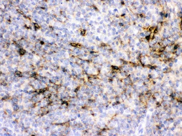
Real IHC data IHC analysis of CRYAA using anti-CRYAA antibody (PB9940). CRYAA was detected in paraffin-embedded section of mouse spleen tissues. Heat mediated antigen retrieval was performed in citrate buffer (pH6, epitope retrieval solution) for 20 mins. The tissue section was blocked with 10% goat serum. The tissue section was then incubated with 1μg/ml rabbit anti-CRYAA Antibody (PB9940) overnight at 4°C. Biotinylated goat anti-rabbit IgG was used as secondary antibody and incubated for 30 minutes at 37°C. The tissue section was developed using Strepavidin-Biotin-Complex (SABC)(Catalog # SA1022) with DAB as the chromogen.
Anti-Alpha A Crystallin/CRYAA Antibody ®
Cat # PB9940
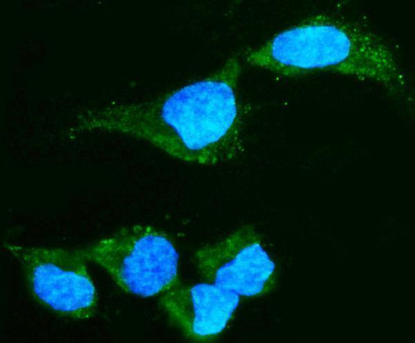
Real IF data IF analysis of Alpha A Crystallin using anti-Alpha A Crystallin antibody (M01900-2). Alpha A Crystallin was detected in an immunocytochemical section of HepG2 cells. Enzyme antigen retrieval was performed using IHC enzyme antigen retrieval reagent (AR0022) for 15 mins. The cells were blocked with 10% goat serum. And then incubated with 5 μg/mL mouse anti-Alpha A Crystallin Antibody (M01900-2) overnight at 4°C. DyLight®488 Conjugated Goat Anti-Mouse IgG (BA1126) was used as secondary antibody at 1:100 dilution and incubated for 30 minutes at 37°C. The section was counterstained with DAPI. Visualize using a fluorescence microscope and filter sets appropriate for the label used.
Anti-Alpha A Crystallin Antibody ® (monoclonal, 10B9)
Cat # M01900-2

PB9940 lists IHC for human, mouse and rat (catalog: PB9940); its IHC captions show paraffin-embedded mouse and rat spleen (PB9940 image captions). M01900-2 lists IF/ICC for human, mouse and rat (catalog: M01900-2); its IF caption shows HepG2 cells (M01900-2 image caption).

Which to pick: Choose PB9940 for tissue IHC: it lists IHC for human, mouse and rat (catalog: PB9940), and its own captions document paraffin sections of mouse and rat spleen (PB9940 image captions). Choose M01900-2 for IF/ICC: it is a mouse monoclonal, clone 10B9, with listed IF/ICC use and human, mouse and rat reactivity (catalog: M01900-2); its image documents HepG2 cells (M01900-2 image caption). For cross-species tissue IHC, start with PB9940 based on its listed reactivity (catalog: PB9940), while checking human tissue performance separately; its IHC captions do not report the fixative (PB9940 image captions).

Each figure is that product's own IHC / IF validation image from its datasheet.

References

  1. UniProt Consortium. UniProt entry P02489 (CRYAA_HUMAN, Alpha-crystallin A chain).
  2. Human Protein Atlas. CRYAA tissue IHC expression (reliability: Supported).
  3. Human Protein Atlas. CRYAA subcellular location (ICC-IF): Mainly localized to the cytosol. In addition localized to the nucleoplasm..
  4. Human Protein Atlas. CRYAA antibody validation summary (2 antibodies).
  5. Novel protein constituents of pathological ocular pseudoexfoliation syndrome deposits identified with mass spectrometry. Molecular vision 2018 — PMC6334984.
  6. Epigenetic regulation of αA-crystallin in high myopia-induced dark nuclear cataract. PloS one 2013 — PMC3849391.
  7. PubMed PMID:817940 — UniProt-cited evidence.
  8. PubMed PMID:8587135 — UniProt-cited evidence.
  9. PubMed PMID:8943244 — UniProt-cited evidence.