This website uses cookies to ensure you get the best experience on our website.


- Table of Contents
Plan chromogenic CRYAB IHC in paraffin sections using cytoplasmic staining as the expected tissue pattern (HPA tissue IHC). Compare heart muscle cardiomyocytes with adrenal gland glandular cells, which have no detected staining (HPA tissue IHC), and start catalog antibody A03496-2 at 2–5 μg/mL (datasheet A03496-2).

Expected localisation, validated protocols, controls and antibodies — the at-a-glance facts below, then the full design guide.
| Expected localisation | Observed cytoplasmic staining (HPA tissue IHC) | |
| Staining pattern | Cytoplasmic staining in glia and cardiomyocytes (HPA tissue IHC) | |
| Antigen retrieval | EDTA pH 8.0 HIER, heat-mediated (datasheet A03496-2) | |
| Positive control | Caudate+4 more · see all | |
| Negative control | Adrenal gland+4 more · see all |
| Fixation | Keep fixation consistent across sections. (standard IHC practice; not target-specific) | |
| Caveat | Heat shock can shift CRYAB to nuclear speckles (UniProt) | |
| Regulation | Expression varies by tissue (HPA tissue IHC) | |
| Isoform / epitope | No listed isoforms; one 1–175 chain (UniProt) |
The catalog antibody uses EDTA pH 8.0 retrieval (datasheet A03496-2). The published IHC methods below cover ovarian paraffin sections (PMC6579977) and frozen cerebral contusions (PMC8963874).
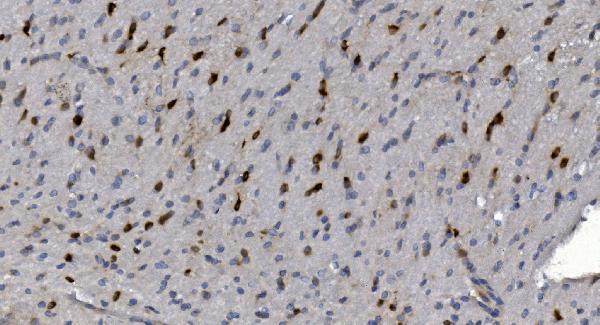
| Sample | Paraffin-embedded rat brain tissue; fixative not specified (datasheet A03496-2) |
| Fixation | Image fixative and duration unreported (datasheet A03496-2); verify before use. |
| Sectioning | 4–5 µm sections on charged slides (standard) |
| Deparaffinisation | Xylene, graded ethanol series to water (standard) |
| Antigen retrieval | Heat retrieval: EDTA pH 8.0 (datasheet A03496-2); 20 min, 95–100 °C (standard) |
| Peroxidase block | 3% H2O2, 10 min, room temperature (standard) |
| Blocking | 10% goat serum (datasheet A03496-2) |
| Primary antibody | Rabbit anti-CRYAB, 2-5μg/ml (datasheet A03496-2) |
| Primary incubation | Overnight at 4 °C (datasheet A03496-2) |
| Detection | Streptavidin-biotin complex (SABC), DAB chromogen (datasheet A03496-2) |
| Counterstain | Hematoxylin, blue, dehydrate and mount (standard) |
| Expected result | CRYAB-positive staining in glial cells of caudate (HPA tissue IHC: High). HPA tissue profile: Cytoplasmic expression in a subset of tissues including heart muscle, brain, thyroid gland, kidney and peripheral nerves. No signal in the no-primary control. |
In paraffin IHC, expect CRYAB mainly in the cytoplasm of selected cells, including cardiomyocytes, lens fiber cells and glia (HPA: Enhanced tissue IHC; High in these cells). CRYAB has no transmembrane segment, so a membrane-only IHC pattern needs scrutiny (UniProt P02511: topology). Nuclear localisation can occur during heat shock (UniProt P02511: subcellular location).
| Strong cytoplasmic staining in cardiomyocytes or lens fiber cells, with identifiable cell boundaries. | This matches reported high staining in both cell types (HPA: High in heart cardiomyocytes and eye lens fiber cells). Assess the named cells rather than assigning one intensity to the whole section, because CRYAB expression is reported in a subset of tissues (HPA: tissue IHC profile). |
| Prominent membrane-only or nuclear-only staining replaces the expected cytoplasmic IHC pattern. | Check whether the compartment assignment is reliable before calling the result positive: tissue IHC is described as cytoplasmic (HPA: tissue IHC profile). A nuclear signal is possible during heat shock (UniProt P02511: subcellular location), while ICC-IF also reports plasma membrane localisation (HPA: subcellular). Neither observation alone validates an unexpected paraffin IHC pattern. |
| Strong signal appears in adrenal glandular cells, appendix glandular cells or marrow hematopoietic cells. | These named cell populations are reported as not detected (HPA: tissue IHC). Review whether the signal follows cellular structure; unexplained staining may reflect antibody cross-reactivity or endogenous detection activity (standard IHC practice). A negative reference is specific to the reported cell type, not every cell in that tissue. |
| Uniform colour covers unrelated cells, empty spaces or the section edge. | Treat this as diffuse background until a cell-specific pattern is visible (standard IHC practice). True tissue staining is reported in selected cytoplasmic cell populations (HPA: tissue IHC profile). Compare with a no-primary control and inspect section edges before scoring weak signal (standard IHC practice). |
| No signal appears in an intact heart muscle or eye section. | Check the assay before concluding CRYAB is absent: cardiomyocytes and lens fiber cells are reported as high (HPA: tissue IHC). Confirm that those cells are present, then review antibody dilution, antigen retrieval and detection controls as general IHC workflow checks (standard IHC practice). |
| Cell type within the section | Compare like cells: glia in caudate, cerebral cortex and hippocampus are high, as are cerebellar Bergmann glia (HPA: tissue IHC). Kidney staining is reported in Bowman's capsule (HPA: tissue IHC); an organ-wide score could hide this distribution. |
| Compartment and biological state | The tissue profile is cytoplasmic (HPA: tissue IHC). CRYAB can enter the nucleus during heat shock and localise to Z-bands and intercalated disks in cardiomyocytes (UniProt P02511: subcellular location). Interpret unusual structure-specific staining in its cellular context. |
| Antibody validation | HPA lists Enhanced IHC validation for HPA057100, CAB002053 and CAB040560 (HPA: antibodies). That supports the reported tissue pattern but does not establish the performance or working dilution of a different catalog antibody. |
| Detection chemistry | Endogenous enzyme activity or nonspecific detection reagents can create colour unrelated to the primary antibody (standard chromogenic IHC practice). Match blocking and a no-primary control to the chosen detection system (standard IHC practice). |
| IF/ICC question: should the same compartment be expected? | HPA reports enhanced plasma membrane and cytosol localisation in ICC-IF, with images from A-431, U-251MG and U2OS (HPA: subcellular). Use that as context for IF interpretation; the tissue IHC profile remains cytoplasmic (HPA: tissue IHC). |
| Situation | Likely cause | Next action |
|---|---|---|
| Known-positive cardiomyocytes remain blank. | The assay may have insufficient detectable signal; cardiomyocytes are reported as high (HPA: tissue IHC). | Verify that cardiomyocytes are present and the detection control works. Review the catalog antibody's IHC-P instructions for dilution and retrieval, then adjust those variables within that workflow (standard IHC practice). No CRYAB-specific retrieval condition is supplied here. |
| The whole section is weakly coloured, including spaces without cells. | Diffuse deposition is consistent with background rather than the selected cellular pattern (standard IHC practice; HPA: tissue IHC profile). | Compare a no-primary control, inspect wash quality and review blocking appropriate to the detection chemistry (standard IHC practice). Score CRYAB only where staining resolves to identifiable cells. |
| An HPA-not-detected cell population stains strongly. | Cross-reactivity or endogenous detection activity is possible (standard IHC practice); HPA reports no signal in the specified population (HPA: tissue IHC). | Confirm the exact cell identity, compare a no-primary control and assess a reported high population in the same run (standard IHC practice; HPA: tissue IHC). Do not extend the negative designation to other cells in that organ. |
| A membrane rim dominates the paraffin IHC slide. | The reported tissue IHC pattern is cytoplasmic, although plasma membrane localisation is reported by ICC-IF (HPA: tissue IHC profile; HPA: subcellular). | Check focus and cellular boundaries, then compare cytoplasmic signal in a high-staining reference population (standard IHC practice; HPA: tissue IHC). Record the rim separately if it persists. |
| Nuclear staining dominates without clear cytoplasmic staining. | CRYAB can translocate to the nucleus during heat shock, but that condition cannot be inferred from staining alone (UniProt P02511: subcellular location). | Record the nuclear pattern and sample context. Check for cytoplasmic staining in an HPA-high population and compare controls before interpreting nuclear signal as CRYAB (HPA: tissue IHC; standard IHC practice). |
| Kidney or brain appears mixed when scored as a whole organ. | High staining is assigned to particular cells: Bowman's capsule in kidney and specified glial populations in brain (HPA: tissue IHC). | Score the named cell populations separately and document their compartment and intensity (HPA: tissue IHC; standard IHC practice). Avoid treating unstained neighbouring cells as a failed positive control. |


Comprehensive Human Protein Atlas IHC scoring per tissue (reliability: Enhanced — High consistency between antibody staining and RNA expression data.). Rows are taken directly from the HPA tissue chart — click any row's HPA link to view the source.
| Tissue | Cell type | Level | Evidence | Source |
|---|---|---|---|---|
| Caudate | Glial cells | High | Protein (IHC) | HPA → |
| Cerebellum | Bergmann glia - cytoplasm/membrane | High | Protein (IHC) | HPA → |
| Cerebral cortex | Glial cells | High | Protein (IHC) | HPA → |
| Epididymis | Glandular cells | High | Protein (IHC) | HPA → |
| Eye | Lens fiber cells | High | Protein (IHC) | HPA → |
| Tissue | Cell type | Level | Evidence | Source |
|---|---|---|---|---|
| Adrenal gland | Glandular cells | Not detected | Protein (IHC) | HPA → |
| Appendix | Glandular cells | Not detected | Protein (IHC) | HPA → |
| Bone marrow | Hematopoietic cells | Not detected | Protein (IHC) | HPA → |
| Breast | Adipocytes | Not detected | Protein (IHC) | HPA → |
| Bronchus | Respiratory epithelial cells | Not detected | Protein (IHC) | HPA → |
Use matched paraffin sections and cell-specific controls to troubleshoot CRYAB staining while keeping retrieval, detection, and scoring consistent.
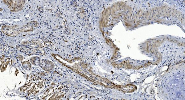
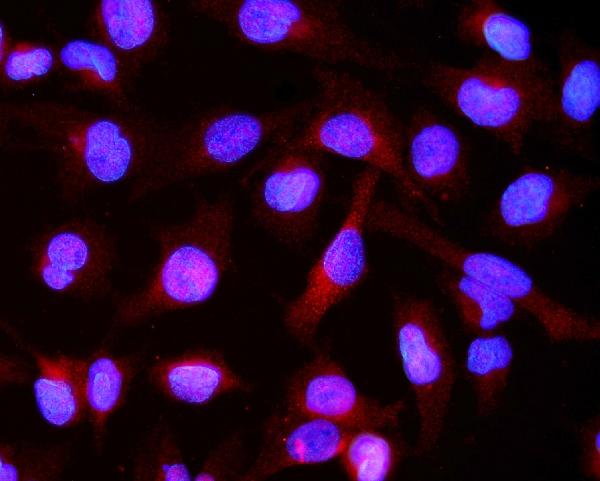
The IHC-validated antibody has paraffin-section images from rat brain and human cancer tissues, plus an IF image from U20S cells (A03496-2 image captions).
A03496-2 has IHC images from paraffin sections of rat brain and human lung, gastric, and skin cancer tissues, plus an IF image from U20S cells (A03496-2 image captions). M03496 lists IHC and ICC/IF applications with human and rat reactivity; no IHC or IF figure is supplied (M03496 catalog payload).
Which to pick: Choose A03496-2 for paraffin-section IHC because its own captions document those samples with 2 μg/ml primary antibody and EDTA retrieval at pH 8.0; the fixative is unreported (A03496-2 IHC image captions). Choose A03496-2 for IF/ICC when an imaged example matters: its U20S-cell IF caption uses 5 μg/ml; M03496 is a rabbit monoclonal listed for IHC and ICC/IF, but has no supplied figures (A03496-2 IF image caption; M03496 catalog payload). For broader listed species coverage, choose A03496-2, which lists human, monkey, mouse, and rat, versus human and rat for M03496; the imaged IHC examples cover human and rat (catalog reactivity; A03496-2 IHC image captions).