This website uses cookies to ensure you get the best experience on our website.


- Table of Contents
Plan chromogenic IHC for CSRP3 in paraffin sections using heart and skeletal muscle as positive tissues (HPA tissue IHC). The guide covers cytoplasmic staining expectations, fixation consistency, and interpretation of the two isoforms (HPA tissue IHC; UniProt).

Expected localisation, validated protocols, controls and antibodies — the at-a-glance facts below, then the full design guide.
| Expected localisation | Muscle cytoplasm observed; Z line expected (HPA tissue IHC; UniProt) | |
| Staining pattern | High cytoplasmic staining in cardiomyocytes and skeletal myocytes (HPA tissue IHC) | |
| Antigen retrieval | Tris-EDTA pH 9.0 HIER, 95–98 °C, 20 min (rule: nuclear antigen) | |
| Positive control | Heart muscle+1 more · see all | |
| Negative control | Adipose tissue+4 more · see all |
| Fixation | Keep fixation consistent across sections. (standard IHC practice; not target-specific) | |
| Caveat | Isoform coverage is unknown without antibody epitope data (UniProt; datasheet) | |
| Regulation | Isoform 2 is higher in muscle disease (UniProt) | |
| Isoform / epitope | Two isoforms; antibody epitope coverage is unspecified (UniProt; datasheet) |
Compare the catalog antibody’s IHC-P protocol (datasheet) with the published chromogenic IHC protocol for rat glioma tissue (PMC13223896).
| Sample | Paraffin-embedded mouse heart tissue; fixative not specified (datasheet A06522) |
| Fixation | Image fixative and duration unreported (datasheet A06522); verify before use. |
| Sectioning | 4–5 µm sections on charged slides (standard) |
| Deparaffinisation | Xylene, graded ethanol series to water (standard) |
| Antigen retrieval | Heat-induced epitope retrieval in Tris-EDTA buffer, pH 9.0, 20 min at 95–98 °C (standard rule: nuclear antigen) |
| Peroxidase block | 3% H2O2, 10 min, room temperature (standard) |
| Blocking | 10% normal serum of the secondary host, 30 min, room temperature (standard) |
| Primary antibody | Rabbit anti-CSRP3, 1:50-1:200 (datasheet A06522) |
| Primary incubation | Overnight at 4 °C (standard) |
| Detection | HRP-polymer secondary, DAB chromogen 5–10 min (standard) |
| Counterstain | Hematoxylin, blue, dehydrate and mount (standard) |
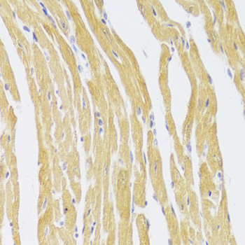
| Expected result | CSRP3-positive staining in cardiomyocytes of heart muscle (HPA tissue IHC: High). HPA tissue profile: Selective cytoplasmic expression in heart and skeletal muscle. No signal in the no-primary control. |
CSRP3 should stain mainly the cytoplasm of cardiomyocytes in heart muscle and myocytes in skeletal muscle; both are reported as high (HPA: tissue IHC, Enhanced reliability). Sarcomeric Z-line association can add an organized striated appearance, while nuclear staining may occur (UniProt P50461: localization). CSRP3 has no transmembrane segment, so a membrane outline is not the expected dominant pattern (UniProt P50461: topology).
| Strong cytoplasmic staining in cardiomyocytes or skeletal myocytes, with an organized pattern where structures are resolved. | This matches the high staining reported in both cell types (HPA: tissue IHC). A Z-line-associated pattern is biologically plausible, but routine chromogenic sections may not resolve individual Z lines (UniProt P50461: localization; general IHC practice). |
| A membrane outline or extracellular-looking signal dominates; alternatively, nuclei stain strongly with little cytoplasmic signal. | Dominant membrane staining conflicts with the absence of a transmembrane segment and mainly cytoplasmic localization (UniProt P50461: topology and localization). Nuclear signal alone is not conclusive evidence of artifact because CSRP3 can shuttle into the nucleus (UniProt P50461: localization); check controls and the full pattern. |
| Strong staining appears in cells reported as negative, such as adipocytes in adipose tissue or glandular cells in adrenal gland. | These cell types were not detected in the HPA tissue survey (HPA: tissue IHC). Unexpected signal can reflect antibody cross-reactivity or endogenous detection activity (general IHC practice); compare the negative tissue, no-primary control and cellular distribution before assigning a cause. |
| Color spreads across tissue or the slide without a clear myocyte-specific cytoplasmic pattern. | Diffuse background does not match the selective cytoplasmic profile (HPA: tissue IHC). Uneven reagent coverage, inadequate blocking or insufficient washing are general IHC possibilities (general IHC practice); background alone cannot establish CSRP3 expression. |
| No convincing staining appears in heart cardiomyocytes or skeletal myocytes. | Those are the supplied high-staining reference cells (HPA: tissue IHC). A negative section therefore needs a technical check before a biological interpretation; inspect tissue integrity, assay controls, retrieval and antibody application (general IHC practice). |
| Tissue and cell selection | Heart cardiomyocytes and skeletal myocytes are high-staining reference cells; the listed adipose adipocytes and adrenal glandular cells were not detected (HPA: tissue IHC). These comparisons help judge selectivity within the stated HPA survey. |
| Subcellular distribution | CSRP3 is mainly cytoplasmic and can associate with sarcomeric Z lines; it also shuttles to the nucleus (UniProt P50461: localization). Score the dominant pattern in its tissue context rather than treating every nuclear signal as impossible. |
| Antibody evidence | HPA042581 has Enhanced IHC validation (HPA: antibody validation). This supports its reported tissue pattern, but does not establish that every unexpected signal in a new section is specific (general IHC practice). |
| Isoforms and epitope | UniProt lists 2 CSRP3 isoforms; isoform 2 is expressed in striated muscle (UniProt P50461: isoforms and tissue specificity). The supplied evidence does not map the antibody epitope, so the IHC pattern cannot identify which isoform contributed signal. |
| IF/ICC Q&A: where should fluorescence appear? | Mainly in cytosol; nucleoplasm is an additional uncertain location in HPA ICC-IF images (HPA: subcellular ICC-IF). That uncertainty should not be converted into a requirement for nuclear staining in paraffin IHC. |
| Situation | Likely cause | Next action |
|---|---|---|
| Heart or skeletal muscle is blank despite an intact-looking section. | A technical failure remains possible because these myocytes are reported high in tissue IHC (HPA: tissue IHC); the image alone does not identify which step failed. | Check positive-control performance, antibody application, retrieval conditions and detection reagents using the established IHC workflow (general IHC practice). Reassess the myocytes before calling the sample CSRP3-negative. |
| Staining is largely nuclear, with weak or absent cytoplasmic signal. | Some nuclear localization is plausible, but CSRP3 is mainly cytoplasmic (UniProt P50461: localization). Counterstain overlap or nonspecific nuclear signal may complicate interpretation (general IHC practice). | Compare nuclear and cytoplasmic signal in the same cells and inspect a no-primary control (general IHC practice). Record limited nuclear signal separately; investigate a dominant nuclear-only pattern before accepting it. |
| Membranes or extracellular spaces look more positive than myocyte cytoplasm. | That distribution conflicts with CSRP3's mainly cytoplasmic localization and lack of a transmembrane segment (UniProt P50461: localization and topology). | Confirm the apparent compartment at suitable magnification and compare positive tissue with a no-primary control (general IHC practice). Treat a persistent membrane-dominant pattern as unexpected rather than scoring it as the expected CSRP3 result. |
| Cells in an HPA-negative tissue show substantial color. | The listed negative cell types were not detected in the HPA survey (HPA: tissue IHC). Cross-reactivity or endogenous detection activity are possible explanations, not diagnoses from appearance alone (general IHC practice). | Compare the staining with a no-primary control and the high-staining reference tissue (HPA: tissue IHC; general IHC practice). If signal persists only with primary antibody, assess whether its cell and compartment pattern is credible. |
| The whole section has diffuse color that obscures cell boundaries. | Diffuse background prevents assessment of the selective cytoplasmic pattern (HPA: tissue IHC). Blocking, washing and reagent handling can affect background in chromogenic IHC (general IHC practice). | Inspect the no-primary control, then review blocking, wash steps and chromogen development under the established assay conditions (general IHC practice). Score CSRP3 only after myocyte cytoplasm is distinguishable from background. |
| A faint positive-looking pattern appears, but the expected striations are unresolved. | Z-line association is reported for CSRP3, while routine section resolution and orientation can limit visible striation (UniProt P50461: localization; general IHC practice). Absence of visible lines alone does not negate cytoplasmic staining. | Judge signal against cardiomyocyte or skeletal-myocyte cytoplasm and matched background (HPA: tissue IHC; general IHC practice). Describe striations only when they are actually visible. |


Comprehensive Human Protein Atlas IHC scoring per tissue (reliability: Enhanced — High consistency between antibody staining and RNA expression data.). Rows are taken directly from the HPA tissue chart — click any row's HPA link to view the source.
| Tissue | Cell type | Level | Evidence | Source |
|---|---|---|---|---|
| Heart muscle | Cardiomyocytes | High | Protein (IHC) | HPA → |
| Skeletal muscle | Myocytes | High | Protein (IHC) | HPA → |
| Tissue | Cell type | Level | Evidence | Source |
|---|---|---|---|---|
| Adipose tissue | Adipocytes | Not detected | Protein (IHC) | HPA → |
| Adrenal gland | Glandular cells | Not detected | Protein (IHC) | HPA → |
| Appendix | Glandular cells | Not detected | Protein (IHC) | HPA → |
| Bone marrow | Hematopoietic cells | Not detected | Protein (IHC) | HPA → |
| Breast | Adipocytes | Not detected | Protein (IHC) | HPA → |
Troubleshoot CSRP3 staining in paraffin sections by checking retrieval, compartment, muscle cell identity, and controls before comparing signal intensity.
A06522 has real IHC data from paraffin-embedded mouse heart (catalog IHC image caption). Its listed reactivity is Human, Mouse and Rat (catalog reactivity); no IF image is supplied (catalog image fields).
A06522 is listed for IHC and WB, with Human, Mouse and Rat reactivity (catalog applications and reactivity). Its IHC image shows paraffin-embedded mouse heart stained at 1:100 after microwave retrieval in 10 mM PBS, pH 7.2 (catalog IHC image caption).
Which to pick: Choose A06522 for paraffin-section IHC because its own image documents that preparation in mouse heart; the fixative is unreported (catalog IHC image caption). No SKU in the payload is listed for IF/ICC or has an IF image, so there is no documented IF/ICC choice here (catalog applications and image fields). A06522 is the cross-species candidate based on listed Human, Mouse and Rat reactivity, although its image documents mouse tissue only; it is rabbit-hosted and its clonality is unreported (catalog reactivity, IHC image caption, host and clone fields).