This website uses cookies to ensure you get the best experience on our website.


- Table of Contents
Plan chromogenic CST1 IHC with salivary gland as a positive tissue and adipose tissue as a negative comparator (HPA tissue IHC). Expect cytoplasmic staining in a subset of glandular cells (HPA tissue IHC), start the catalog antibody at 1:100–1:300 (datasheet), and account for CST1 secretion when interpreting staining (UniProt).

Expected localisation, validated protocols, controls and antibodies — the at-a-glance facts below, then the full design guide.
| Expected localisation | Cytoplasm in a subset of salivary gland cells (HPA tissue IHC) | |
| Staining pattern | Subset of salivary glandular cells shows cytoplasmic staining (HPA tissue IHC) | |
| Antigen retrieval | Citrate pH 6.0 HIER, 95–98 °C, 20 min (rule: cytoplasmic / membrane antigen) | |
| Positive control | Salivary gland | |
| Negative control | Adipose tissue+4 more · see all |
| Fixation | Keep paraffin-section fixation consistent (standard IHC practice; not target-specific) | |
| Caveat | Secreted protein may appear away from its source cells (HPA tissue IHC) | |
| Regulation | Expression regulation not specified (UniProt) | |
| Isoform / epitope | No annotated isoforms; signal peptide 1–20 is cleaved (UniProt) |
The catalog antibody’s IHC-P protocol is accompanied by published CST1 IHC methods from four articles (PMC5777761; PMC10704982; PMC3741298; PMC6767558).
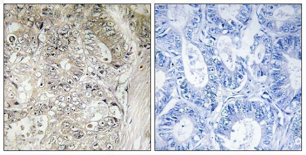
| Sample | Paraffin-embedded human colon carcinoma tissue; fixative not specified (datasheet A30630) |
| Fixation | Image fixative and duration unreported (datasheet A30630); verify before use. |
| Sectioning | 4–5 µm sections on charged slides (standard) |
| Deparaffinisation | Xylene, graded ethanol series to water (standard) |
| Antigen retrieval | Heat-induced epitope retrieval in citrate buffer, pH 6.0, 20 min at 95–98 °C (standard rule: cytoplasmic / membrane antigen) |
| Peroxidase block | 3% H2O2, 10 min, room temperature (standard) |
| Blocking | 10% normal serum of the secondary host, 30 min, room temperature (standard) |
| Primary antibody | Rabbit anti-CST1, 1:100 - 1:300 (datasheet A30630) |
| Primary incubation | Overnight at 4 °C (standard) |
| Detection | HRP-polymer secondary, DAB chromogen 5–10 min (standard) |
| Counterstain | Hematoxylin, blue, dehydrate and mount (standard) |
| Expected result | CST1-positive staining in glandular cells of salivary gland (HPA tissue IHC: Medium). HPA tissue profile: Cytoplasmic expression in a subset of cells in salivary gland. No signal in the no-primary control. |
CST1 is secreted and has no transmembrane segment (UniProt P01037 topology). In tissue IHC, expect cytoplasmic staining in a subset of salivary glandular cells, with medium staining reported in the positive cells (HPA tissue IHC). HPA rates the tissue pattern Enhanced for consistency with RNA expression, while cautioning that secreted proteins can have different RNA and protein locations and that the target may involve protein from more than one gene (HPA tissue IHC).
| A subset of salivary glandular cells shows cytoplasmic chromogenic staining (HPA tissue IHC). | This matches the reported CST1 tissue pattern; medium staining is the HPA reference level (HPA tissue IHC). Score the stained cells and their location, rather than expecting every glandular cell to stain (HPA tissue IHC). |
| Strong nuclear or crisp plasma membrane staining dominates the section. | That compartment pattern does not match the reported cytoplasmic staining or secreted topology (HPA tissue IHC; UniProt P01037 topology). Treat it as possible artefact and check controls before assigning it to CST1 (standard IHC practice). |
| Cells outside the expected salivary glandular subset stain prominently. | The pattern needs specificity review: cross-reactivity or endogenous detection activity may contribute (standard IHC practice). HPA also cautions that the tissue target may involve protein from more than one gene (HPA tissue IHC). |
| Color spreads broadly across cells, stroma, or the whole section without a clear cellular pattern. | Diffuse staining is difficult to score as CST1 when the reference pattern is cytoplasmic staining in a subset of glandular cells (HPA tissue IHC). Compare a reagent omission control and the distribution of stain (standard IHC practice). |
| No glandular cell staining is visible in a salivary gland section. | This conflicts with the reported positive tissue pattern, though only a subset of cells is expected to stain (HPA tissue IHC). Check that the sampled area contains glandular cells, then review detection and staining controls (standard IHC practice). |
| Secretion and topology (UniProt P01037) | CST1 is secreted, with no transmembrane segment (UniProt P01037 topology). Interpret a cellular signal against the reported cytoplasmic glandular pattern; do not require membrane outlining (HPA tissue IHC; UniProt P01037 topology). |
| Protein processing (UniProt P01037) | The precursor has a signal peptide at residues 1–20, and the annotated chain spans residues 21–141 (UniProt P01037 processing). The payload gives no antibody epitope, so it cannot predict which form an antibody detects. |
| Tissue and fluid distribution (UniProt P01037; HPA tissue IHC) | UniProt reports protein in submandibular and sublingual saliva, but not parotid saliva; it also reports saliva, tears, urine, and seminal fluid (UniProt P01037 tissue specificity). HPA's tissue IHC reference is salivary glandular cells (HPA tissue IHC). |
| IHC evidence and specificity (HPA tissue IHC; HPA antibodies) | HPA rates the tissue profile Enhanced and lists IHC status as Supported for HPA043706 and HPA044763, and Enhanced for CAB026769 (HPA tissue IHC; HPA antibodies). Its multi-gene target caution still matters when interpreting unexpected staining (HPA tissue IHC). |
| IF/ICC: what localization can be expected? (HPA subcellular) | HPA calls CST1 secreted but provides no main subcellular location or ICC-IF cell images (HPA subcellular). Use the tissue IHC cytoplasmic pattern as context, not as an established IF/ICC staining pattern (HPA tissue IHC; HPA subcellular). |
| Situation | Likely cause | Next action |
|---|---|---|
| Salivary gland appears entirely negative. | The sampled area may miss the reported positive subset, or the staining run may have failed (HPA tissue IHC; standard IHC practice). | Inspect glandular areas and a positive control; review antibody dilution, retrieval, and detection against the run's validated procedure (standard IHC practice). No CST1-specific retrieval condition is supplied. |
| An HPA-listed negative tissue shows prominent color. | HPA reports no detection in adipocytes of adipose tissue and glandular cells of adrenal gland (HPA tissue IHC). Unexpected color may reflect nonspecific staining or endogenous activity (standard IHC practice). | Compare tissue morphology and a reagent omission control; review blocking and detection controls before calling the color CST1 (standard IHC practice). |
| The whole slide has diffuse background. | Diffuse color lacks the reported subset-of-glandular-cells pattern (HPA tissue IHC); nonspecific reagent binding or detection background are possible (standard IHC practice). | Check a reagent omission control, blocking, washing, and detection development under the validated workflow (standard IHC practice). |
| Only nuclei or cell borders show strong staining. | This differs from HPA's cytoplasmic glandular pattern and UniProt's secreted, non-transmembrane annotation (HPA tissue IHC; UniProt P01037 topology). | Recheck morphology and controls, then compare with the expected cytoplasmic pattern before scoring the section (HPA tissue IHC; standard IHC practice). |
| Staining appears in many cell types across a section. | Broad cellular staining exceeds HPA's reported salivary glandular subset and warrants specificity review (HPA tissue IHC). Endogenous activity or cross-reactivity may contribute (standard IHC practice). | Compare the pattern with an omission control and a documented positive section; assess cell identity before assigning positivity (standard IHC practice). |
| An IF/ICC image is being used to confirm the IHC pattern. | HPA supplies no ICC-IF images or main subcellular location for CST1 (HPA subcellular). | Judge the paraffin IHC section against HPA's tissue pattern and IHC antibody validation; treat the IF/ICC image as separate evidence (HPA tissue IHC; HPA antibodies; HPA subcellular). |


Comprehensive Human Protein Atlas IHC scoring per tissue (reliability: Enhanced — High consistency between antibody staining and RNA expression data. Caution, targets protein from more than one gene. Secreted protein, tissue location of RNA and protein is expected to differ.). Rows are taken directly from the HPA tissue chart — click any row's HPA link to view the source.
| Tissue | Cell type | Level | Evidence | Source |
|---|---|---|---|---|
| Salivary gland | Glandular cells | Medium | Protein (IHC) | HPA → |
| Tissue | Cell type | Level | Evidence | Source |
|---|---|---|---|---|
| Adipose tissue | Adipocytes | Not detected | Protein (IHC) | HPA → |
| Adrenal gland | Glandular cells | Not detected | Protein (IHC) | HPA → |
| Appendix | Glandular cells | Not detected | Protein (IHC) | HPA → |
| Bone marrow | Hematopoietic cells | Not detected | Protein (IHC) | HPA → |
| Breast | Adipocytes | Not detected | Protein (IHC) | HPA → |
Use cell distribution and secretion biology to evaluate CST1 staining in paraffin sections, with antibody specificity and tissue processing assessed separately.
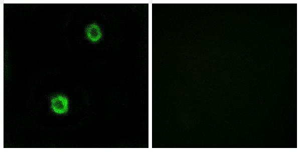
Anti-CST1 antibody A30630 has real IHC data from paraffin-embedded human colon carcinoma tissue and IF data from MCF7 cells (catalog IHC/IF captions); listed reactivity covers human, mouse and rat (catalog reactivity).
A30630 will render with an IHC image of paraffin-embedded human colon carcinoma tissue and a peptide-blocked comparison (catalog IHC image caption). Its IF image shows MCF7 cells and a peptide-blocked comparison; IHC, IF and ICC are listed applications (catalog IF image caption; catalog applications).
Which to pick: For tissue IHC, choose A30630: it is a rabbit polyclonal antibody with a listed IHC dilution of 1:100–1:300 and an image from paraffin-embedded human colon carcinoma tissue (catalog host/clonality/dilution; catalog IHC image caption). For IF/ICC, the same SKU lists both applications and a dilution of 1:200–1:1000, with IF shown in MCF7 cells (catalog applications/dilution; catalog IF image caption). For mouse or rat samples, A30630 lists reactivity to both species; the supplied IHC caption identifies human tissue, and its fixative is unreported (catalog reactivity; catalog IHC image caption).